Neuromuscular

Home, Search, Index, Links, Pathology, Molecules, Syndromes,
Muscle, NMJ, Nerve, Spinal, Ataxia, Antibody & Biopsy, Patient Info

MUSCULAR DYSTROPHY SYNDROMES 2

Limb Girdle Dystrophy
  Dominant (LGMDD)

    1: DNAJB6; 7q36
    2: TNPO3; 7q32
    3: HNRPDL; 4q21
    4: Calpain-3; 15q15
    5: Bethlem 1
      A. COL6A1: 21q22
      B. COL6A2: 21q22
      C. COL6A3: 2q37
    Bethlem 2
      COL12A1; 6q13
    MMCKR2: DTNA: 18q12
    Previous classification
Myopathies, Dominant
  1H: 3p23
  Ankle contract
  Bethlem 2: COL12A1; 6q13
  Central core: RYR1; 19q13
  Cytoplasmic body
  Distal myopathies
    MPD2: MATR3; 5q31
  Emery-Dreifuss
    Lamin A/C; 1q21
    SYNE1; 6q25
    SYNE2; 14q23
  Facioscapulohumeral
    1A: DUX4; 4q35
    1B: DUX4; 10qter
    2: SMCHD1; 18p11
  Myofibrillar (Desmin Δ)
  Myoglobin: MB; 22q12
  CMYP7A: MYH7; 14q11
  Myotonic
    DM1: DMPK; 19q13
    DM2: ZNF9; 3q21
  OPMD: PABP2; 14q11
  Oculopharyngodistal
  Tremor: MYBPC1; 12q23
Limb Girdle Dystrophy
  Recessive (LGMDR)

  1 (2A): Calpain-3; 15q15
  2 (2B): Dysferlin; 2p13
  3 (2D): α-Sarcoglycan; 17q21
  4 (2E): β-Sarcoglycan; 4q12
  5 (2C): γ-Sarcoglycan; 13q12
  6 (2F): δ-Sarcoglycan; 5q33
  7 (2G): Titin-Cap; 17q12
  8 (2H): TRIM32; 9q33
  9 (2I; MDDGC5): FKRP; 19q13
  10 (2J): Titin; 2q24
  11 (2K; MDDGC1): POMT1; 9q34
  12 (2L): ANO5; 11p14
  13 (2M; MDDGC4): Fukutin; 9q31
  14 (2N; MDDGC2): POMT2; 14q24
  15 (2O; MDDGC3): POMGnT1; 1p32
  16 (2P; MDDGC9): DAG1; 3p21
  17 (2Q): Plectin 1f; 8q24
  18 (2S): TRAPPC11; 4q35
  19 (2T): GMPPB; 3p21
  20 (2U): ISPD; 7p21
  21 (2Z): POGLUT1; 3q13
  22: COL6A2; 21q22
  23: LAMA2; 6q22
  24: POMGNT2; 3p22
  25: POPDC1; 6q21
  26: POPDC3; 6q21
  27: JAG2; 14q32
  28: HMGCR; 5q13
  29: SNUPN; 15q24
  Other
    Pompe (2V): GAA; 17q25
    2W: LIMS2; 2q14
    2Y: TOR1AIP1; 1q25
    Caveolin-3
    MDDGC12: POMK; 8p11
    Merosin (Laminin α2)
      23 (Absent): 6q22
      Reduced
      Abnormal: LGMD 2I
    MFM (2R): Desmin; 2q35
    PYROXD1: 12p12
  Myopathy +
    Cardiomyopathy
      Arrhythmia: POPDC1; 6q21
      Dilated: DPM3; 1q12
      Hypertrophic: TRIM63; 1p36
        Digenic: + Murf3 heterozygous
      Triangle tongue (2W): LIMS2; 2q14
    Contractures
      29: SNUPN; 15
      TOR1AIP1; 1q25
    Epilepsy: DPM2; 9q34
    Infant stiffness: CRYAB; 11q22
    Lipodystrophy: PTRF; 17q21
    MR & Eye (2T): GMPPB; 3p21
    Ophthalmoplegia: MYH2; 17p13
    Short stature & Ptosis: RAB3GAP2
Limb Girdle Dystrophy
  X-linked

  Barth: Tafazzin; Xp28
  Dystrophin; Xp21
    Becker
    Duchenne
  Emery-Dreifuss
    Emerin; Xq28
    FHL1: Xq26
  Manifesting carriers
    Dystrophinopathy
    Myotubularin
  McLeod: XK; Xp21.1
  Vacuolar
    Danon: LAMP-2; Xq24
      Scapuloperoneal
    XMEA: VMA21; Xq28

LGMD features
  General
  Muscle pathology

Muscle proteins
  Connective tissue
  Dystrophin & related
  Intermediate filaments
  Neuromuscular junction
  Nuclear envelope
  Structural & Contractile
Inherited myopathies: Other
  α-Dystroglycan Δ (MDDGC)
  Aggregates
    Cytoplasmic body
    Hyaline body: MYH7; 14q11; Dom
    KFS4: MYO18B; 22q12; Rec
    Myofibrillar (Desmin)
    Reducing body
    Spheroid body (Myotilin)
    Tubular
    Tubular arrays
    VMCQA: CASQ1; 1q23; Dom
  APECED: AIRE; 21q22; Rec
  Autophagy
    XMEA: VMA21; Xq28
    Multisystem: CLN3; 16p11; Rec
    Other
  Barnes myopathy: Dom
  Cardiac + Myopathy
    Cardiomyopathy-associated
    Cardiomyopathy (? LGMD1B)
    LGMD 1E: Desmin; 2q35; Dom
  Congenital
    Myopathies: Late-onset
    Muscular dystrophies (MDDG)
    EOM Δ: MYH2; 17p13; Rec
  Distal myopathies
  Ehlers-Danlos: Recessive
    TNXB; 6p21
    FKBP14; 7p14
  Fatigue & CK high: PACSIN3
  FSH dystrophy: Dom or Digenic
  Glycogenoses
  Glycosylation
  Hearing loss & Ovary Δ: GGPS1
  Inclusion Body (IBM)
    HMERF: TTN; 2q31; Dom
    IBM1: Desmin; 2q35; Dom
    IBM2: GNE; 9p12; Rec
    IBM3: MYH2; 17p13; Dom
    IBM4: 7q22; Dominant
    LGMD 1D: DNAJB6; 7q36; Dom
    Multisystem Proteinopathy (MSP)
      IBM + Paget
    TDP-43: 1p36; Dom
  Lipid
  Lysosomal
  Protein degradation
  Mitochondrial
  Myasthenia gravis, Familial
  Myotonic dystrophy
  Other dystrophies
  Respiratory failure
  Satellite cell Δ
    CFZ: MYMK
    LGMDR21: POGLUT1
    MYOSCO: PAX7
  Scapuloperoneal syndromes
  Skeletal + Myopathy: Dom
    Bone fragility: MTAP; 9p21
    MSP
      VCP; HNRNPA2B1; HNRNPA1
    Dysplasia
      Diaphyseal: TGFB1; 19q13
      Epiphyseal
        COL9A3; COL9A2; COMP
  Strongman: DCST2; 1q22; Dom


LGMD: General features

Inheritance
Myopathy: General features
Presenting features
Prevalence
Proteins: Subcellular location
Sarcoglycans
Weakness

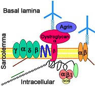
alpha-Sarcoglycan: LGMD 2D gamma-Sarcoglycan: LGMD 2C delta-Sarcoglycan: LGMD 2F beta-Sarcoglycan: LGMD 2E Dystrophin: Duchenne & Becker MD Laminin-alpha2: Congenital muscular dystrophy Integrin, alpha7: Congenital muscular dystrophy Connective tissue Agrin alpha-Dystroglycan beta-Dystroglycan alpha-Dystrobrevin Syntrophins Sarcospan Filamin 2 Filamin 2 Integrin, beta1 Dystrophy-associated proteins DAG complex +

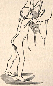
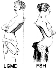
From Bramwell:
Atlas of Clinical Medicine


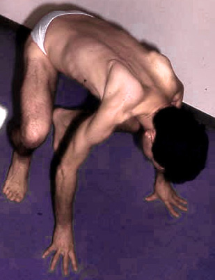
MD: Toe walking


  • Definitions
    • Dystrophy (Classic)
      • Tissue involved: Myopathy
      • Course: Progressive
      • Etiology: Genetic
    • LGMD 112
      • Tissue involved: Muscle
      • Etiology: Genetic
      • Weakness: Predominantly proximal
      • Serum CK: High
      • Muscle histology: "Dystrophic"
      • Course: Progressive
      • Atypical LGMD
        • 1A (Myotilin): Distal weakness
        • 1B (LMNA): Cardiac arrhythmias
        • 1C (CAV3): Mainly rippling muscles & Myalgias
        • 1E & 2R (Desmin): Distal weakness; Cardiomyopathy
        • 2V: Glycogen storage disease
  • Myopathies: General features
  • LGMD: Previous classification
    • LGMD1: Dominant
      • 1A: Myotilin; 5q31; Dysarthria
      • 1B: Lamin A/C; 1q21; + Cardiac
      • 1C: Caveolin-3; 3p25; Child onset
      • 1D: DNAJB6; 7q36
      • 1E: Desmin; 2q35
      • 1F: TNPO3; 7q32
      • 1G: HNRPDL; 4q21
      • 1H: ? 3p23
      • 1I: Calpain-3; 15q15
    • LGMD2: Recessive
  • Prevalence
    • LGMD general: 1 to 6 per 100,000
    • Recessive muscular dystrophies
      • Réunion Island, Guipuzcoa, North Indiana, Russia: LGMD 2A
      • Tunisia: LGMD 2C
      • US, Europe & Brazil: LGMD 2D
      • Brazil: LGMD 2E (8%); 2F (6%); 2G
      • Manitoba Hutterites of Canada: LGD 2H
      • Denmark & England: LGD 2I
      • Finland: LGD 2J
      • French-Canadian: LGD 2L; OPMD
      • Asia: OPDM
  • Genetics
    • Inheritance
    • Mutations: Copy number variants common 161
      • Types: Exons, 1 or Multiple; Whole gene
      • DMD, EMD, CAPN3, ANO5, SGCG,
          COL6A2, DOK7, LAMA2


LGMDD (LGMD 1): Dominant inheritance


LGMD D1 82
  DNAJ/HSP40 Homolog, subfamily B, Member 6 (DNAJB6) ; Chromosome 7q36.3; Dominant
LGMD D2 35
  Transportin 3 (TNPO3) ; Chromosome 7q32.1; Dominant
LGMD D3 128
  Heterogeneous nuclear ribonucleoprotein D-like protein (HNRNPDL; HNRPDL) ; Chromosome 4q21.22; Dominant
Myofibrillar myopathy 3 (MFM3)
  Myotilin (Titin immunoglobulin domain protein (TTID); MYOT) 19; Chromosome 5q31.2; Dominant or Sporadic

Myopathy, Dominant 17
  Lamin A/C ; Chromosome 1q21.2; Dominant
LGMD 1C (RMD2) 27
  Caveolin-3 ; Chromosome 3p25.3; Dominant, Also Recessive

Caveolin protein
LGMD 1C
  Allelic disorders
  Clinical
  Genetics
  Laboratory
  Mouse models
Caveolin-3: Located on
muscle fiber sarcolemma

Familial Dilated Cardiomyopathy with Conduction Defect & Muscular Dystrophy 81
  Desmin; Chromosome 2q35; Dominant
Myopathy with Sarcoplasmic Inclusions & Dilated Cardiomyopathy (MYOSB) 118
  Myoglobin (MB) ; Chromosome 22q12.3; Dominant
Myopathy with Myalgia, High CK ± Rhabdomyolysis 2 (MMCKR2), Dominant 155
  α-Dystrobrevin (DTNA) ; Chromosome 18q12.1; Dominant
Myopathy, Dominant 160
  MAM Domain-containing protein 2 (MAMDC2) ; Chromosome 9q21.12; Dominant
Myopathy, Dominant 77
  ? Chromosome 3p25.1–p23; Dominant

LGMDR (LGMD2): Recessive Inheritance


LGMD R1
  Calpain-3 (CAPN3; p94 protein) ; Chromosome 15q15.1; Recessive or Dominant

Recessive
  Genetics
  Calpain-3 protein
  Clinical
  Laboratory
  Muscle pathology

Dominant

From: C Angelini MD

  • Nosology: LGMD 2A
  • Epidemiology
    • 30% to 40% of LGMD
    • Most common recessive LGMD worldwide
      • Especially: Eastern Europe, Spain, Italy
      • Denmark: Less common than 2I (FKRP) & 2L (ANO5)
  • Genotype & Phenotype
    • Mutations: > 500 identified 7
      • Most common type: Missense
      • Mutation patterns
        • Private variant mutations in 70%
        • Recurrent mutations
          • Founder effect 1.5x more frequent than than recurrent
      • Single base pair: 60% to 70%
        • Missense: Most, ~80%
        • Stop codons: Few
        • Splicing defects: ~15%
      • Small insertions or deletions
        • Frequency: 30% to 40%
        • Most cause frameshift & stop codon
      • 1 Large genomic deletion
        • 2° to unequal recombination between two intragenic Alu elements
      • Overall ~45% of mutations are truncating
      • Location: Present through most of gene
        • Excess of mutations: Exon 21
        • Excess of point mutations
          • Exons 5, 11 (11 amino acid span with multiple CpG sites) & 21
        • Excess of deletions/insertions: Exons 15, 17, & 22
        • R490Q mutation
          • Located in catalytic site
          • Protein
            • Autolytic activity: Lost
            • CAPN3 protein present by Western blot
              • CAPN3 remains after incubation of muscle without EDTA 130
          • LGMD phenotype: Weakness severity = Variable
        • G222R mutation 54
          • Mutations: Compound heterozygote for R110X & G222R
          • Loss of function: Size & Abundance normal
          • Early onset weakness
      • Disease foci: Geography
        • Europe
          • Réunion island
            • 60% have IVS6-1G>A
            • Amish & Basque populations
            • Digenic inheritance: ??
          • Guipuzcoa (Basque Country, Spain)
            • Exon 22, 2362AG[→]TCATCT in 76%
          • Italy
            • Mocheni, Italian Alps: c.1193+6T>A
            • Venetian lagoon (Chioggia): Arg490Gln
          • Central & Eastern Europe
            • c.1746-20C>G; Mutation frequency 1%
          • Russia, Eastern Mediterrananean & North Italy
            • 550delA: Mutation frequency 1/150
        • North America
          • US Northern Indiana Amish: Arg769Gln
          • Tlaxcala, Mexico: Ala116Asp
        • Asia
          • China: c.2120A>G
          • Japan: c.1795_1796insA
          • Northern India: Asp780His, c.2099-1G>T, c.2051-1G>T, c.2338G>C
        • Large (31,012-bp) deletion (Exons 2-8) (c.309+4469_c.1116-1204del)
          • Occurs in different ethnic backgrounds
      • Clinical correlations
        • Severe phenotype
          • Homozygous null mutations: Often but not always
          • NS1 domain mutations; S86F
        • Least severe phenotype: Homozygous missense mutations
    • Allelic disorder: LGMD, Dominant
    • Mutation Databases
  • Calpain-3 (p94) protein 145
    • Localization
      • Expressed prominantly in skeletal muscle: N2-line region
      • Associates with
        • C-terminus of titin (connectin) in muscle
        • Filamin C
        • Calmodulin kinase IIβ (CaMKIIβ).
      • Subcellular: Nucleus ± Cytosol; Not membrane related
    • Substrates
      • Metabolic
      • Myofibrils (MLC1)
    • Functions
      • General
        • Ca++ activated non-lysosomal cysteine thiol-protease
        • Relies on Na+ activation
        • Cleaves proteins into short polypeptides, not single amino acids
        • Activity may not be Ca++ dependent
        • Use cysteine as active site residue
        • Limited proteolytic cleavage of substrate
      • Related to location in cell
        • Nuclear: Cell survival
        • Cytoplasm: Cell motility; Skeletal plasticity
      • Cytoskeletal remodeling
        • Assembly & remodelling of contractile proteins in sarcomere
        • Can bind & cleave titin
      • Control of Ca++-efflux from sarcoplasmic reticulum
      • Membrane repair
      • Muscle regeneration
      • Mutations: Reduced affinity of calpain-3 for titin
    • Half life
      • Short (< 10 minutes)
      • Rapid turnover related to insertion sequences (IS) not present in other calpains
      • Autolysis: Not dependent on Ca++ activation
    • Disease mutations: Effects on calpain function vary, may be selectve for
      • Proteolytic activity vs fodrin substrate: Reduced
      • Autolysis capacity: Reduced
      • Titin binding: Reduced
      • Altered maintenance & remodelling of contractile apparatus
        • Abnormal calpain-3 proteolysis & cleavage of titin
    • Expression in other disorders
      • Increased: FSHD; Melanoma, Vitiligo, Cataract (Age-related)
      • Reduced: Dystrophies (2B, 2J, Tibial, Duchenne, Ullrich); IBM; Rhabdomyolysis
  • LGD 2A: Clinical features 21
    • Onset
      • Early motor milestones: Most normal; Toe walking common
      • Range: 2 to 65 years; Median = 14 to 20 years; 71% between 6 to 18 years
      • Weakness: Proximal legs; Rectus abdominus
      • No clear sex differences; ? Females slightly younger than males
    • Disease patterns: Various phenotypes
      • General pattern
        • Weakness: Scapula (Symmetric), Pelvic girdle
        • Normal: Face
      • Differential Diagnosis: FSH; LGMD 2I
      • HyperCKemia, asymptomatic (14%): Male > Female 3:1
      • Limb girdle types (76%)
        • Early onset: < 12 year
          • Group with most homogeneous progression
          • Contractures more common
          • Null mutations: Common
        • Leyden-Mobius type
          • Onset 13 to 29 years
          • Weakness
            • Early: Pelvic-Femoral girdle
            • Later: Shoulders
        • Late onset
          • Age: > 30 years
          • Weakness: Pelvic girdle
      • Erb dystrophy type (10%): Scapular-Humeral phenotype
        • Onset age: 16 to 37 years
        • Weakness early: Shoulder girdle
        • Male = Female
        • Calpain-3 protein in muscle: Often normal
      • Miyoshi muscular dystrophy phenotype
    • Weakness
      • General
        • Symmetrical
        • Proximal + Milder Distal
        • Legs > Arms at onset: Most patients
        • May be asymmetric
        • Progressive
        • Severity
          • Heterogeneous: Mild to Early onset severe
          • Mild phenotype in majority
          • Male = Female
      • Trunk
        • Peri-Scapular
          • Latissimus dorsi; Serratus magnus
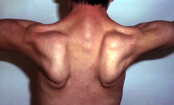
          • Scapular winging (83%): Symmetric
        • Rectus abdominus
        • Pelvic girdle
        • Gluteus maximus
      • Legs
        • Most severe: Adductors; Knee flexion
        • Ankle dorsiflexion: Some patients
      • Arms
        • Most severe: Shoulder adduction; Elbow flexion
        • Wrist extension: Some patients
      • Respiratory
        • Vital capacity: Declines over time; Rarely < 80%
        • No nocturnal hypoventilation
      • Quadriceps: May be selectively spared
      • Gait
        • Inability to walk on heels
        • Lumbar lordosis
        • Loss of walking: Mean late 2nd or 3rd decade
      • Normal: Face; Ocular & Bulbar
      • Some patients are asymptomatic with elevated serum CK levels
    • Muscle size
      • Atrophy
        • Early in posterior compartments of limbs
          • Different from Quadriceps wasting in Sarcoglycanopathies
          • May be apparent on CT
        • Most affected: Gluteus maximus & Thigh adductors
        • Pelvic; Shoulder; Proximal limbs; Trunk; May be scapuloperoneal
      • Hypertrophy
        • Some patients
          • Earlier in disease course
          • Especially in Brazil
        • Most common: Calf (Not prominent); Proximal
    • Contractures
      • Onset: May occur early in disease course
      • Calf: Toe walking may be presenting sign
      • Other: Elbow, Wrist & Finger flexion
      • Common late in disease progression: Rigid spine
      • May be severe & require surgery
      • Differential diagnosis: Emery-Dreifuss
    • CNS
      • Normal, or
      • Mild mental retardation: Average IQ = 77
    • Cardiac: No involvement
    • Progression
      • Slow: Earlier onset more rapid
      • Loss of walking
        • 10 to 30 years after onset
        • 50% in 3rd decade
        • Most by age 40 years
      • Many patients can still stand, even when in wheelchair
    • Syndrome variation
      • Intrafamilial
        • Similar onset age & severity in some families with null mutations
          • Onset at 15 years
        • Variation in others with
          • 2 Missense mutations or
          • Compound heterozygotes for missense & null mutation
          • Onset 2.5 to 45 years
      • Interfamilial: Prominent
  • LGD 2A: Lab features
    • Serum CK: Normal to 80 times normal; 190 to 11,000
    • MRI
      • Thigh: Varies with degree of function
        • Prominent in most patients: Posterior
        • Early involvement: Adductors; Semimembranosus
        • Restricted ambulation
          • Diffuse involvement: Posterolateral thigh & Vastus intermedius
          • Sparing: Vastus lateralis, Sartorius & Gracilis
      • Calf
        • Involvement: Soleus; Gastrocnemius, Medial head
        • Relative sparing: Gastrocnemius, Lateral head
    • Muscle Biopsy
      • Myopathic
        • Muscle fibers
          • Necrosis & Regeneration
            • Active myopathic changes
            • Eosinophils in some associated cell foci
            • Myopathic groups
              • May involve clusters or whole fascicles early in disease 34
          • Size: Variable; Small rounded to Hypertrophic
          • Internal architecture
            • Usually unremarkable
            • Regenerating fibers: Coarse internal architecture
            • Lobulation of Type I fibers: Later in disease course
            • Nuclei: Internal; Apoptosis with altered IκBα/NF-κB pathway
            • Groups of atrophic (type 2) muscle fibers
        • Endomysial fibrosis: Increased with longer disease course
      • Fiber types
        • Type 2 hypertrophy: Some patients
        • Type I predominance: With increasing weakness
        • Type 2C: Common
      • Inflammation
        • Some biopsies
        • May be perivascular or endomysial
      • Dystrophy-related proteins
        • Calpain-3: Western blot patterns
          • Loss of all calpain-3 bands (94 kDa, 60 kDa & 30 kDa)
            • High specificity for LGMD2A: Frequency 23%
            • May have null or missense mutations
          • Absence or reduction of 60 kDa bands
            • High specificity (94%)
            • Sensitive (64%)
          • Absence or reduction of 30 kDa bands
            • Specific (88%)
            • Sensitive (58%)
          • Increased lower MW 60 kDa region bands
            • No mutations (92%)
            • Suggests protein degradation artifact
          • Normal full-sized calpain-3 protein
            • Occurs in 23% of patients with 2 calpain-3 mutations
            • Often have loss of autocatalytic function
          • Abnormal Western blot with only 1 calpain-3 mutation
            • Similar phenotype to patients with 2 mutations found
          • Calpain-3 reduction may occur in other myopathies
        • Dysferlin: Reduced or absent in some patients; Western blot normal
        • Sarcoglycans & Dystrophin: Normal
      • Ultrastructure: Patchy damage to myofibrillar Z, I & A bands
    • Diagnosis 41
      • Genetics
        • Difficult due to many different mutations
        • More common in some regions of gene
          • 87% of mutant alleles in exons 1, 4, 5, 8, 10, 11 & 21
        • Some common mutations: 61% have one of eight mutations
        • Directed mutation analysis: Geographic groups
        • Some patients with only 1 detected mutation have
      • Western blot
      • Calpain-3 enzyme activity: Reduced
  • Mouse model: Calpain-3 knockout 42
    • Misaligned A-bands
    • Abnormal sarcomere formation
  • CAPN3 variant syndrome: LGMD, Dominant 4 (LGMD D4) 104
    • Nosology: LGMD 1I; LGMD D4
    • Epidemiology: 37 families
    • Genetics
      • Inheritance: Dominant
      • Mutations
        • In-frame deletions: c.643_663del21; c.598_612del15; c.759–761delGAA [Lys254del]
        • Missense: c.1333G>A [p.(Gly445Arg)]
        • S86F heterozygotes: Mildly high serum CK; Normal strength
        • Gly234Arg heterozygotes: Exertional myalgia
    • Clinical: Milder than recessive CAPN3 mutations
      • Onset age
        • Mean 34 years: 16 years later than Recesive LGMD 2A
        • Range: 13 to 84 years
      • Pain (50%)
        • Muscle
        • Back
        • Exertional
        • May occur without weakness
      • Weakness
        • Severity: Milder than LGMD 2A
        • Pattern: Similar to LGMD 2A
          • Paraspinal: Lumbar; Camptocormia
          • Leg: Proximal
          • Distal leg: Gastrocnemius, medial
          • Arms: Proximal
        • Normal strength: Some patients
        • Course: Variable
          • Severe disability: Some patients
          • Asymptomatic: Some patients
      • Muscle wasting: Proximal arms
    • Laboratory
      • Serum CK: High in 90%; Range 169 to 9,000
      • Muscle MRI: Fat replacement
        • Trunk: Paraspinal
        • Pelvis: Glutei
        • Thigh: Hamstring
        • Leg: Gastrocnemius, medial
      • Muscle pathology
        • Myopathic
          • Internal nuclei
          • Fiber size: Varied
          • Internal architecture: Lobulated
          • Necrosis: Scattered
          • Endomysial connective tissue: Increased
        • Calpain-3 protein: Reduced (< 15% of control levels) or Normal
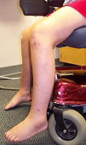
LGD 2A: Erb phenotype


From: C Angelini


LGMD R2
  Dysferlin ; Chromosome 2p13.2; Recessive

Gene
Protein
Clinical
  Variants
Laboratory
Pathology
Mouse models

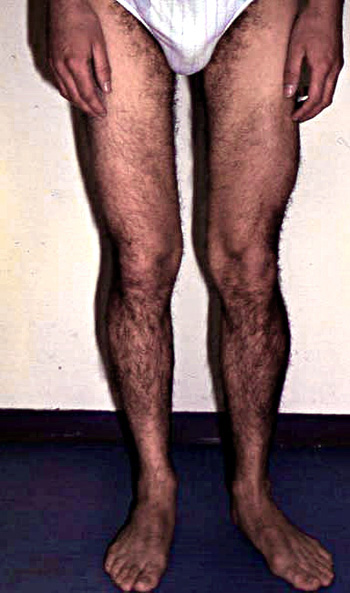
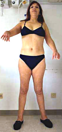
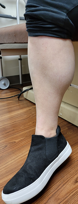
No calf hypertrophy

From: C Angelini

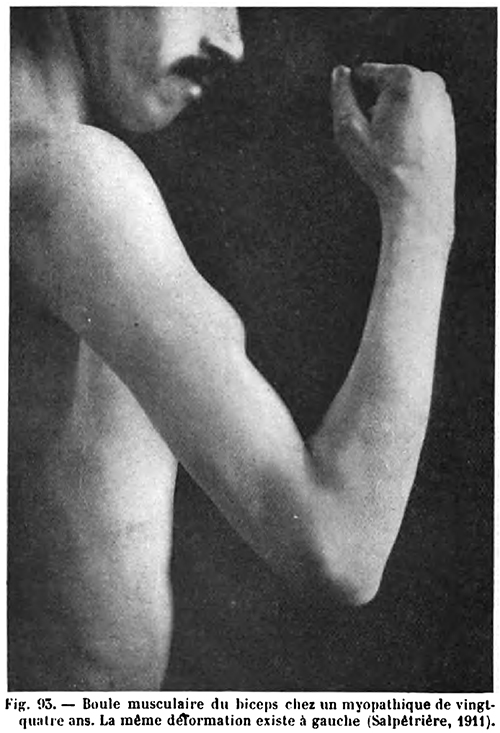
Boule du Biceps
From: Dejerine, J. Semiologie des affections du systeme nerveux.
  Paris: Masson et Cie (pub.) 1914
  • Nosology: LGMD 2B
  • Epidemiology
  • Genetics 25
    • Gene: 55 exons
    • > 450 different mutations described
      • Dysferlin deficiency: 18% with only 1 (heterozygous) mutation identified on 1 allele
    • Mutation types
      • Types: Missense (26% to 46%); Nonsense (18% to 26%); Deletions; Insertions; Intron; Splice
      • Distributed over entire coding sequence + Introns: Most mutations private variants
      • Most introduce stop codons or premature truncations of the dysferlin protein
      • Splice site (in-frame) mutation (A3443-33G) 53
        • May allow some residual dysferlin protein production
        • Clinical phenotype: Milder
    • Mutation locations in gene: Widely dispersed over coding sequence
    • Genotype-Phenotype
    • Geographic distribution
      • Mutations present at low frequency in many populations: Typically
        • 1% of recessive LGMD
        • 33% of distal myopathies
      • Common in Libyan Jews: 1624delG; Carrier frequency up to 10%
      • Japanese
        • G2997T (Trp999Cys); G3370T: Mild disease phenotype; Later onset
        • G3510A: More severe disease; Earlier onset; High CK
        • Other common mutations: 3746delG; 4870delT
        • 3373delG: Japanese Miyoshi
      • Sueca, Spain: Arg1905X 50
    • Allelic disorders
    • External links
  • Protein: Dysferlin 166
    • Tissues
      • Skeletal muscle > Heart
      • Ubiquitously expressed at low levels
    • Size & Forms
      • 237 kDa + Multiple lower weight bands
      • 55 exons
      • Isoforms: Alternative splicing at exon 40a
        • Inclusion of exon 40a: Produces calpain-cleavable site
        • Calpain cleavage at exon 40a produces: Mini-dysferlinc72
          • Regulates membrane repair
          • Damage-dependent production
    • Subcellular localization
      • Plasma membrane: Present at periphery of muscle fibers
      • T-tubules
      • Cytoplasmic vesicles: Trans-Golgi network secretory vesicles
      • Injury: Translocated to cytoplasm by endocytosis
    • Homology to
      • Nematode spermatogenesis factor (fer-1): Required for cell fusion
      • Myoferlin: Plasma membrane & Nuclear envelope
      • Otoferlin
    • Domains
      • Transmembrane: Short, C-terminal, hydrophobic
      • C2
        • 7 domains
        • Hydrophilic
        • Ca2+-dependent membrane binding
        • Bind lipids
      • Most of dysferlin protein intracellular
      • Extracellular domain: Small
    • Interactions 62
      • Protein complex
        • Caveolin-3
        • Mitsugumin 53 (MG53; TRIM72) : May play role in membrane repair
        • No direct interaction between Dysferlin & Caveolin-3
      • T-tubules
      • AHNAK
        • Interaction mediated by calpain-3
        • Levels reduced in dysferlinopathies
      • Annexins A1 & A2 : Regulate dysferlin localization
      • S100 Calcium binding protein A10
      • Calpain 3 (CAPN3)
      • β-Parvin
      • Tetraspanin CD82
      • Other: Ca++; Lipids
    • Functions
      • Membrane repair: Sarcolemma & T-tubules
        • General membrane actions
        • Ca++-triggered fusion of nearby vesicles
        • Provides source of membrane: To restore membrane barrier
        • Facilitates aggregation of exocytotic repair vesicles into repair patches
      • Ca++-mediated muscle membrane processes
        • Homeostasis processes
        • Signal transduction events
        • Excitation-Contraction coupling
        • Modulated molecules: L-type Ca++ channels; RyR
        • Buffers Ca++ in triadic cleft via C2A domain
      • T-tubule growth & morphology
        • May act synergistically with BIN-1
      • Tissue growth
      • Microtubule dynamics
    • Dysferlin turnover & levels
    • Dysferlin: LGMD 2B patients
      • Immunohistochemistry (IHC)
        • Sarcolemma: Absent or Reduced
        • Cytoplasm: Reduced or Increased
      • Western blot
        • Absence has more specificity for LGMD 2B than IHC changes
        • Dysferlin may be normal or increased
          • Some mutations produce cytoplasmic aggregates of normal sized dysferlin protein
      • Disease mechanism: Reduced sarcolemmal membrane repair
  • Clinical: Milder LGMD phenotype 85
    • Onset
      • Age
        • Mean 19 to 27 years
        • Range
          • General: 10 to 39 years
          • Congenital & 8th decade onsets reported
        • Heterogeneity in single family may occur
      • Leg weakness
        • Proximal
        • May be associated with period of exercise
    • Weakness
      • Mild
      • Asymmetry: Common
      • Legs
        • Distal: Posterior muscles
        • Proximal
          • Anterior & Posterior muscle groups
          • Especially adductors, also psoas
          • "Diamond on Quadriceps": Asymmetric 164
        • Lower limbs involved 9 years before upper limbs
      • Arms
        • Especially biceps
        • Deltoid: Often spared
      • Trunk: Glutei, Erector spinae, Shoulder girdle involved
      • Face: Generally normal
      • Loss of ambulation: > 30 years
      • Respiratory 167
        • More with longer disease duration
        • Overall: 50% have VC < 80%
      • Wide inter- & intrafamilial variation
      • Progression: Slow; Most walk until > 33 years, some into 6th decade
    • Calf size
      • Hypertrophy: Some patients
        • Deltoid hypertrophy with biceps atrophy
        • Posterior leg (Calf): May be early in disease course
      • Occasional distal onset patients
        • Subacute, painful enlarged calves early in course 60
    • Cramps & muscle discomfort: Some patients
    • No cardiomyopathy: Clinically preserved; EF generally > 50%
    • Same mutations may also produce Miyoshi distal myopathy
      • ? Modifier genes determine phenotype
  • Laboratory features
    • CK
      • Typical
        • Very high
        • Range: 10x to 72x normal; Up to 27,000
        • Lowest: Normal
      • Presymptomatic: 343 to 3,000
      • ? Age-dependent increase
    • EMG: Myopathic
      • Motor units: Small amplitude; Short duration
      • No spontaneous activity
    • Imaging
      • Early involvement on MRI or CT
      • Late or minimal involvement: Gracilis; Sartorius; Glutei
    • Skeletal Muscle
      • Pathology: Dystrophic Muscle
        • Necrosis & degeneration of muscle fibers
        • Endomysial connective tissue: Increased in more involved muscles
        • Muscle fibers: Size variation, may appear bimodal; Splitting
        • No vacuoles
        • Inflammation: Some mutations 22
          • Perimysial & perivascular cell infiltrates: Some muscles
          • T-lymphocytes
          • No MHC class I antigen on muscle fibers
        • Complement: Membrane attack complex (but not C3) on muscle fiber surface
        • Amyloid
          • Present in muscle in some patients
            • Mutation locations: N-terminal or other regions
          • Location: Muscle fiber surface; Endomysial connective tissue; Perivascular
          • Amyloid may contain dysferlin
        • Ultrastructure
          • Sarcolemmal defects
          • Replacement of membrane by layers of small vesicles 23
      • Dysferlin tissue staining 30
        • Levels
          • Absent, Reduced or Normal
          • Absent staining: More specific for LGMD 2B than reduced, but present, staining
          • Retained staining: May be related to splice site mutations in dysferlin gene
          • No correlation between level of staining and clinical severity
        • Staining may be patchy among muscle fibers
        • Reduced dysferlin staining: Differential diagnosis
          • Not specific for dysferlinopathy: 30% to 60% with no DYSF mutation
          • Other disorders: CAPN3 (10%); CAV3 (2%); GNE; MYH2; ANO5
      • Dysferlin Western blot
        • Common & specific change in LGMD 2B: Dysferlin reduced to 0% to 20% of normal
        • Absent dysferlin on Western blot: Dysferlin gene mutations very common
        • Good correlation on WB between results in
          • Skeletal muscle (234 kDa)
          • Monocytes (doublet 224 to 234 kDa)
        • Increased dysferlin expression: Mutatons in exons 36 or 37
      • Other molecules
        • Calpain-3: Reduced especially with C2 domain mutations
        • Sarcoglycans & Dystrophin: Present
        • Aquaporin-4: Reduced
    • Cardiac 167
      • EKG
        • P-wave abnormalities (58%)
        • Indicates delayed trans-atrial conduction
      • Left ventricle function: Normal
  • Variant syndromes: Different phenotypes may occur in same family
    • General
      • Late in disease course: Varying weakness phenotypes merge
    • Distal myopathy: Miyoshi
    • Distal myopathy: Distal Myopathy Anterior Tibial, Dysferlinopathy (DMAT; DACM)
      • Genetics
        • Mutation: Homozygous; 5966delG
      • Clinical
        • Onset: Foot drop; Steppage gait
        • Weakness
          • Legs
            • Anterior: Ankle > Knee
            • Distal > Proximal
          • Arms: Proximal
      • Muscle pathology: Absent dysferlin
    • Myopathy: Proximal + Distal
      • Onset: Proximal weakness; Calf atrophy
      • Weakness: More rapidly progressive
      • Muscle pathology: Inflammation more common
    • Myopathy: Later onset, Proximal, Arms > Legs 85
      • Genetics
        • Inheritance: Recessive
        • Mutation: Trp999Cys; Homozygous
      • Onset
        • Age: Mean 38 years; 16 years later than others average
        • Arm symptoms
      • Weakness: Proximal
      • Serum CK: High but lower than average
    • Pain syndrome
      • Onset: Distal leg (calf) painful swelling without weakness or atrophy
        • Unilateral or Bilateral
      • No weakness or atrophy
    • Asymptomatic with elevated serum CK levels
      • Frequency: 5% of dysferlinopathies
      • Mutations: Missense or Stop
      • Age: 28 & 50 years
      • MRI: Mild gastrocnemius pathology
    • Dysferlinopathy: Congenital myopathy 70
      • Genetics
      • Clinical
        • Onset age: Congenital
        • Weakness
          • Birth: Hypotonia
          • Legs & Neck flexor ± Arms
          • Walking: At 19 to 30 months
          • Motor milestones: Delayed
        • Contractures: Ankles & Hips; Onset 3 years
        • CNS: Normal
      • Laboratory
        • Serum CK: Up to 5000; Normal or mildly high early
        • MRI: Pathology (STIR) in hamstring & gastrocnemius
        • Muscle
          • Myopathic: Fiber size varied; Endomysial connective tissue increased
          • Dysferlin absent; α-dystroglycan normal
    • Dysferlin variant: Late onset MD with High CK 162
      • Epidemiology: 1 family, 4 patients
      • Genetics
        • Inheritance: Dominant
        • Mutation: c.6207del
      • Clinical
        • Onset age: 4th to 6th decade
        • Weakness: Legs, Distal & Posterior
        • Discomfort: Myalgias, Fatigue
      • Laboratory
        • Serum CK: 800 to 3,400
        • MRI: soleus, Gastrocnemius, medial
        • Muscle pathology
          • Immature fibers, scattered
          • Fiber sizes: Varied
          • Internal nuclei
          • Dysferlin staining: Reduced
    • Dysferlin: Mutation carriers 74
      • May be symptomatic
      • Mutations: G519R; D625Y
      • Clinical
        • Weakness: Legs; Proximal or Distal; Progressive
        • Calf myalgias: 1 patient
      • Serum CK: High; 600 to 3000
      • Muscle
        • Myopathy
        • Dysferlin expression: Reduced & Patchy
  • Mouse model: SJL
    • Mutation
      • Genomic: 141 bp intronic deletion involving tandem repeat
      • Effects of mutation
        • Alters 3' splicing site
        • Causes deletion of exon 45 in cDNA: 171-bp in-frame deletion
      • Protein results
        • Deletion of 57 amino acids
        • Reduced amount to 15% of normal
    • Pathology: Myopathy ± Inflammation
  • Mouse model: A/J
    • Retrotransposon insertion in intron 4 (5' end) of dysferlin gene
    • Progressive myopathy: Proximal > Distal
    • Perivascular inflammation
    • Dysferlin: Absent by Western blot


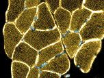
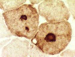
Dysferlin staining
in normal muscle

LGMD R5
  γ Sarcoglycan ; Chromosome 13q12.12; Recessive
LGMD R3
  α-Sarcoglycan (Adhalin; SGCA) ; Chromosome 17q21.33; Recessive

LGMD R4
  β-Sarcoglycan ; Chromosome 4q12; Recessive
LGMD R6
  δ-Sarcoglycan (SGCD) ; Chromosome 5q33.3; Recessive
LGMD R7 157
  Telethonin (Titin-Cap; TCAP) ; Chromosome 17q12; Recessive
LGMD R8: Manitoba Hutterite Dystrophy 113
  Tripartite-motif containing gene 32 (TRIM32) ; Chromosome 9q33.1; Recessive Sarcotubular myopathy 46
  Tripartite-motif containing gene 32 (TRIM32) ; Chromosome 9q33.1; Recessive
LGMD R9 (MDDGC5) 18
  Fukutin-related protein gene (FKRP) ; Chromosome 19q13.32; Recessive Myopathy with abnormal merosin (Laminin-2) 4
    Fukutin-related protein gene (FKRP) ; Chromosome 19q13.3; Recessive
LGMD R10
    Titin ; Chromosome 2q31.2; Recessive
LGMD R11 (MDDGC1) : with Mental retardation and Reduced α-dystroglycan 45
    O-mannosyltransferase 1 (POMT1) ; Chromosome 9q34.13; Recessive
LGMD R12 67
    Anoctamin 5 (ANO5, TMEM16E, GDD1) ; Chromosome 11p14.3; Recessive
LGMD R13 (MDDGC4) 59
    Fukutin (FCMD; FKTN) ; Chromosome 9q31.2; Recessive
Myopathy, Early-onset with Ophthalmoplegia 78
    Myosin, heavy chain 2, skeletal muscle, adult (MYH2) ; Chromosome 17p13.1; Recessive
Early-onset Myopathy with External ophthalmoplegia 78
  Myosin, heavy chain 2, skeletal muscle, adult (MYH2) ; Chromosome 17p13.1; Recessive
Myosclerosis (LGMD R22) 69
    Collagen, type VI, subunit α2 (COL6A2) ; Chromosome 21q22.3; Recessive
Limb Girdle Dystrophy + Mental Retardation (MDDGC9; LGMD R16) 79
  Dystrophin-associated glycoprotein 1 (Dystroglycan 1; DAG1) ; Chromosome 3p21.31; Recessive

LGMD R18: Limb Girdle Dystrophy with Movement Disorder & Intellectual Disability 89
  Transport (Trafficking) protein particle complex, Subunit 11 (TRAPPC11) ; Chromosome 4q35.1; Recessive

Other Dominant Myopathies


Bethlem Myopathies 1
  Bethlem Myopathy 1A (BTHLM1A) ; Collagen, type VI, α1 (COL6A1) ; Chromosome 21q22.3; Dominant
  Bethlem Myopathy 1B (BTHLM1B) ; Collagen, type VI, α2 (COL6A2) ; Chromosome 21q22.3; Dominant or Recessive
  Bethlem Myopathy 1C (BTHLM1C) ; Collagen, type VI, α3 (COL6A3) ; Chromosome 2q37; Dominant

Bethlem myopathy 2 (BTHLM2) 90
  Collagen, type XII, subunit α-1 (COL12A1) ; Chromosome 6q13-q14; Dominant

Multiple epiphyseal dysplasia with mild myopathy (EDM3) 13
  Collagen, type IX, subunit α-3 (COL9A3) ; Chromosome 20q13.3; Dominant
  Collagen, type IX, subunit α-2 (COL9A2) ; Chromosome 1p34.2; Dominant
  Cartilage oligomeric matrix protein (COMP) ; Chromosome 19p13.1; Dominant

Dominant Myopathy with Bone Fragility 51
  Methylthioadenosine phosphorylase (MTAP) ; Chromosome 9p21.3; Dominant
Hereditary Inclusion Body Myopathy 4 (HIBM4) 83
  Chromosome 7q22.1-31.1; Dominant
Dominant Myopathy with Cardiomyopathy 1
  Autosomal dominant
Dominant myopathy with Ankle contractures & High CK 10
  Autosomal Dominant
Barnes's Myopathy

Other Recessive Myopathies


Familial Limb-Girdle Myasthenia
    Dok-7 (C4ORF25) ; Chromosome 4; Recessive


MDDGC15: Myopathy ± Dilated Cardiomyopathy 75
  Dolichyl-phosphate mannosyltransferase 3 (DPM3) ; Chromosome 1q22; Recessive
LGMD 2W: Myopathy with Cardiomyopathy & Triangular Tongue (MDRCMTT) 91
  Lim and senescent cell antigen-like domains 2 (LIMS2; PINCH2) ; Chromosome 2q14.3; Recessive
LGMD R25: Myopathy with Cardiac Arrhythmias 103
  Popeye domain–containing 1 gene (POPDC1; BVES) ; Chromosome 6q21; Recessive
LGMD R26: Myopathy 121
  Popeye domain–containing gene 3 (POPDC3) ; Chromosome 6q21; Recessive
Myopathy with Contractures (LGMD 2Y; MRRSDC) 125
  Torsin A-interacting protein 1 (TOR1AIP1; LAP1B) ; Chromosome 1q25.2; Recessive
Myopathy with Contractures (LGMD R29) 163
  Snurportin-1 (SNUPN; RNUT1) ; Chromosome 15q24.2; Recessive
LGMD R21: Myopathy with Satellite cell loss 105
  Protein O-glucosyltransferase 1 (POGLUT1) ; Chromosome 3q13.33; Recessive
CMYO19: Myopathy with Scoliosis & Satellite cell loss 119
  Paired Box Gene 7 (PAX7) ; Chromosome 1p36.13; Recessive
Cardiomyopathy + Myopathy 115
  Tripartite motif-containing protein 63 (TRIM63; Murf1) ; Chromosome 1p36.11; Recessive
LGMD R27: Myopathy 142
  Jagged 2 (JAG2) ; Chromosome 14q32.33; Recessive Limb-Girdle Dystrophy R28 (LGMDR28; MYPLG) 154
  3-Hydroxy-3-Methylglutaryl-CoA Reductase (HMGCR) ; Chromosome 5q13.3; Recessive
Congenital disorder of glycosylation, Type Iu (CDG1U; DPM2-CDG): Myopathy with Epilepsy & Microcephaly
  Dolichyl-phosphate mannosyltransferase 2 (DPM2) ; Chromosome 9q34.13; Recessive
Myopathy with Lipodystrophy (Congenital generalized lipodystrophy, Type 4; CGL4) 76
  Polymerase I and transcript release factor (PTRF; Cavin-1; FKSG13) ; Chromosome 17q21.31; Recessive
Ehlers-Danlos with Myopathy 88
  Tenascin-XB (TNXB) ; Chromosome 6p21.33; Recessive
LGMD 4
    Autosomal Recessive
Metabolic myopathies
Childhood autophagic vacuolar myopathy 57
    Autosomal Recessive or X-linked carrier
Adult-onset Autophagic Vacuolar Myopathy with Multiorgan involvement 58
    Sporadic
Cardiomyopathy, Retinal degeneration & Epilepsy (CLN3) 93
    CLN3 Lysosomal/Endosomal transmembrane protein, Battenin (CLN3) ; Chromosome 16p11.2; Recessive
Mucolipidosis IV 140
    Mucolipin 1 (MCOLN1; ML1) ; Chromosome 19p13.2; Recessive

X-linked myopathies


X-linked Myopathy with Excessive Autophagy (MEAX; XMEA) 5, 16
  VMA21 vacuolar H+-ATPase homolog (VMA21; ATP6V0E1; LOC203547) ; Chromosome Xq28; Recessive
X-linked Vacuolar Cardiomyopathy & Myopathy (Danon disease) 122
  Lysosome-associated membrane protein 2 (LAMP-2) ; Chromosome Xq24; Dominant, more severe in males

Myofibrillar (Desmin -storage) Myopathies 3,11

MFM: Hereditary types
  Congenital
    Cap
      TPM2: 9p13
      TPM3: 1q22
      ACTA1: 1q42
    MD: SEPN1; 1p36
    MD + JEB: Plectin; 8q24
    Myopathy: CCDC78; 16p13
  Childhood
  MFM
    1 (LGMD 1E): Desmin; 2q35
    2: αB-crystallin; 11q22
    3 (LGMD 1A): Myotilin; 5q31
    4: ZASP; 10q23
    5: Filamin C; 7q32
    6: BAG3; 10q25
    7: KY; 3q22; Recessive
    8: PYROXD1; 12p12
    9: Titin; 2q31
    10: SVIL; 10p11; Recessive
    11: UNC45B; 17q12
    12: MYL2; 12q24
    MFM: ACTA1; 1q42
  Other
    Distal myopathy: HSPB8; 12q24
    DMRV: SQSTM1 + TIA1
    LGMD
      1B; LMNA; 1q21
      1D: DNAJB6; 7q36
      2Q: Plectin; 8q24
    Polyglugosan body: RBCK1; 20p13
    PYGM, Dominant: 11q13
    Rod myopathy: NEB; 2q23
    Respiratory Δ: 2q21
    Respiratory + Rigid spine: DNAJB4; 1p31
    Restrictive cardiomyopathy
    Scapuloperoneal: FHL-1; Xq26
MFM
  Clinical features
  Pathological features

Also see
  Contractures + Weakness: KY; 3q22
  Desmin knock-out mouse
  Inclusion body myositis
  Intermediate filament disorders


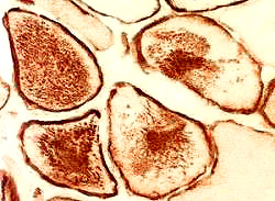
Desmin aggregates


Myofibrillar myopathies: General 114


Myofibrillar myopathies: Specific syndromes


Myofibrillar myopathy 1 (MFM1)
  Desmin ; Chromosome 2q35; Dominant, de novo or Recessive
  • Nosology
  • Gene mutations
  • Desmin protein features 43
    • Class III intermediate filament protein
    • Vimentin-like intermediate filament
      • Appearance follows vimentin in development
      • 53 kDa monomer
      • Domains: Head; Rod; Tail
      • Assembles into 10nM diameter filaments
      • Non-polar filament
    • Locations
      • Cytoplasm: Subsarcolemmal
      • Cell: Z-band encircled; NMJ; Myotendinoous junctions
    • Tissues: Muscle specific
      • Skeletal
        • 0.35% of total cell protein
        • Major intermediate filament in muscle
        • Increased amounts at
          • Myotendinous junctions
          • Neuromuscular junctions (Postsynaptic)
        • Development
        • Mutations
          • Levels of mutant protein: Vary within regions of single fibers
      • Cardiac
        • 2% of total cell protein
        • Location: Intercalated disks
      • Smooth
        • Location: Especially resistance-sized mesenteric microarteries
    • Functions
      • Overall: Links myofibrils to sarcolemma, nucleus & mitochondria
      • Contractile apparatus in muscle
        • Role in maintenance of structural & mechanical integrity
        • Force transmission
      • Mitochondrial function
        • Influences: Position, Movement & Respiratory activity
      • Myogenesis
        • Modulates cell adhesion & migration
        • Mechanochemical signalling
          • Between cell components, including mitochondria
      • Maintains structural integrity
        • Provides myofiber resistance to external applied mechanical forces
    • Desmin interactions: Binding to
    • Inclusions
      • Cytoplasmic, Spheroid, Mallory, Lewy bodies
      • Associated with: Actin; Ubiquitin;
          Heat shock proteins (α-B crystallin); Amyloid proteins
    • Mutant desmin
      • Abnormal assembly of intermediate filaments
      • Produces fragility of myofibrils
  • Myopathy + Cardiomyopathy (MFM1)
    • Genetics
      • Inheritance: Dominant
      • Mutations: Heterozygous; Ala337Pro; Leu345Pro; L370P
      • Mechanism: ? Dominant negative effect on filament formation
    • Clinical
      • General
        • Syndromes may involve skeletal muscle, heart or both
        • Smooth muscle involved in some syndromes
        • Inter- and intra-familial variability with same mutation
      • Skeletal myopathy
        • Onset: 20's & 30's
        • Early: Distal progressing to Proximal weakness
        • Patterns of weakness
        • Progression of weakness
          • Limbs; Neck; Pectoral; Bulbar & Facial
          • Disability: Wheelchair, walker or braces in 75%
          • Respiratory failure in some families
      • Cardiac involvement (60%)
        • EKG: Right bundle branch block; ST segment elevation
        • Syncopal episodes: Pacemaker often required (40%)
    • Laboratory
      • Serum CK: Mildly elevated in 50%
      • EMG: Myopathic; Fibrillations
      • MRI: Early involvement of peroneus lateralis, sartorius, gracilis, semitendinosus
      • Muscle pathology
        • Variable fiber size
        • Internal nuclei: Increased
        • Storage: Desmin, Vimentin & Nestin accumulate in muscle fibers
          • Location
            • Cytoplasmic
            • Subsarcolemmal & some sarcoplasmic aggregates
          • Accumulations also contain dystrophin
          • Aggregate shapes
            • May be different in type 1 & 2 muscle fibers
          • Ultrastructure: Granulofilamentous material
        • Occasional: Rimmed vacuoles; Small rods
  • Desmin variant syndrome: Distal myopathy, Cardiomyopathy & Sarcolemmal Vacuoles 98
    • Epidemiology: 2 families
    • Genetics
      • Inheritance: Dominant
      • Mutations: R454W (Tail domain); I402N
    • Clinical
      • Onset age: 3rd to 5th decades
      • Myopathy
        • Weakness
          • Distal > Proximal
          • Respiratory
          • Symmetric
        • Atrophy: Distal
      • Cardiac
        • Conduction block
        • Cardiomyopathy: Dilated
    • Laboratory
      • Serum CK: Mildly high; 280 to 810
      • EMG: Myopathic; Fibrillations, distal
      • NCV: Normal
      • Muscle
        • Fiber size: Varied; Clustered atrophy
        • Internal nuclei
        • Ensomysial connective tissue: Increased
        • Aggregates: Desmin
        • Cytoplasmic bodies
        • Vacuoles: Autophagic with Sarcoplasmic features (AVSF)
  • Desmin variant syndrome: LGMD 2R
    • Genetics
      • Inheritance: Recessive
      • Mutations: Compound heterozygosity (Type 3); Ala360Pro & Asn393Ile
      • Heterozygote carriers: Not involved
    • Clinical
      • Cardiac
        • A-V conduction block: Require pacemaker at ages 2 to 10
        • Cardiomyopathy onset: Childhood to 20's
      • Weakness
      • Skeletal
        • High arched palate
        • Scoliosis
    • Pathology
      • Hyaline/desmin plaques (Mallory body-like)
      • Amorphous subsarcolemmal material
        • Desmin- & dystrophin-immunoreactive
  • Desmin variant syndrome: Myopathy + Cardiomyopathy, Early onset 73
    • Epidemiology: 2 families
    • Genetics
      • Inheritance: Recessive
      • Mutations: In-frame deletion; Splice-altering
    • Desmin: Mutated protein
      • No assembly of desmin filaments
      • Reduced ability to form intermediate filament network
    • Clinical
      • Onset
        • Age: 6 months to 2 years
        • Syncope
      • Cardiomyopathy
      • Weakness: Progressive
      • Tendon reflexes: Reduced or Absent
      • Course: Progressive
    • Laboratory
      • CK: Up to 5,000
      • EMG: Complex repetitive discharges; Myotonic discharges; Fibrillations
      • EKG: Ventricular tachycardia, QT interval prolonged; 2nd degree AV block
      • Muscle
        • Desmin: Reduced; Aggregates, subsarcolemmal (Cap-like)
        • Small vacuoles
        • Fiber sizes: Varied
  • Desmin variant syndrome: Myopathy, Adult onset 87
    • Epidemiology: Turkish family
    • Genetics
      • Inheritance: Recessive
      • Mutation: c.1289-2A>G (Tail domain; Lamin B interaction)
      • Homozygous splice site mutation
    • Clinical
      • Onset age: 2nd or 3rd decade
      • Weakness
        • Distribution: Proximal; Symmetric; Face, mild
        • May be severe
        • Scapular winging
        • Course: Wheelchair in 2 decades
      • Contractures: Elbow
    • Laboratory
      • Muscle pathology
        • Fiber size: Varied
        • Necrosis: Occasional
        • Endomysial connective tissue: Increased
        • Type 2 predominance
        • Desmin staining: Present; No aggregates
      • Serum CK: Normal
      • EKG: Right bundle branch block
      • Cardiac echo: Normal
  • Desmin variant syndrome: Myopathy + NMJ disorder 120
    • Epidemiology: 5 families
    • Genetics
      • Inheritance: Recessive
      • Mutations: Loss-of-function; Splice site
    • Clinical
      • Onset age: Infancy (Fetal movements reduced) & Early Childhood
      • Weakness
        • Early onset: Motor milestones delayed
        • Generalized: Proximal > Distal
        • Face & Bulbar
        • Respiratory
      • Eye: Ptosis; Ophthalmoparesis
      • Fatigue
      • Cardiomyopathy: Left ventricular Hypertrophy; Some patients
      • Face: Long; Hypertelorism
      • Skeletal: Contractures; Scoliosis
      • Course: Slow progression
      • Treatment: Salbutamol (2 mg tid), Pyridostigmine; Partial response
    • Laboratory
      • EMG: Myopathic
      • RNS: Decrement
      • NCV: Normal or Motor axonopathy
      • Muscle
        • Myopathy: Internal nuclei, Abnormal structure, Clumped
        • Desmin: Absent or Reduced
        • Ultrastructure
          • Myofibril: Degeneration & Disorganization
          • Autophagic vacuoles
          • Rods
      • Serum CK: Up to 7,000
      • Muscle MRI: Gracilis sparing
  • Desmin variant syndromes: Cardiomyopathies
    • Types
    • Other features
      • Atrioventricular conduction abnormalities: Require pacemaker
      • Sudden death
      • Heart disease eventually in > 60% of desminopathies
      • Earlier onset in cases without skeletal myopathy

From C Weihl MD

Myofibrillar myopathy 2 (MFM2) (Type 1) 39
  αB-crystallin (CRYAB; HSPB5) ; Chromosome 11q23.1; Dominant or Recessive
Myofibrillar myopathy 4 (MFM4) 44
  ZASP (Lim domain-binding 3 (LDB3)) ; Chromosome 10q23.2; Dominant
Myofibrillar myopathy with arrhythmogenic right ventricular cardiomyopathy (ARVD7) 14
  Desmin (DES) ; Chromosome 2q35; Dominant

Desmin-associated restrictive cardiomyopathy 28
  Autosomal Dominant
Cardioneuromyopathy with Hyaline masses and Nemaline rods
  Autosomal Recessive
Desmin myopathy (Type 2)
  Autosomal Dominant
Congenital muscular dystrophy with Desmin inclusions
  Selenoprotein N, 1 (SEPN1) ; Chromosome 1p36-p35; Recessive
Myofibrillar myopathy 5 (MFM5) 47
  Filamin C (Filamin 2; FLNC) ; Chromosome 7q32.1; Dominant
Myofibrillar myopathy 6 (MFM6) 68
  BCL2-associated athanogene 3 (BAG3) ; Chromosome 10q25.2-q26.2; Dominant
Myofibrillar myopathy 10 (MFM10)
  Supervillin (SVIL) ; Chromosome 10p11.23; Recessive
CMYO21: Myofibrillar myopathy + Respiratory Failure & Rigid spine 152
  DNAJ/HSP40 homolog, Subfamily B, Member 4 (DNAJB4) ; Chromosome 1p31.1; Recessive
Desmin disorders: Other

Intermediate filaments & Disorders 20

General features
Proteins & Mutations


CYTOPLASMIC BODY MYOPATHIES (Also see Distal myopathies)

Severe limb-girdle: ? Recessive
Mild limb-girdle: Dominant
Desmin myopathies
Myopathy + Respiratory weakness
  Type 1: Titin; 2q31; Dominant
  Type 2: 2q21; Dominant
Distal: Dominant
  Gowers-Laing: MYH7; 14q12
Plectin deficiency
Congenital
  ACTA1: 1q42

Pathology
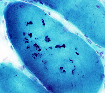
  Cytoplasmic bodies

Gomori trichrome

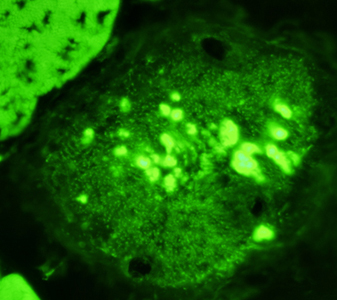
Phalloidin

Myopathies with Tubular Aggregates 172


General features
Pathology
Syndromes
  Myopathy, Proximal (Ca++ Homeostasis)
    TAM1: STIM1; 11p15; Dominant
    TAM2: ORAI1; 12q24; Dominant
    TAM (VMCQA): CASQ1; 1q23; Dominant
  Congenital Myasthenia (CMS)
    7B: SYT2; 1q32
    N-linked Glycosylation-related
      12: GFPT1; 2p13; Recessive
      13: DPAGT1; 11q23; Recessive
      14: ALG2; 9q22
      15: ALG14; 1p21
    Slow channel MG: AChR mutations
  Ion Channels
    Periodic paralysis
      Hypokalemic: CACNA1S
      Andersen: KCNJ2
    Exercise intolerance, CK high: RYR1; 19q13
    Myotonia: SCN4A; 17q23
    Paramyotonia congenita: Some patients
  Rhabdomyolysis & Cramps
    PGAM2; 7p13; Recessive
  Neuromuscular, Other
    Gyrate atrophy: OAT; 10q26; Recessive
  Occasional: Differential Diagnosis

Gomori trichrome

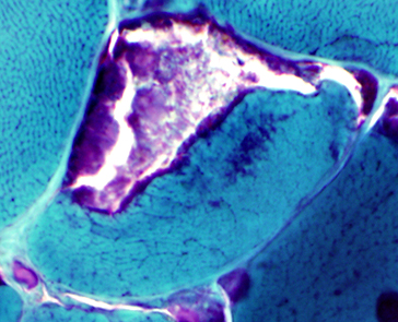
Tubular aggregates: General features Tubular aggregates: Proximal myopathy syndromes Tubular aggregates: Other Neuromuscular Disorders Tubular aggregates: Other occasional associations Tubular Aggregates: Mouse models
  • SLC6A8-/y male mouse 137: Creatine deficiency
    Myopathy with Tubular Arrays 48
    Myopathy, vacuolar, with CASQ1 aggregates (VMCQA) 94
      Calsequestrin 1 (CASQ1) ; Chromosome 1q23.2; Dominant

    Myopathy + Hearing loss & Ovarian Failure (MDHLO) 127
      Geranylgeranyl Diphosphate Synthase 1 (GGPS1) ; Chromosome 1q42.3; Recessive

    Myopathy, Childhood onset with Fatigue & High CK 168
      Protein kinase C and casein kinase substrate in neurons 3 (PACSIN3; Syndapin III) ; Chromosome 11p11.2; Recessive

    Congenital Myopathy Syndromes: Late onset or progressive
    Animal models with Myopathy
    Also see: Other Dystrophies


    Return to Myopathy & NMJ Index

    References
    1. Ann Neurol 1996;39:636-642
    2. Current Opinion in Neurology 2000;13:511-517
    3. Current Opinion in Neurology 1997;10:426-429
    4. Brain 1998;121:581-588
    5. Ann Neurol 1988;23:258-65; Neuromuscular Disorders 2001;11:376-388
    6. AJHG 1999;64:788-792
    7. AJHG 1999:64:1524-1540, Hum Mutat 2022;43:1347-1353
    8. Human Molecular Genetics 1999;8:1329-1336
    9. Neuromuscular Disorders 1999;9:308-312
    10. Acta Neurol Scand 1999;100:199-201
    11. Ann Neurol 1999;46:681-683
    12.Neurology 1999;52:861-863
    13. PNAS 2000;97:1212-1217, Am J Med Genet Part A 152A:863–869, Hum Mutat 2005;25:593-594
    14. Ann Neurol 1999;46:684-692, European Journal of Human Genetics 2012; Mar
    15. FASEB J 2000;14:761-768, Ann Neurol 2008;64:294–303
    16. Europ J Hum Genet 2000;8:125-129, Neurology 2002;59:596–601, Acta Neuropathol 2013 Jan 12
    17. Human Mol Genet 2000;9:1453-1459, Hum Mutat 2003;21:473-481
    18. Neuromuscular Disorders 2000;10:240-246, Hum Mol Genet 2001;10:2851-2859, Ann Neurol 2003;53:537-542, J Neuromuscul Dis 2023;10:615-626, Neuromuscul Disord 2023;33:119-132
    19. Hum Mol Genet 2000;9:2141-2147; Brain 2005;128:2315–2326
    20. Curr Opin Cell Biol 2000;12:79-90, N Engl J Med 2004;351:2087-2100
    21. Neuromuscular Disorders 2001;11:287-296, Pediatric Neurology 2001;24:235-237, Brain 2005;128:732-742, Brain 2007;130:3237-3249
    22. Am J Med Genet 2000;91:305-12
    23. Neurology 2001;56:1472-1481
    24. Acta Neuropathologica 2001;102:27-35
    25. Neurology 2001;57:271-278, Neurology 2003;60:1799–1804, Clin Neuropathol 2008;27:289-294, Hum Mutat 2008 Oct 13
    26. Nature Genet 2001;28:September
    27. Trends Molec Med 2001;7:435-441, Ann Neurol 2003;Online January
    28. Clin Genet 2001;59:248-256
    29. Neurology 2002;58:1773–1778
    30. Acta Neuropathologica 2002; On-Line July, PlosOne 2011; 6:e29061, Neurol Genet 2015;1:e36
    31. Eur J Paediatr Neurol. 2002;6:309-314, 2002:6:305-307
    32. Neuromuscular Disorders 2002;12:984–993
    33. Glycobiology 2003;13:67R-75R
    34. J Clin Pathol 2003;56:624–626R
    35. Neurology 2001;56:450–454, Neurology 2003;61:404–406, PLoS Pathog 2019 Aug 29;15(8):e1007958
    36. Neuromuscular Disorders 2003; Online September
    37. Eur J Paediatr Neurol. 2002;6:309-314
    38. Ann Neurol 2003;54:674–678
    39. Ann Neurol 2003;54:804-810
    40. Neurology 2004;62:1363–1371
    41. Hum Mutat 2004;24:52–62
    42. Hum Mol Genet 2004; Online May
    43. Experimental Cell Research 2004;301;1–7
    44. Ann Neurol 2005;57:269–276, J Am Coll Cardiol
    45. Neuromuscul Disord. 2005;15:271-275, Mutat Res 2018;778:45-50
    46. Ann Neurol 2005;57:591–595
    47. Am J Hum Genet 2005; Online May, Neuromuscular Disorders 2010; Online April, Hum Mutat 2020 Feb 29
    48. Ann Neurol 1999; 45:512-515
    49. Acta Neuropathol (Berl) 2004;107:546-552.
    50. Arch Neurol 2005;62:1256-1259
    51. Hum Genet. 2005; Oct 22
    52. Neurology 2005;65:1936–1940
    53. Neurology 2006;66:1114-1116
    54. Dev Med Child Neurol 2006;48:304-306
    55. Neuromuscular Disorders 2006;16:S109
    56. Neurology 2001;57;903-905
    57. Neuropathology and Applied Neurobiology 2006;32:253–259
    58. Neurology 2003;61:128–131
    59. Ann Neurol 2006; Online Oct 16, Neuromusc Disord 2009;19:182-188
    60. Neuromuscular Disorders 2006; Online December
    61. JNNP 2006; Online November 6
    62. FASEB 2006; Online December 21
    63. Muscle Nerve 2006;34:656–658
    64. Neuromuscular Disorders 2006;16:432–436
    65. Circulation 2006;114:2104-2112
    66. Human Mutation 2005;25:82-89
    67. Brain 2007;130:368–380, Am J Hum Genet 2010; Online Jan, Brain 2011;134:171-182, Neuromuscular Disorders 2011 Online Aug, Neurol Sci 2020;41:2967-2971, Muscle Nerve 2021;64:743-748
    68. Ann Neurol 2008;64 (S12): 569, Neuromuscular Disorders 2020; July
    69. Neurology 2008;71:1245–1253
    70. Neuromuscular Disorders 2009;19:21-25, Journal of Neuromuscular Diseases 2015;2:87–92, Clin Genet 2020;97:789-790
    71. Cell 2009;137:235–246, Cell 2010;142:984
    72. JAMA 2009;301:1253-1259
    73. Neuromuscular Disorders 2009; Online May, HGG Adv 2024;5:100274
    74. Neurology 2007;68:1284–1289
    75. Am J Human Genet 2009; Online July, Neuromuscular Disord 2017; Online July
    76. J Clin Invest 2009; Online Aug
    77. European Journal of Human Genetics 2010; Online Jan
    78. Brain 2010:133;1451–1459, Brain 2005;128:42-51
    79. NEJM 2011;364:939-946
    80. Neuromuscular Disorders 2011; Online Apr
    81. Ann Neurol 2011; On line October
    82. Ann Neurol 2011; On line November, Nat Genet 2012;44:450-455, Ann Clin Transl Neurol 2023;10:181-194
    83. PLoS One 2012;7:e39288
    84. Muscle Nerve 2013;47:135-137
    85. JNNP 2012; Online December
    86. Am J Human Genet 2013; On-Line Jan, J Med Genet 2014; On-line Oct
    87. J Med Genet 2013 May 18
    88. Neuromuscular Disorders 2103; Online June
    89. Am J Human Genet 2013; On-Line July, Neuropathol Appl Neurobiol 2021 Oct 14, Mol Genet Metab 2024;142:108469
    90. Neuromuscular Disorders 2013;23:739, Hum Mol Genet 2014 Jan 12
    91. ASHG 2013
    92. Human Molecular Genetics 2014; Online March
    93. Neurology 2014; Online May
    94. Brain 2006;129:2085-2092, Hum Mutat 2014 Aug 12, Neurology 2018 Sep 26, Clin Case Rep 2022;10:e6689
    95. Neurogenetics 2014 Jun 14
    96. Neurology 2014; Online Dec, Eur J Hum Genet 2024 Jan 4
    97. Clin Genet 2014 Dec 22
    98. Neuromuscul Disord 2014; Dec 12
    99. Acta Neuropathol Commun 2015 Jul 25;3(1):44
    100. G3 (Bethesda) 2015 Oct 4
    101. Neuromuscul Disord 2015; Sept
    102. Neurology 2015; Online November
    103. Journal of Clinical Investigation 2015; Online December, Neuromuscul Disord 2022 May 17
    104. Brain 2016; Online June, Neuromuscul Disord 2021 Feb 14
    105. EMBO Mol Med 2016;8:1289-1309, Acta Neuropathol 2020 Jan 3
    106. Muscle Nerve 2017 Feb 10
    107. Cold Spring Harb Perspect Biol. 2017 Apr 3;9(4), Nat Rev Neurol 2024 Apr 12
    108. J Neurol Neurosurg Psychiatry 2017 Jul 28, Neuromuscul Disord 2020 Aug 1
    109. Eur J Hum Genet 2017;25:572-581
    110. JAMA Neurol 2018 Feb 12
    111. Am J Med Genet A 2018;176:1001-1005
    112. Neuromuscular Disorders 2018; Online May
    113. J Neurol Neurosurg Psychiatry 2018 Jun 19, Bosn J Basic Med Sci 2020 Dec 29
    114. J Appl Genet 2018 Sep 10
    115. Hum Mol Genet 2015;24:3638-3650
    116. J Neuromuscul Dis 2018; Oct 25
    117. Neuromuscul Disord 2018 Nov
    118. Nat Commun 2019;10:1396
    119. Genet Med 2019 May 16
    120. Neurology 2016;87:799-805, Neuromuscul Disord 2024 Jun;39:10-18
    121. Ann Neurol 2019 Oct 14
    122. Neuropathol Appl Neurobiol 2019 Nov 7
    123. Acta Neuropathol 1989;78:615-20, Methods Mol Biol 2024 Sep 25
    124. Hum Genet 2020 Feb 13
    125. Clin Imaging 2019;58:108-113
    126. Neurology 2020;94:83-84
    127. Ann Neurol 2020 May 13
    128. J Neurol 2019;266:2524-2534, J Neurol Sci 2020;414:116875
    129. J Clin Neuromuscul Dis 2020;21:230-239
    130. Eur J Hum Genet 2009;17:598-603
    131. Acta Neuropathol Commun 2015;3:44, Neuromuscul Disord 2020;30:38-46
    132. Hum Mutat 2020 Jun 9
    133. J Neuropathol Exp Neurol 2020 Jun 30
    134. Neuromuscular Disorders 2020 July
    135. Brain 2020 Sep 1
    136. Trends Cell Biol 2020 Dec 1
    137. J Inherit Metab Dis 2021 Jan 2
    138. Skelet Muscle 2021;11(1):2
    139. Nat Commun 2019;10(1):605
    140. Neuromuscul Disord 2021 Jan 6
    141. Acta Neuropathol Commun 2021 Feb 8;9(1):23
    142. Am J Hum Genet 2021 Apr 9, Front Pediatr 2024:12:1414465
    143. Nat Commun 2021;12(1):2951
    144. Nat Rev Mol Cell Biol 2021 Jul 13
    145. Int J Mol Med 2021;48:203
    146. J Neuropathol Exp Neurol 2020;79:1257-1264
    147. Neurol Genet 2021;7:e612
    148. Cell Calcium 2022;103:102551, Cell Calcium 2024 Nov;123:102926
    149. Eur J Neurol 2022 May 31, Front Neurol 2022;13:833507
    150. Clin Genet 2022 Aug 6
    151. Eur J Hum Genet 2022 Oct 17
    152. Acta Neuropathol 2022 Oct 20
    153. Acta Neuropathol Commun 2023;11:4
    154. Proc Natl Acad Sci U S A 2023;120:e2217831120, Am J Hum Genet 2023 May 8
    155. Acta Neuropathol 2023 Feb 17
    156. Zhejiang Da Xue Xue Bao Yi Xue Ban 2022;51:640-646
    157. Hum Mol Genet 2023 May 22
    158. Neuromuscul Disord 2023;33:605-609
    159. Mol Biol Rep 2023 Jul 17
    160. Brain 2023 Jul 28
    161. Ann Clin Transl Neurol 2023 Sep 8
    162. Neuropathol Appl Neurobiol 2022;48:e12846
    163. Brain 2024 Feb 15, Nat Commun 2024;15:1758
    164. Neurology 2008;70:322
    165. Nat Genet 2024 Mar 1
    166. J Physiol 2024 Apr 14
    167. Muscle Nerve 2022;65:531-540
    168. Brain 2024 Apr 18
    169. J Cell Physiol 2024;239:e31254
    170. BMC Med Genomics 2024;17:1
    171. Clin Genet 2023;104:705-710
    172. Ann Clin Transl Neurol 2022;9:4-15

    11/15/2024