Hydroxychloroquine Myopathy
|
Muscle Aggregates Lipid/Lipofuscin-like material Lysosomal accumulations Ultrastructure Autophagic material Lipid-like structures Curvilinear bodies Membranous whorls Vacuoles Nerve |
Hydroxychloroquine Myopathy
Scattered muscle fibers with: Vacuoles; Aggregates; Basophilic cytoplasm; Small size
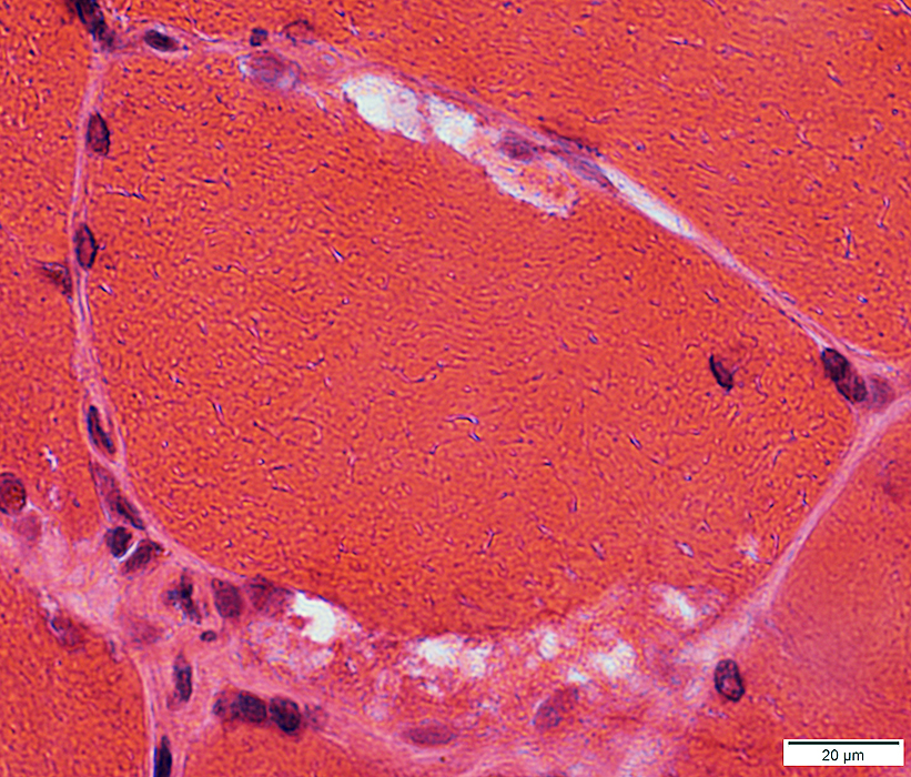
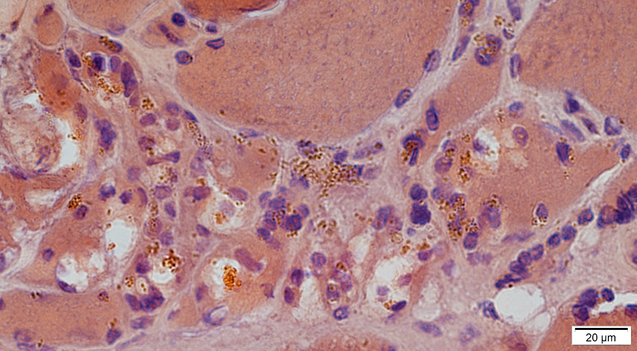
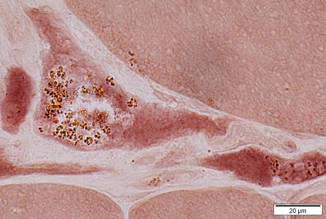
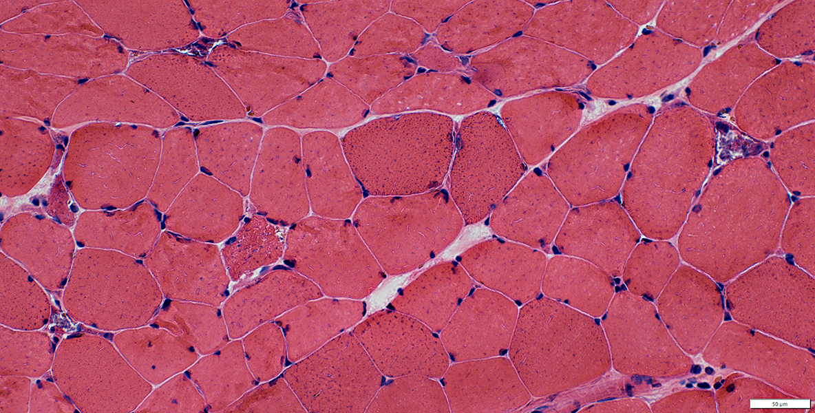 H&E stain |
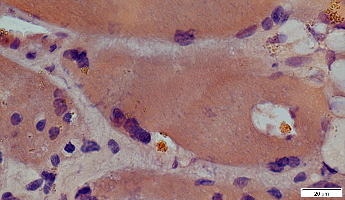
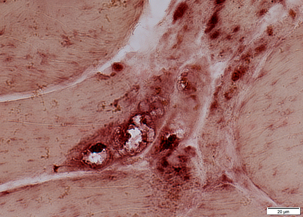
 H&E stain |
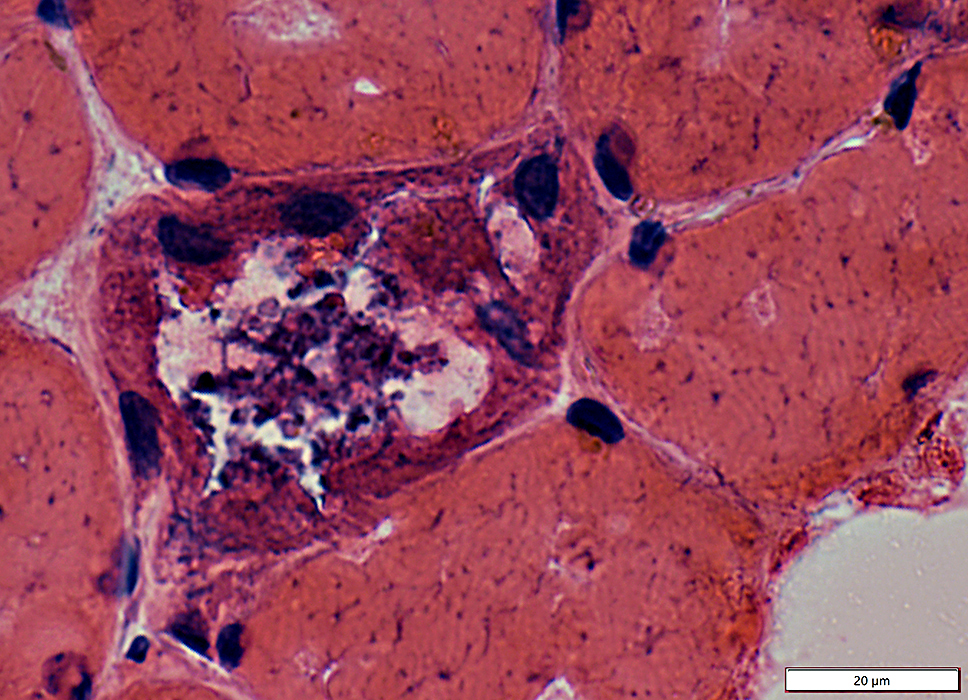
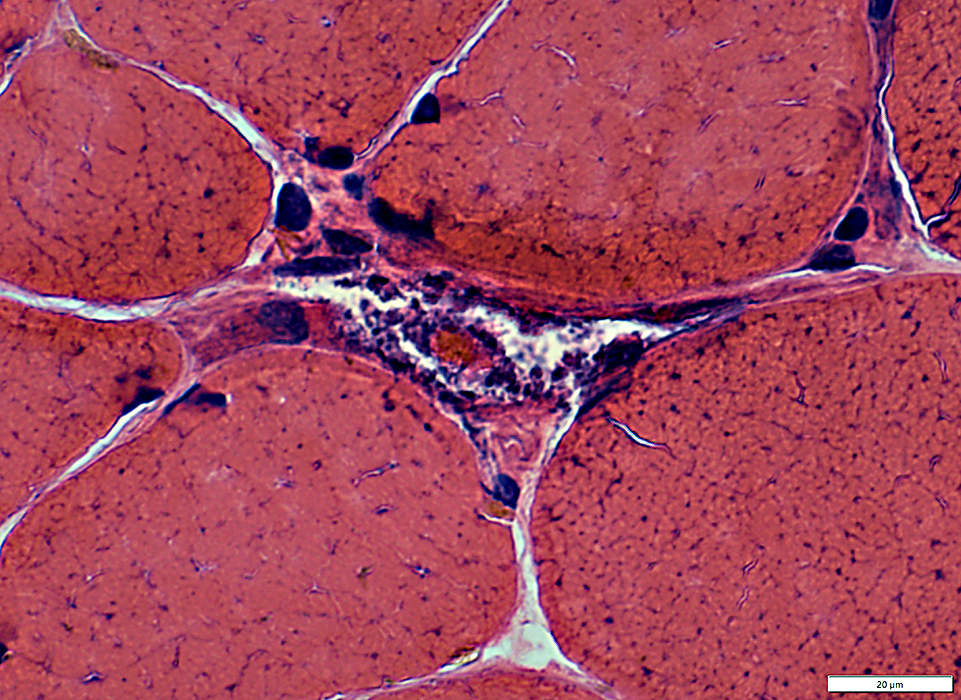
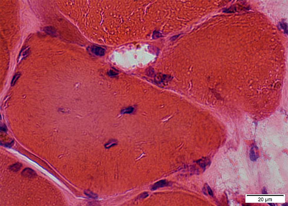
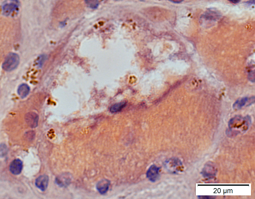
Vacuoles
May be internally clear, or contain basophilic aggregatesd material May be Acid phosphatase positive
May contain: Lipofuscin-like granules; Granular material
Internal nuclei
LAMP2 granules
 H&E stain |
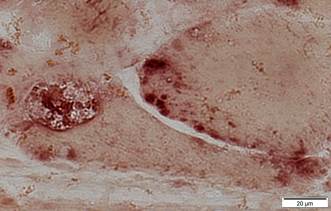
 H&E stain |
Vacuoles
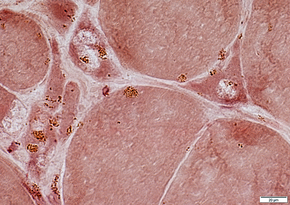
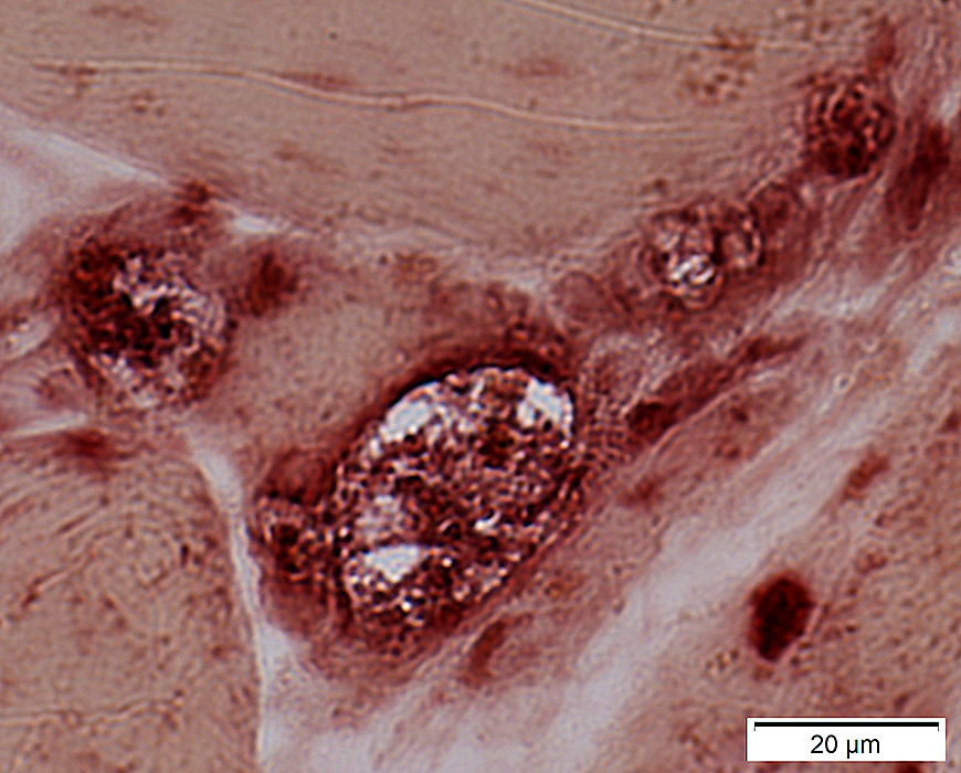
 H&E stain |
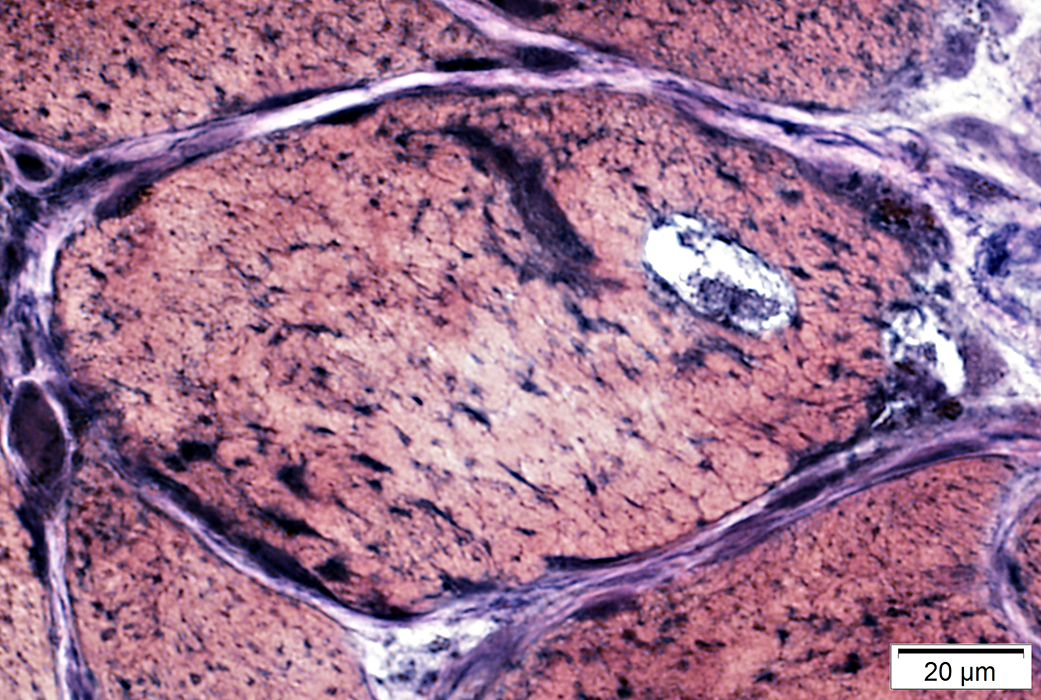
Muscle fibers
3 small muscle fibers with vacuoles & red staining of cytoplasm.
Fiber sizes: Intermediate to Very small
Vacuoles
Borders: Irregular
May contain: Aggregates; Clustered & lipofuscin-like material
 Gomori Trichrome stain |
 VvG stain |
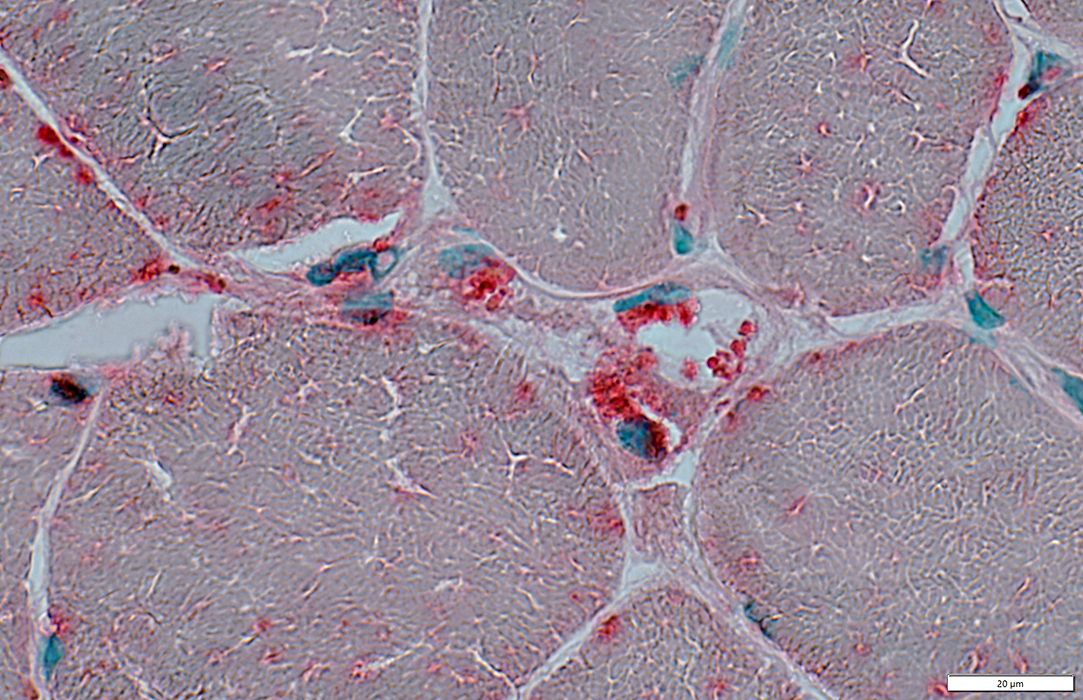
Vacuoles: Gray-Black rims & Aggregates
 VvG stain |
Lipofuscin-like material: In vacuoles & cytoplasm
 Congo red stain |
 Congo red stain |
Vacuoles: Contain lipofuscin-like material
 Congo red stain |
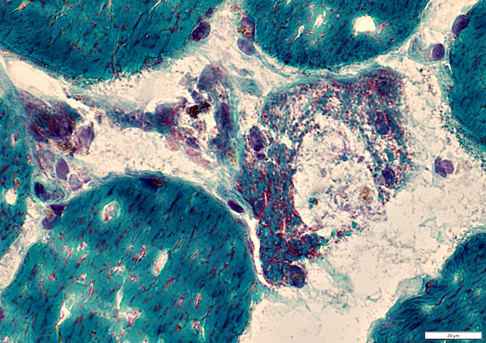
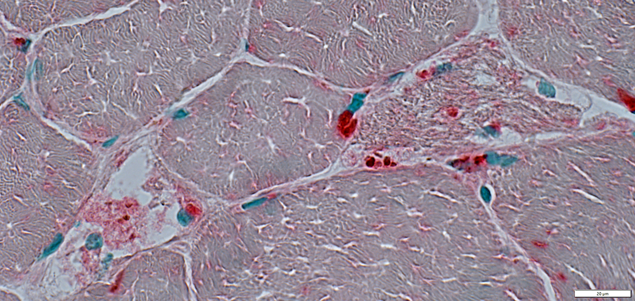
 Esterase stain |
Vacuoles: Contain lipofuscin-like material
 Esterase stain |
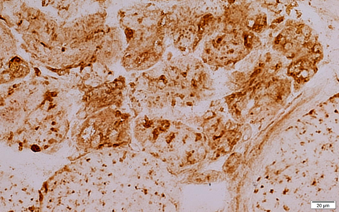
Lysosomal material in muscle fibers: Acid phosphatase & LAMP-2 staining
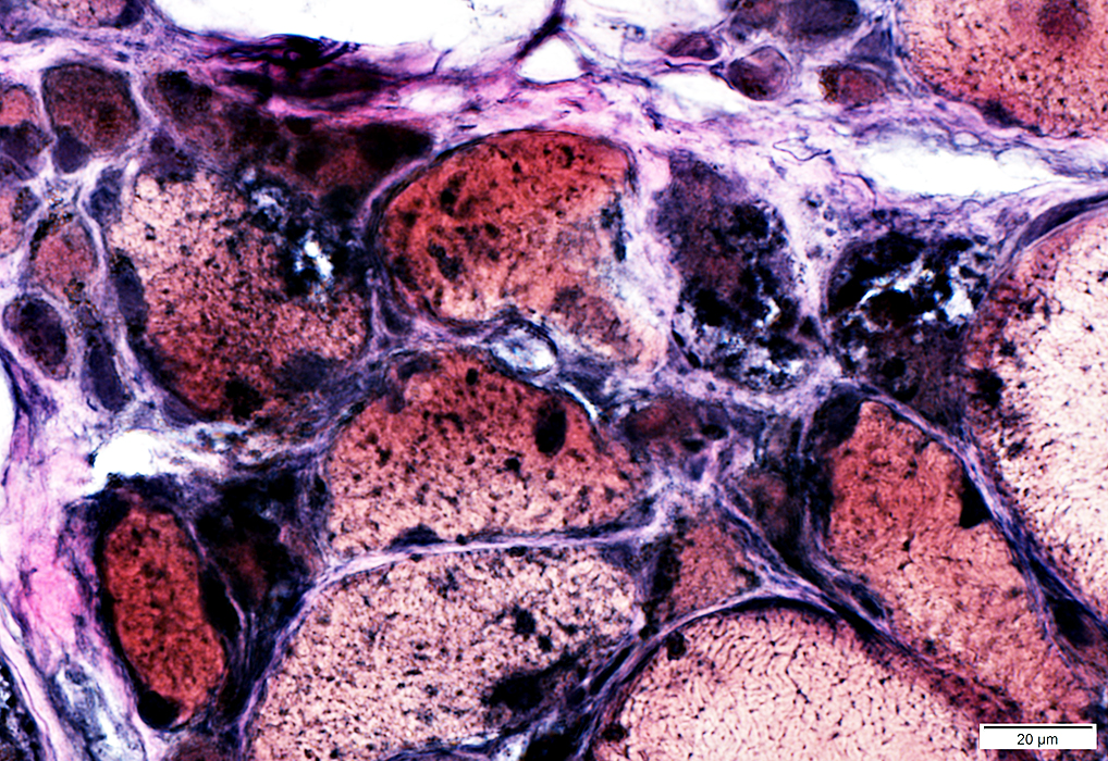
 Acid phosphatase stain |
 Acid phosphatase stain |
 Acid phosphatase stain |
 Acid phosphatase stain |
Location: Present in vacuoles & cytoplasm
 Acid phosphatase stain |
LAMP-2 stained organelles
 LAMP2 stain |
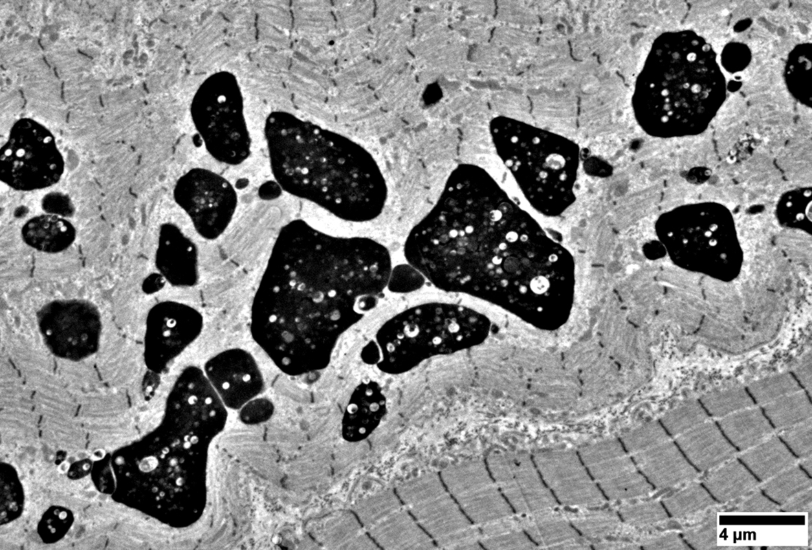
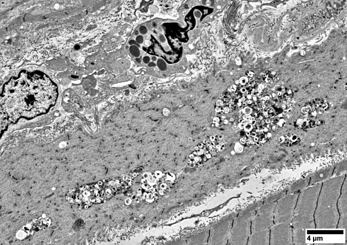
Aggregates in muscle fibers
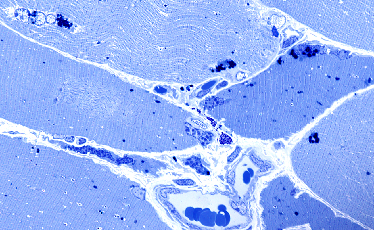 Toluidine blue stain |
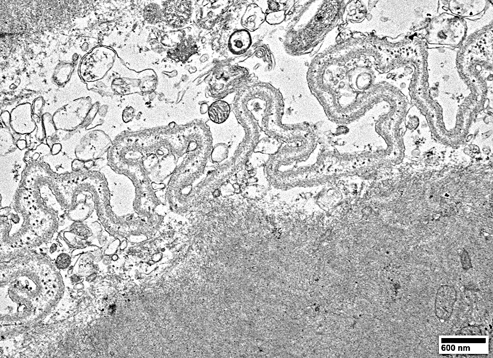
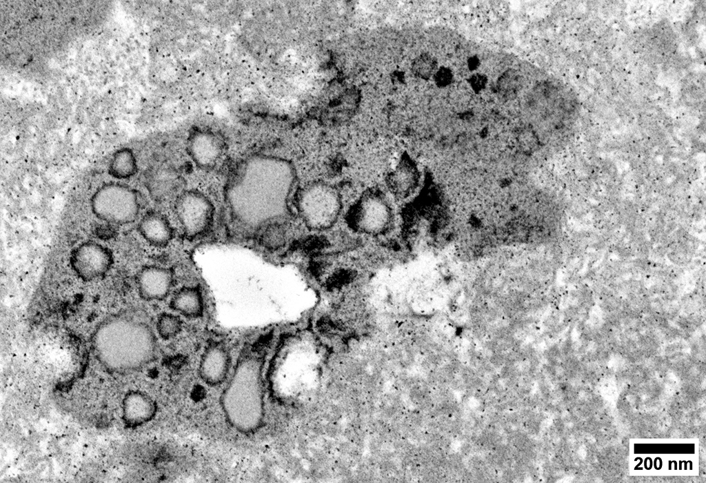
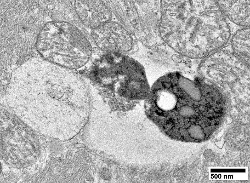
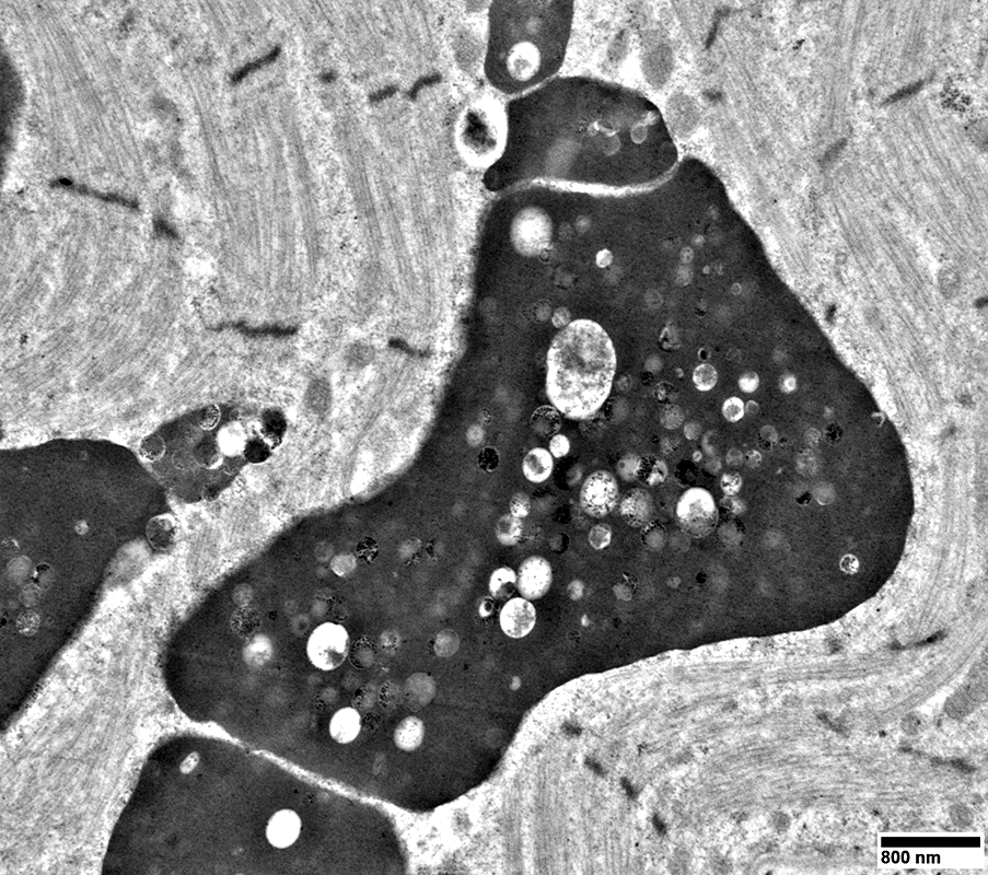
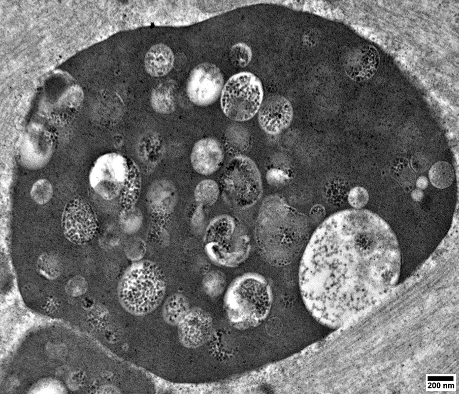
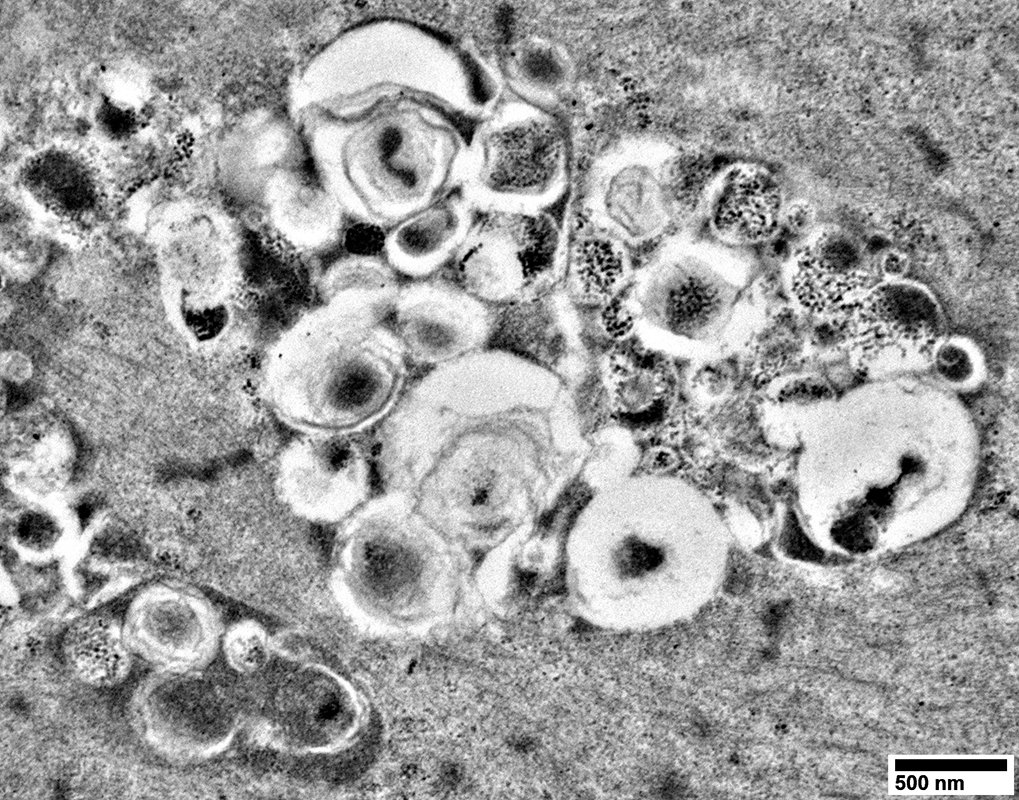
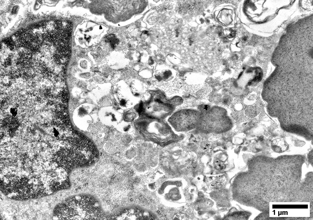
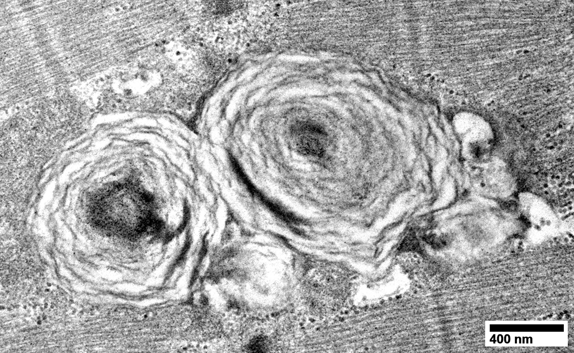
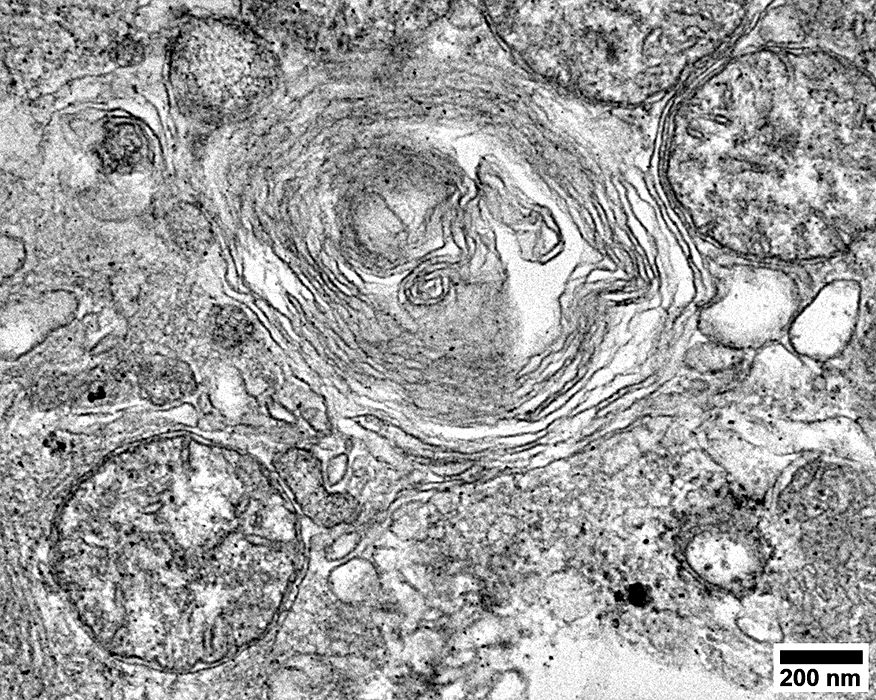
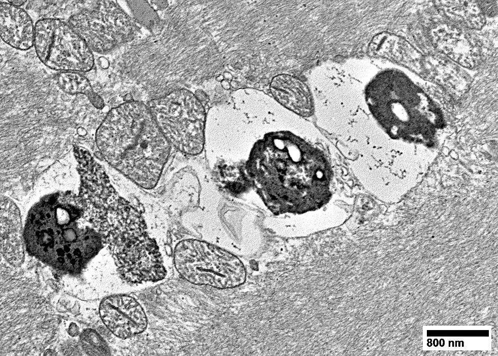
Hydroxychloroquine Myopathy: Ultrastructure
Aggregates: Contain Heterogeneous MaterialMembrane-bound vacuoles
Curvilinear bodies
Degradation products
Lipid & Lipofuscin structures
Membranous whorls
Mitochondria
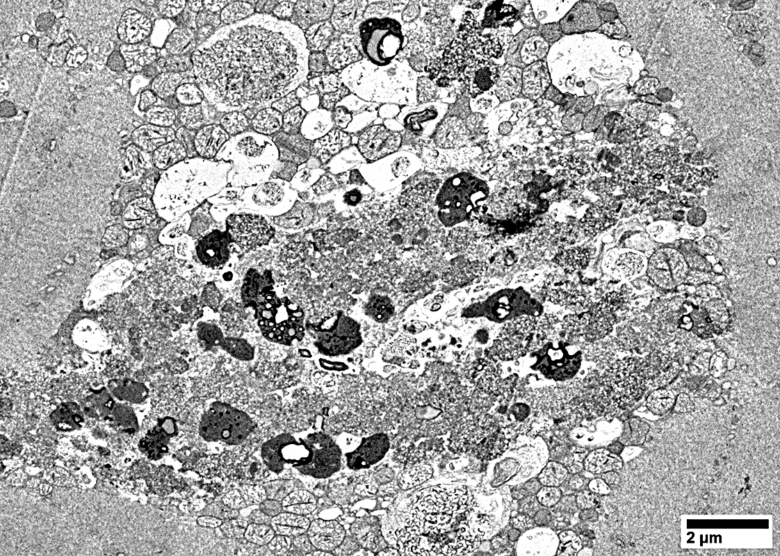 From: R Schmidt |
Curvilinear bodies (Arrow)
Lysosome-derived cytoplasmic inclusions
Also occur in ceroid lipofuscinosis
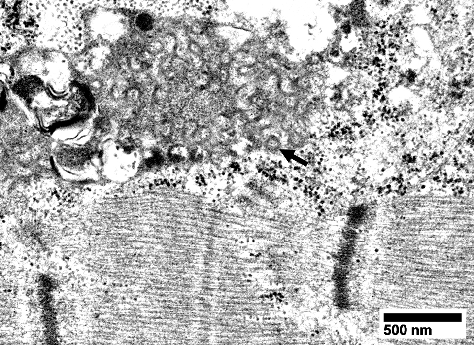 From: J Chen |
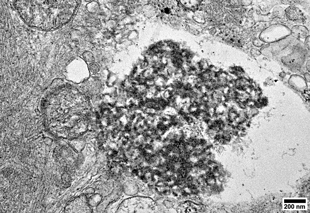
Curvilinear bodies & Complex lipid structures with globular elements
 From: J Chen |
 From: R Schmidt |
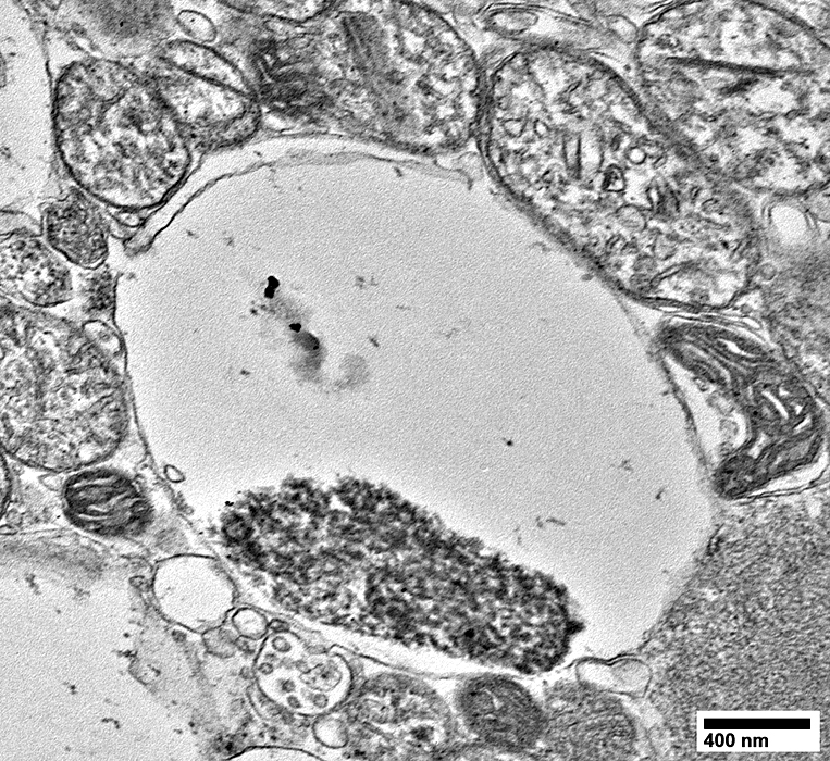
Extracellular Vesicles
 From: R Schmidt |
Complex Lipid, Lipopigment & Osmophilic Structures
 From: R Schmidt |
 From: R Schmidt |
Near normal, large & abnormal mitochondria
 From: R Schmidt |
 From: R Schmidt |
 From: R Schmidt |
Autophagic Degradation Products
 From: R Schmidt |
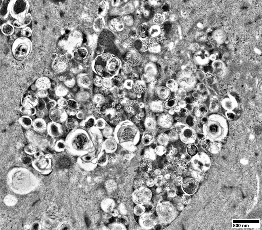 From: R Schmidt |
 From: R Schmidt |
 From: J Chen |
Membranous Whorls
 From: J Chen |
 From: R Schmidt |
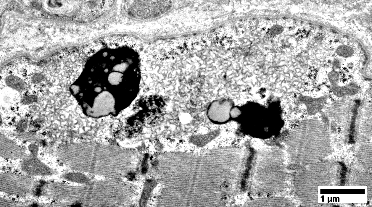
Membrane-bound vacuoles
Contents: Membranes; Lipopigment
? Lysosomal
 From: R Schmidt |
 From: R Schmidt |
 From: R Schmidt |
Hydroxychloroquine Myopathy: Mitochondria
Scattered tubular cristae
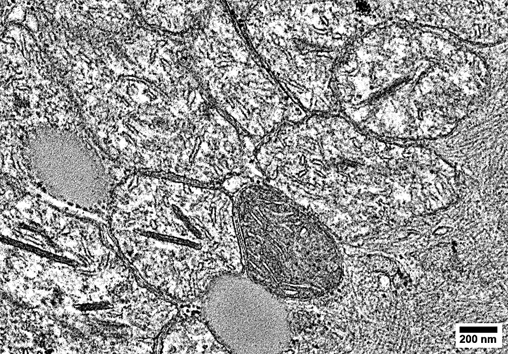 From: R Schmidt |
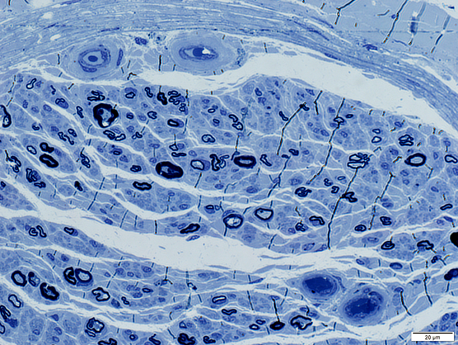
Neuropathy: Hydroxychloroqine-associated
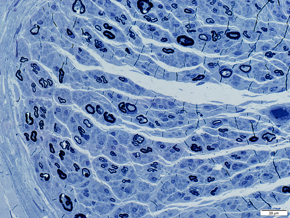 Toluidine blue stain |
Large axons
Loss
Thinly myelinated
Regenerating axon clusters
Endoneurial vessels: Thick walls
 Toluidine blue stain |
Return to Hydroxychloroquine myopathy
Return to Muscle biopsies
1/12/2026