
TUBULAR AGGREGATES
|
Tubular aggregates Differential Diagnosis Pathology
|
 |
 |

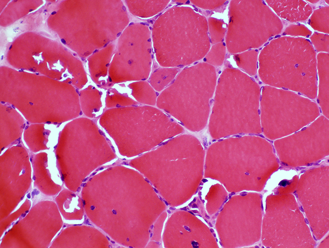 H & E stain |
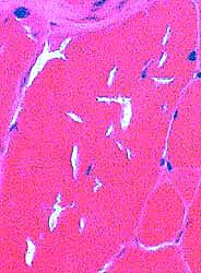
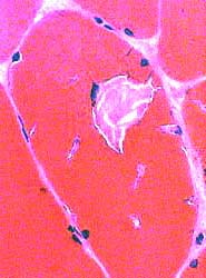
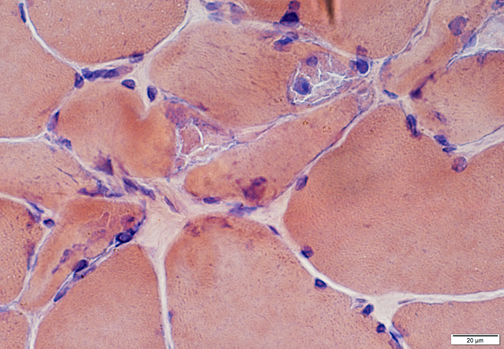
Pink cytoplasmic areas: May contain nuclei
Irregular-shaped clear regions
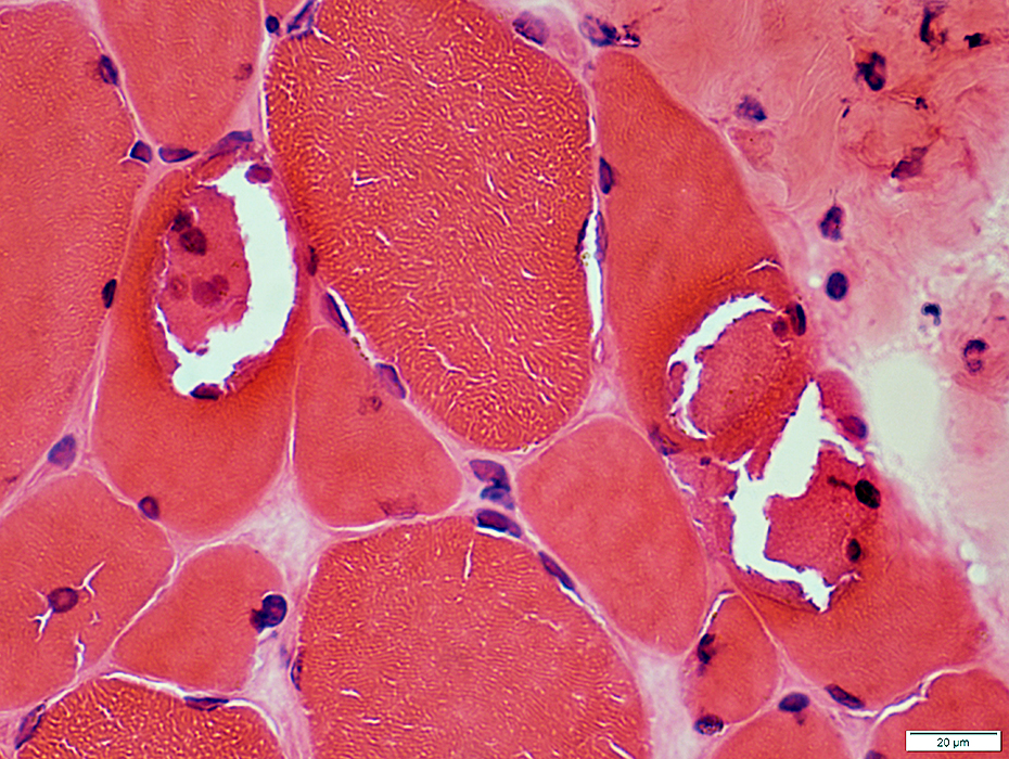 H & E stain |
 Congo red stain |
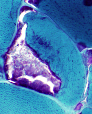 Gomori trichrome stain Tubular aggregates Stain red on Gomori trichrome |
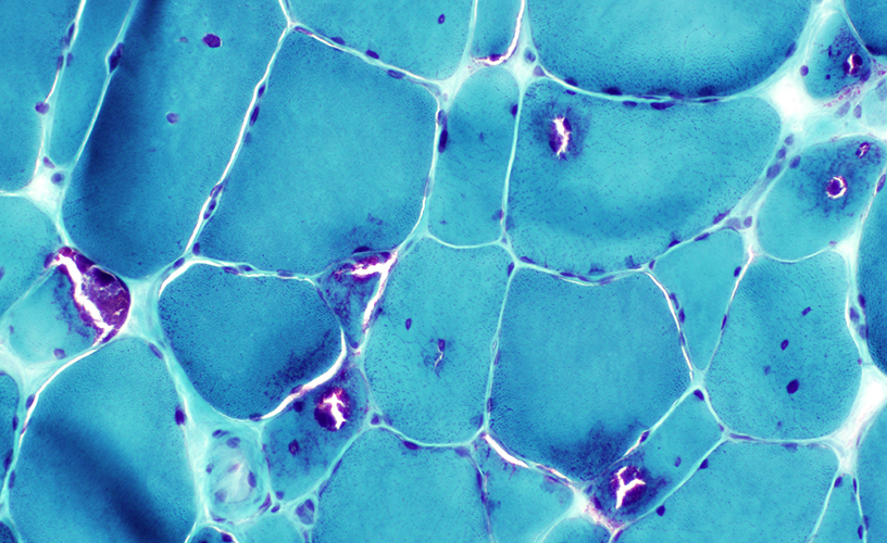
|
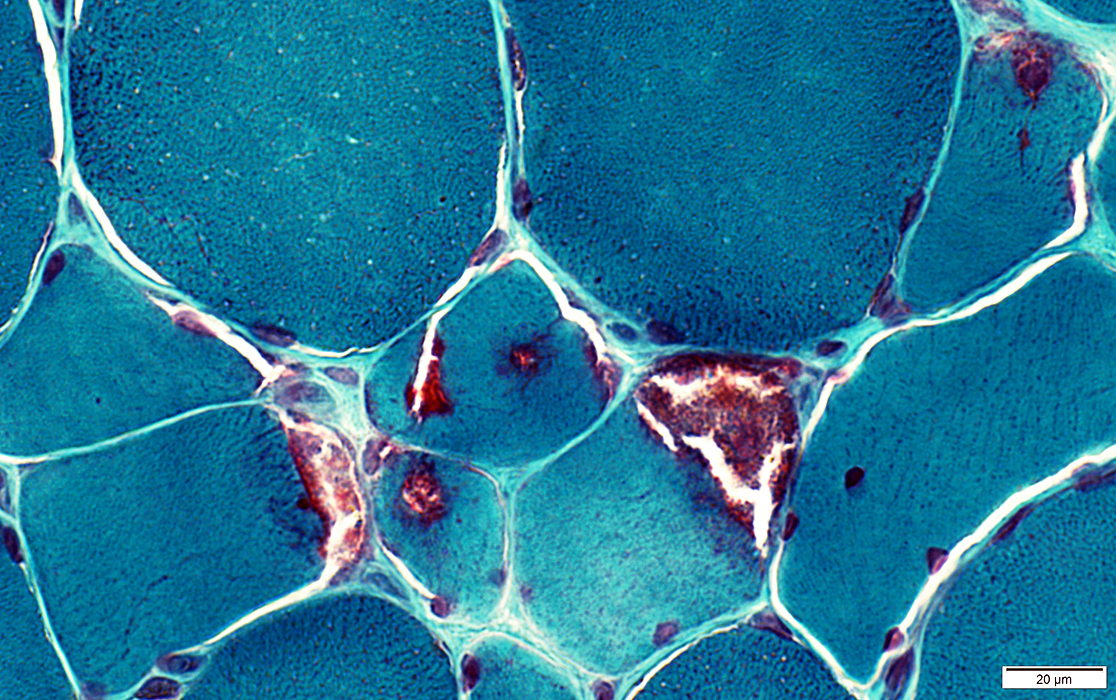 Gomori trichrome stain |
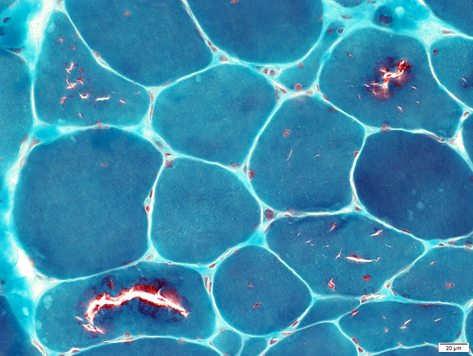 Gomori trichrome stain |
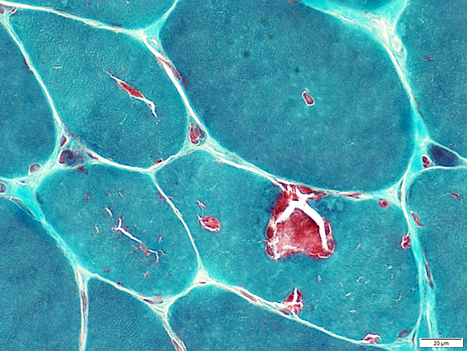 Gomori trichrome stain |
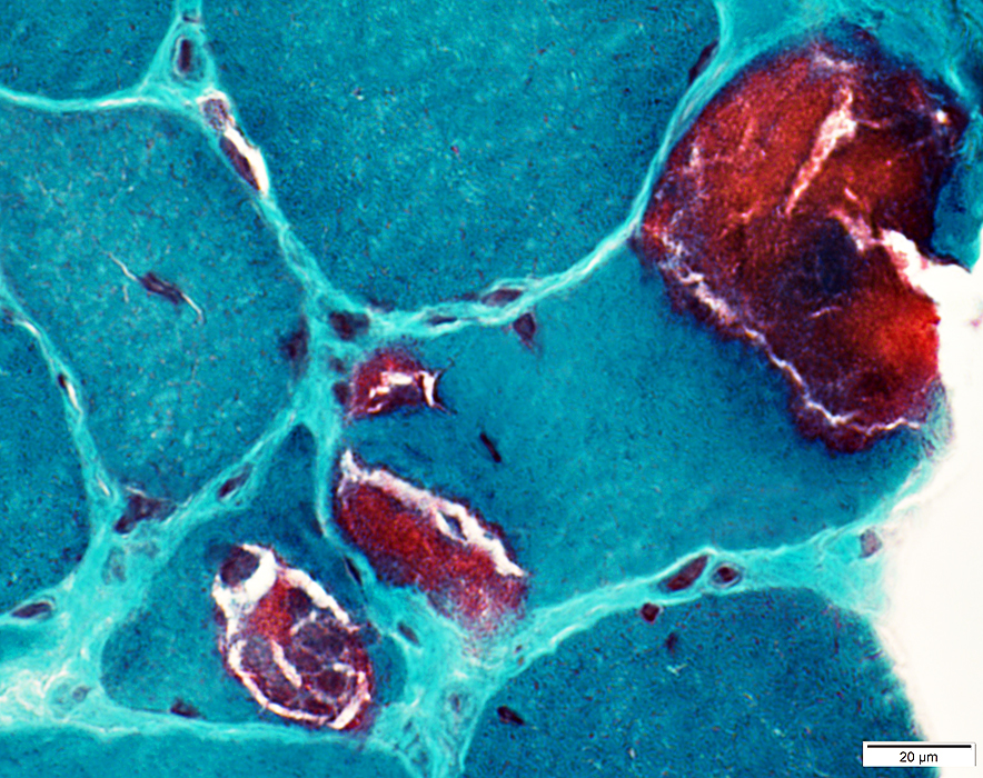 Gomori trichrome stain |
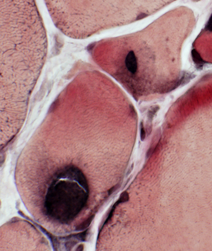
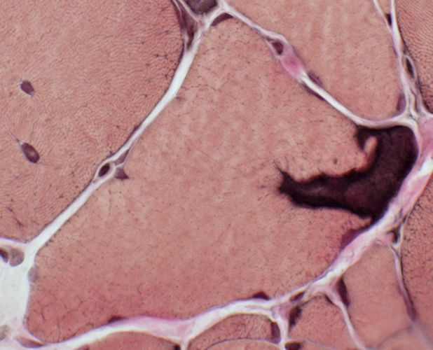
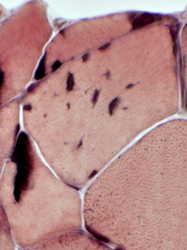
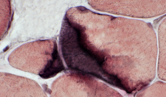
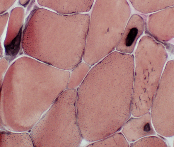 VvG stain Tubular aggregates Stain gray-black on VvG |
|
|
|
|
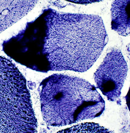
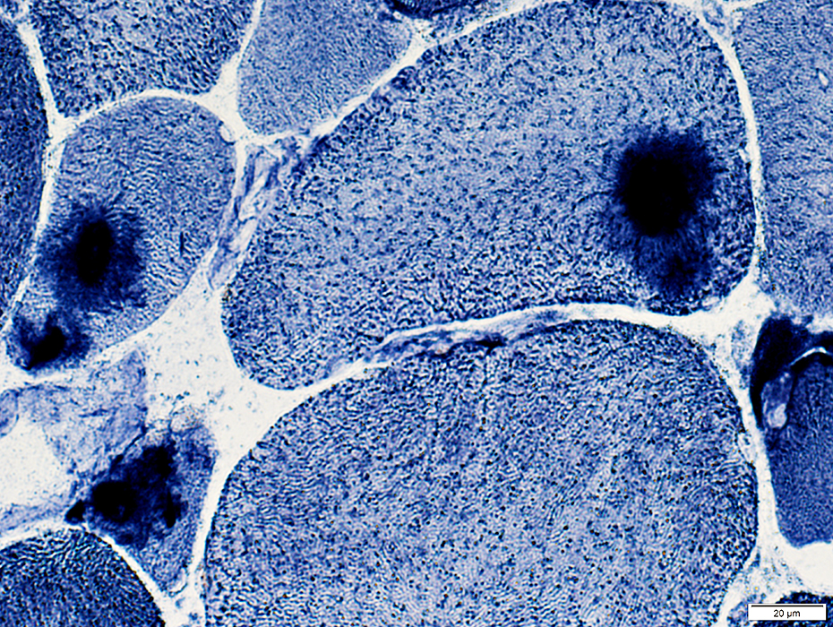
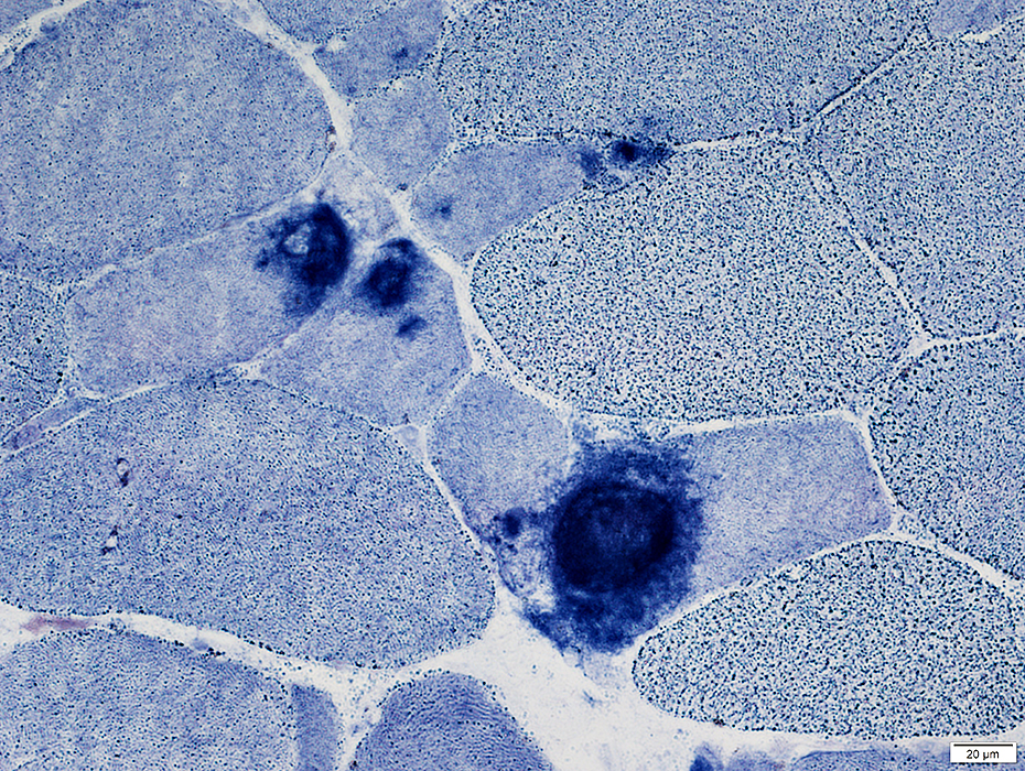
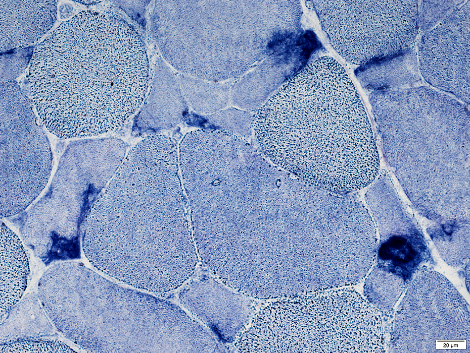
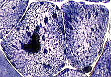 NADH-TR reductase (NADH) stain 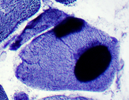 Tubular aggregates: Stain dark on NADH |
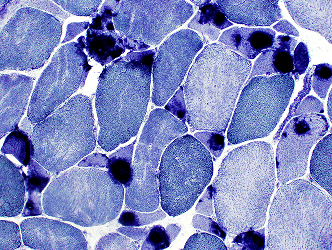
|
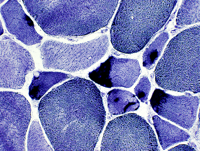
|
|
 NADH stain |
 AMPDA stain |
 AMPDA stain |
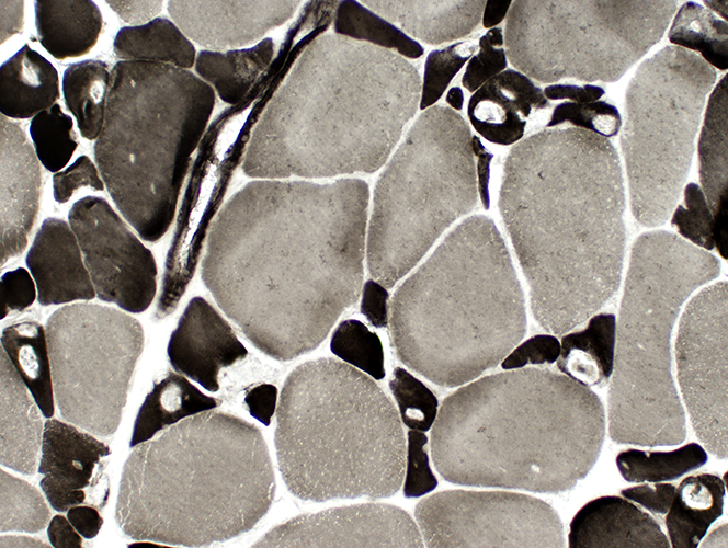 ATPase pH 9.4 stain |
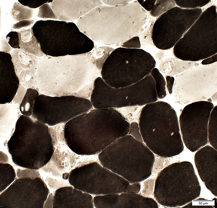 ATPase pH 4.3 stain |
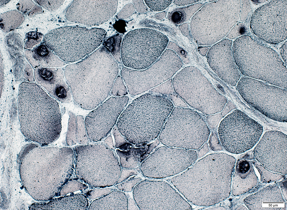 Sudan black stain |
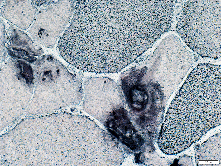 Sudan black stain |
Tubular aggregates
May have mild or patchy staining for Acid Phosphatase
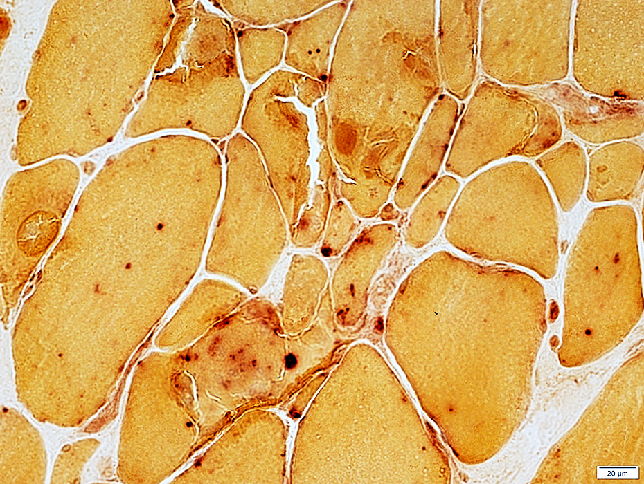 Acid Phosphatase stain |
Tubular aggregates: Ultrastructure 2
Tubules: 3 types
- I: 50 to 70 nm diameter tubules with 20 to 40 nm inner central tubule
- II: 70 to 400 nm tubules with moderately dense material in center
- III: 130 to 400 nm diameter tubules containing several 25 to 40 nm tubules
- Ordered Tubules
- Tubule contents: Varied
- Inner smaller tubule (Double-walled)
- Flocculent, granular materal
- Tubule sizes: Similar for most; May contain scattered moderately larger tubules
- May be surrounded by larger sarcoplasmic reticulum vacuoles
- Tubule contents: Varied
- Clustered/Disordered Tubules
- Tubule sizes: Varied
- Tubule variants
- Locations
- Entire tubular aggregates
- Similar regions may surround ordered tubular aggregates
- Associated Structures
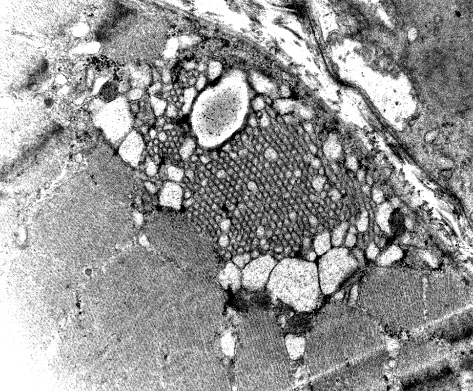 From: R Schmidt |
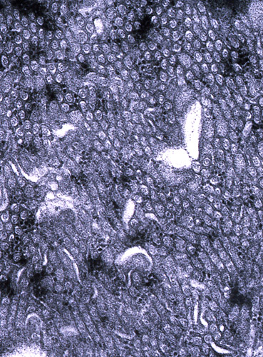
Arrays of 50 nm tubules: May range from to 80 nm
Single-walled
Contents: Clear or Granular; May contain 20 to 30 nm inner tubules
Locations
Often sub sarcolemmal
May be in central regions of muscle fibers
Some are peri-nuclear
Associations
May connect with lateral cisternae of Sarcoplasmic reticulum
May be surrounded by dilated SR sacs
Variants: Irregular clusters of varied sized tubules
Common syndrome associations: Periodic paralysis; Congenital MG syndromes; Cramp/Myalgia syndromes
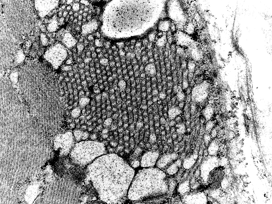 From: R Schmidt |
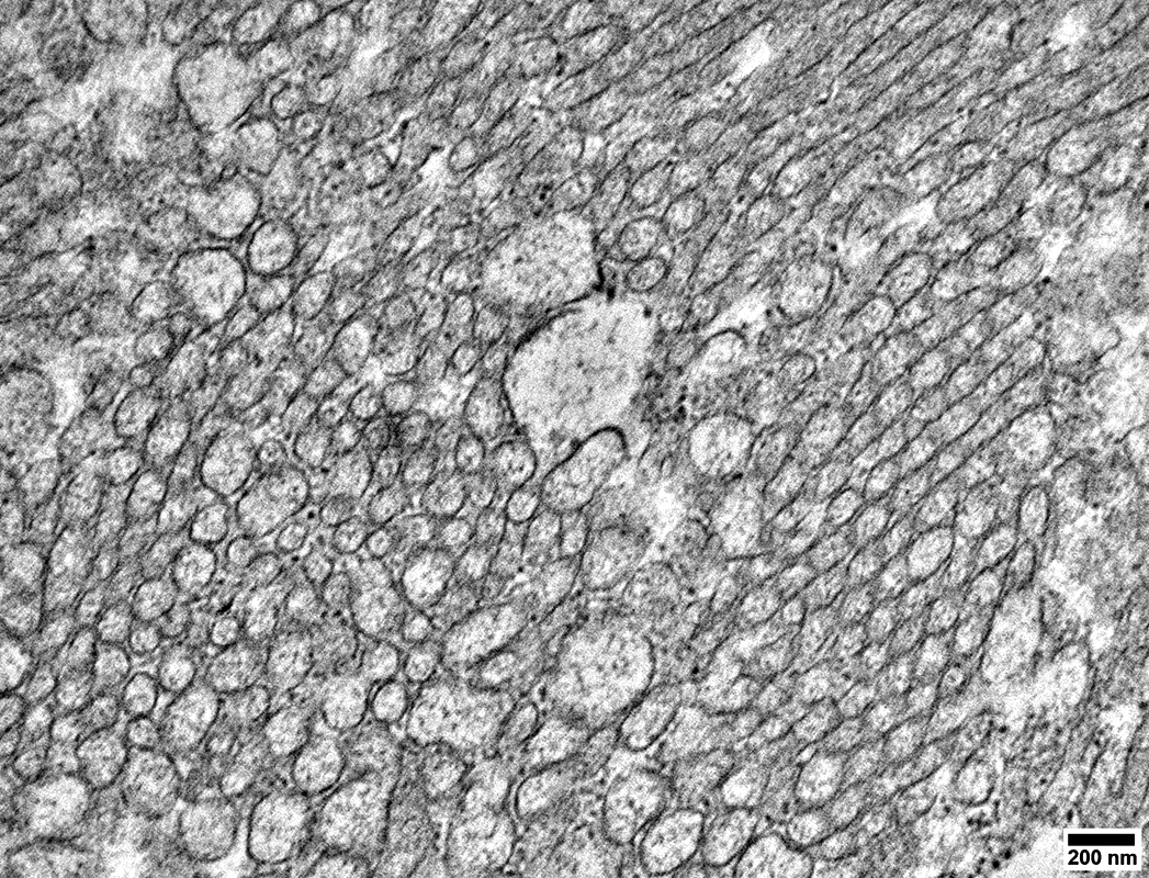 From: R Schmidt |
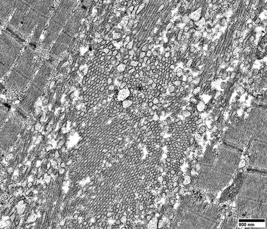
Arrays of 20 to 50 nm tubules
Single-walled
Some larger 150 to 500 nm sacs are intermixed within the tubular aggregates
 From: R Schmidt |
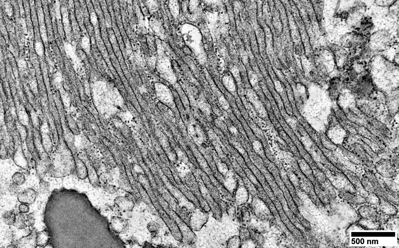
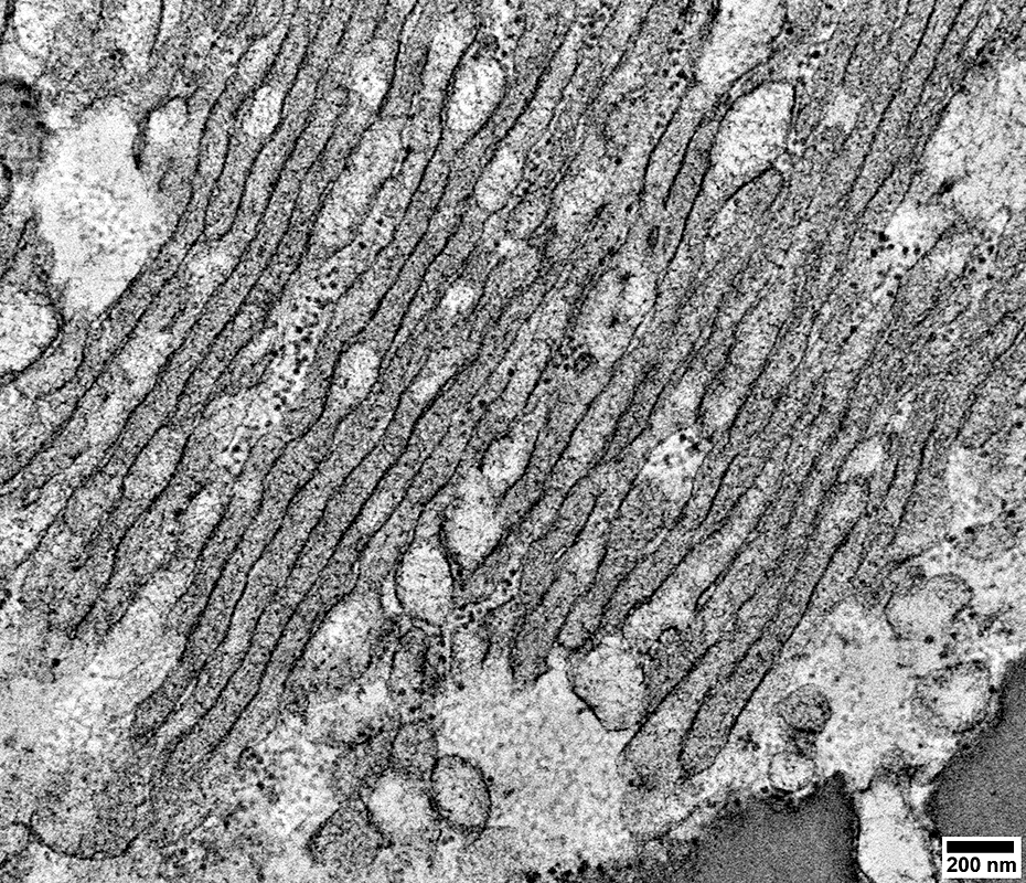
Long parallel membranes
General contents: Granular; Moderately dark-stained
Interruptions: Contain pale rounded swellings in regions along their length
 From: R Schmidt |
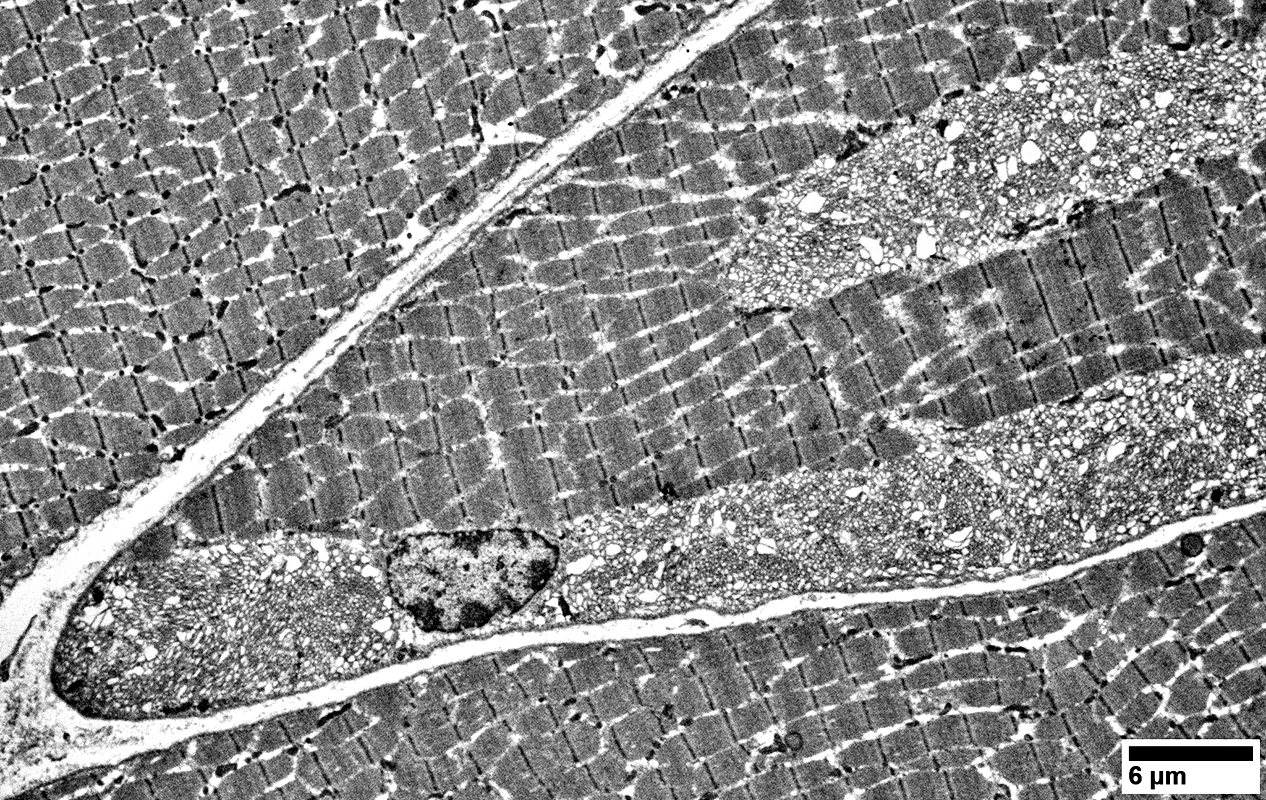
Tubular aggregates: Regional associations
 From: R Schmidt |
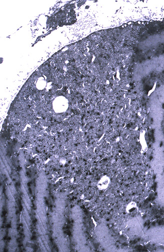
Locations: 1 subsarcolemmal; 1 in central region of fiber
Fiber type: Likely type 2 with fewer mitochondria & paler Z-bands than type 1 fiber at upper left
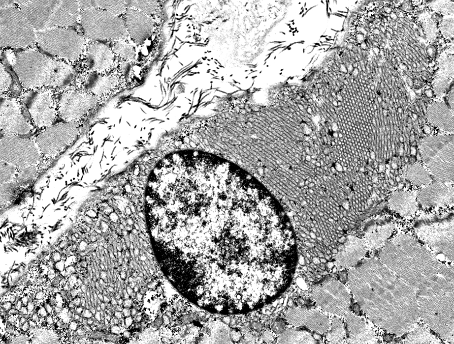
Nucleus is within subsarcolemmal region of tubular aggregates
 From: R Schmidt |
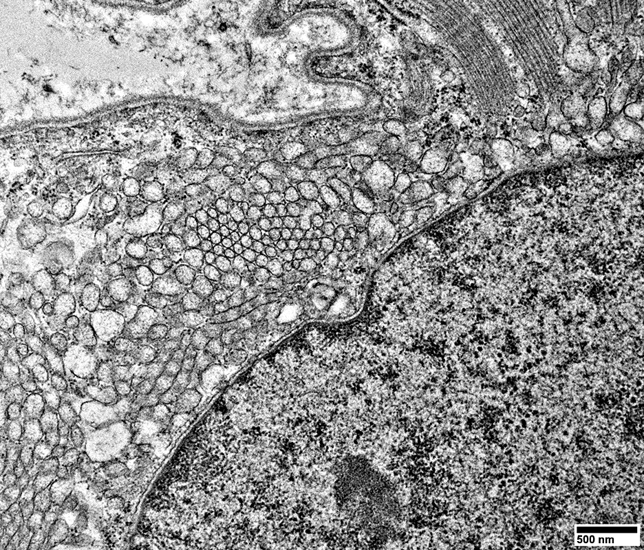 From: R Schmidt |
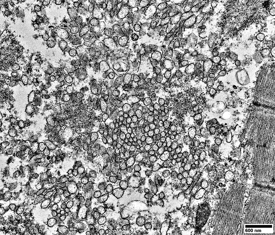 From: R Schmidt |
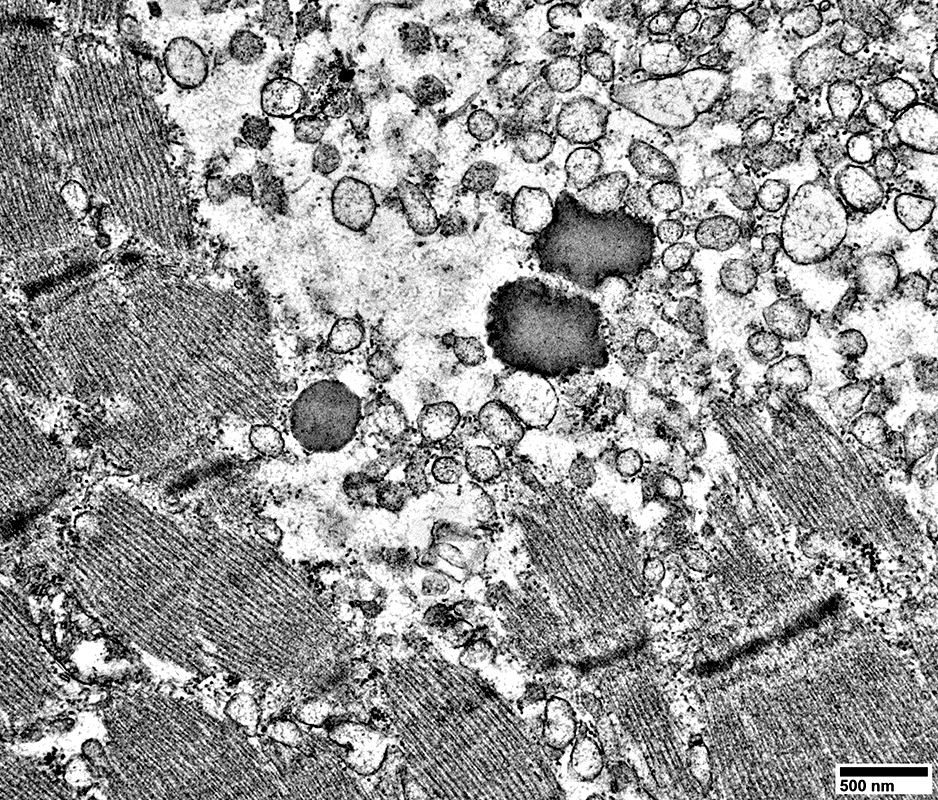 From: R Schmidt |
Tubule sizes vary widely
Larger tubules: Contents are often pale-stained
Small tubules: Contents are often darker-stained
Lipopigment may be scattered in these areas
Tubules may be located between sarcomeres
Tubular Aggregates: Unordered
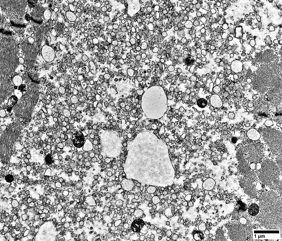 From: R Schmidt |
Tubule sizes vary widely
Larger tubules
Some have thick walls
Contents are often pale-stained
Small tubules: Contents are often darker-stained
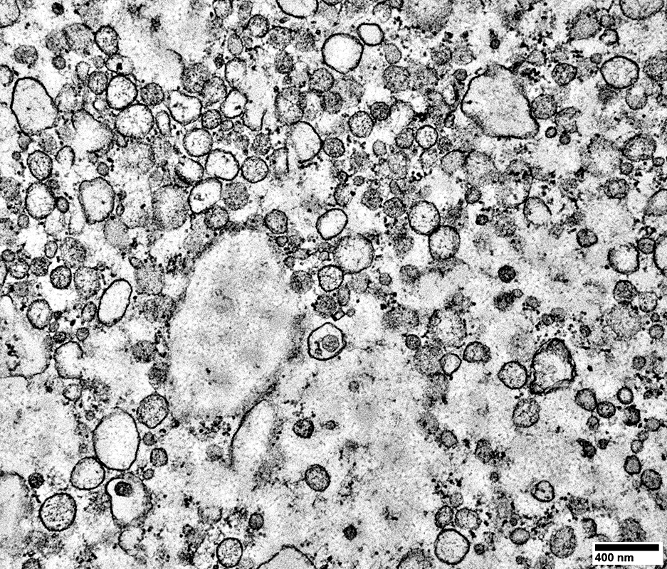 From: R Schmidt |
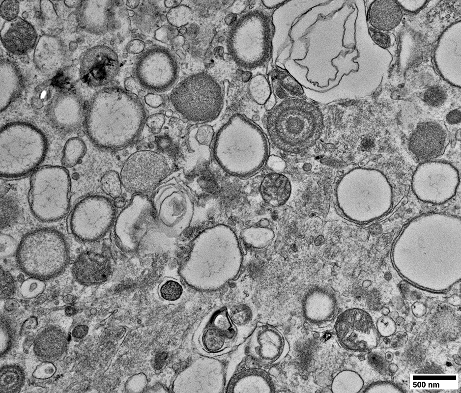
Tubule Variants
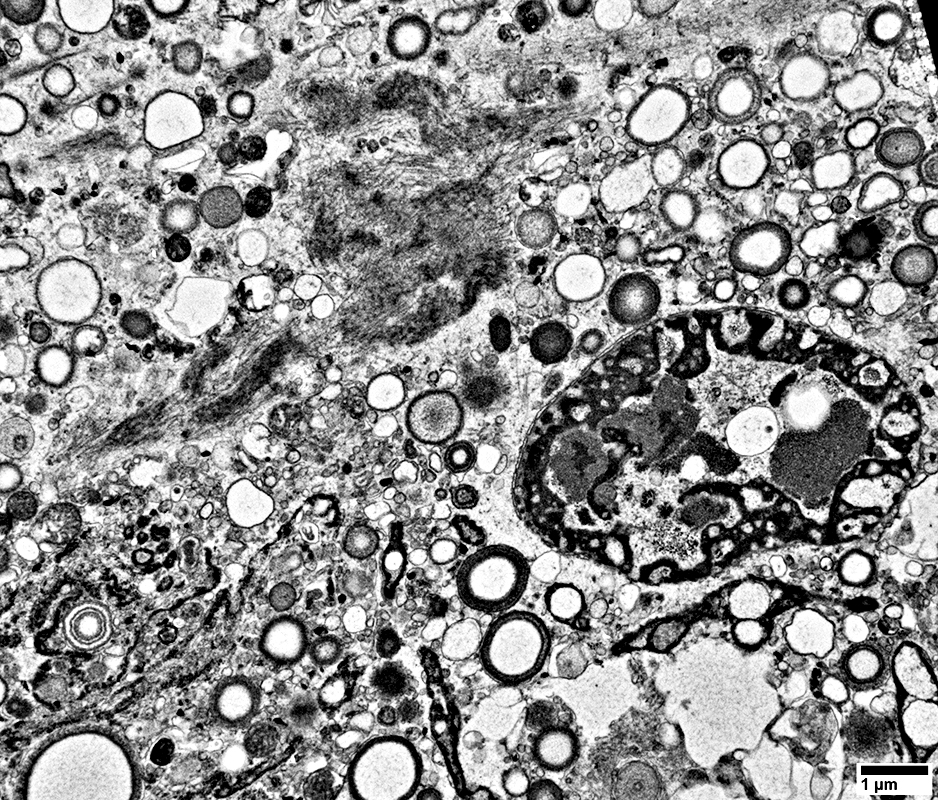 From: R Schmidt |
Thick walls
Layered walls
Dark-stained internal regions
Thin membrane contents
Cylindrical Spirals
Large size
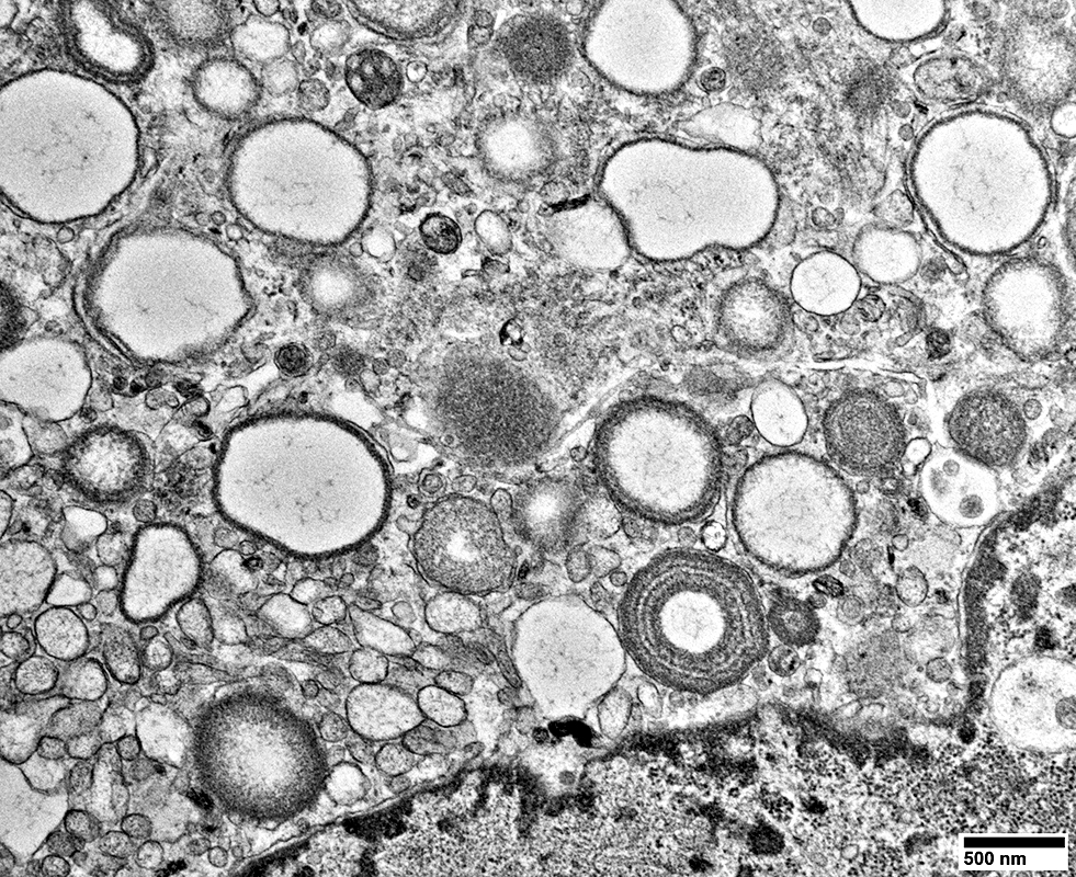 From: R Schmidt |
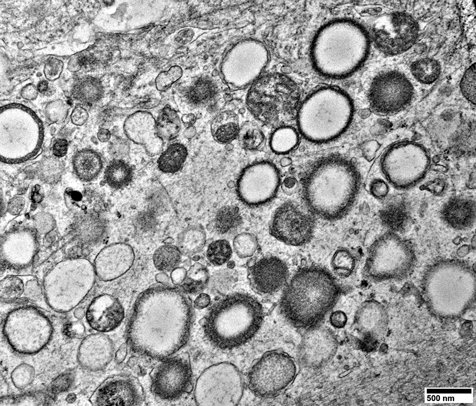 From: R Schmidt |
Thick walls
Layered walls
Dark-stained internal regions
Thin membrane contents
Cylindrical Spirals
Large size
 From: R Schmidt |
 From: Tahseen Mozaffar |
 From: Oliver Ni |
Tubular Aggregates: Mouse Muscle
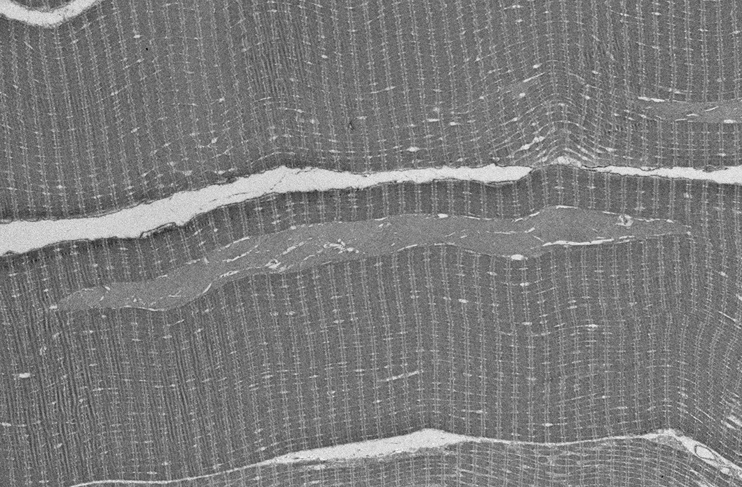 From: Andrew Findlay |
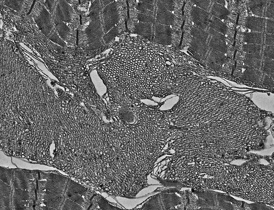 From: Andrew Findlay |
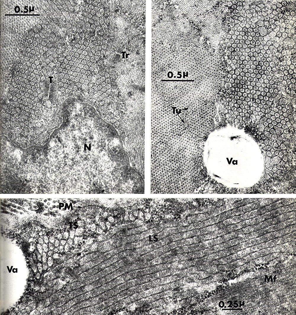 Mair & Tome |
Return to Myopathies with tubular aggregates
References
1. J Neuropathol Exp Neurol 2016;75:1171-1178
2. Gen Physiol Biophys 2003;22:425-40
7/19/2026