Neuromuscular

Home, Search, Index, Links, Pathology, Molecules, Syndromes,
Muscle, NMJ, Nerve, Spinal, Ataxia, Antibody & Biopsy, Patient Info

MUSCLE FIBER ACTIVITY & CRAMPS

Syndromes
Brody's syndrome
Complex repetitive discharges
Contracture: Muscle
Cramps
  Cramp-Fasciculation
  Dominant
  Electrically silent
  Familial
Electrical activity: No clinical Δ
Endplate Spikes
Fasciculations
Fibrillations
Miniature endplate potential
Minipolymyoclonus
Muscle pain
Muscle spasms
Myoedema

Myokymia
  Diffuse: Isaac's Syndrome
  Hereditary
  Localized: Radiation
Myopathy
Myotonic dystrophies
Neural origin
Neuromyotonia
Paramyotonia
Periodic paralyses
  Hyper K+
Positive sharp waves
Rippling muscles
Satoyoshi Syndrome
Schwartz-Jampel
Spinal stenosis
Stiff-Person syndrome
Tetanus
Tremor, Myopathic
Myotonia
  Comparative features
  General features
  Ion channels

Acquired
Chondrodystrophic: 1p34
Congenita
  Acetazolamide-responsive: Na+
  Becker's (Recessive): Cl-
  Thomsen's (Dominant): Cl-
Cylindrical spirals
Dystrophy
  Type I (DM1): 19q13
  Type II (DM2; PROMM): 3q21
Fluctuans: Na+
Levior: Cl-
Paramyotonia: Na+; Cl-
Permanens: Na+
Muscle fiber activity
Neural origin
Amyotrophic Lateral Sclerosis
Black widow spider
Blepharospasm
Complex repetitive discharges
Crisponi syndrome
Dystonia variants
Fasciculations
Firing patterns
Geniospasm
Hemifacial spasm
Hyperexcitability
  Differential diagnosis
Isaac's Syndrome
Motor neuron disorders
Motor neuropathies
Myokymia
Neuromyotonia
Palmaris brevis spasm
Polyneuropathy
Spinal:
  Vascular; Compression
Startle syndrome (Hyperekplexia)
Strychnine
Stiff-Person Syndrome
Superior oblique myokymia
Tetanus
Tetany
Tremor
Whipple's


Myotonia

Comparative features
General features

Acquired
Chondrodystrophic: Perlecan; 1p34
Congenita
  Acetazolamide-responsive: Na+
  Becker's (Recessive): Cl-
  Thomsen's (Dominant): Cl-
Dystrophy
  Type I (DM1): DMPK; 19q13
  Type II (DM2; PROMM): ZNF9; 3q21
  Type III (DM3): VCP
Fluctuans: Na+
Levior: Cl-
Paramyotonia: Na+; Cl-
Permanens: Na+
Potassium activated myotonia: Na
Proximal Myopathy (PROMM): ZNF9; 3q21


Myotonia: General features 56


Myotonia Congenita, Dominant (Thomsen)

  Muscle Chloride Channel (CLCN1) ; Chromosome 7q34; Dominant

Myotonia Congenita, Recessive (Becker)

  Muscle Chloride Channel (CLCN1) ; Chromosome 7q34; Recessive


Myotonia levior

  Muscle Chloride Channel (CLCN1); Chromosome 7q34; Dominant


Paramyotonia syndromes 64


  Sodium Channel - α subunit (SCN4A) ; Chromosome 17q23.3; Dominant
  Muscle Chloride Channel (CLCN1) ; Chromosome 7q34; Dominant


Myotonia Fluctuans

  Sodium Channel - α subunit (SCN4A; Nav1.4) ; Chromosome 17q23.3; Dominant

Myotonia Permanens

  Sodium Channel - α subunit (SCN4A; Nav1.4) ; Chromosome 17q23.3; Dominant or Sporadic


Acetazolamide-responsive Myotonia Congenita

  Sodium Channel - α subunit (SCN4A; Nav1.4) ; Chromosome 17q23.3; Dominant

Myotonic Dystrophy (DM1)

  Myotonin protein kinase; Chromosome 19; Dominant


Proximal Myotonic Myopathy (PROMM; DM2)

  CCHC-type Zinc finger Nucleic acid-Binding Protein (CNBP; Zinc finger protein 9 (ZNF9)) ; Chromosome 3q21.3; Dominant
  • DM2: Epidemiology
    • Most families European, especially German/Polish
    • Anticipation 6
      • Mild: 0.5 to 1.9 decades (vs 2.9 decades in myotonic dystrophy)
      • ? More prominent with paternal transmission
  • Genetics: Mutation in CCTG repeat sequence in intron 1 of ZNF9 gene 12
    • Normal CCTG repeat sequence
      • Complex repeat motif: (TG)n(TCTG)n(CCTG)n
      • Size: 104 to 176 base pairs
      • Interruptions: GCTG + TCTG motifs, or one or two TCTG motifs
    • CCTG expansion
      • (CCTG)n repeat size: Range 75 to ~11,000; Mean ~5000
      • Interruptions: None
      • No clear relation of CCTG repeat size with: Onset age; Disease severity
      • Somatic instability
        • Different repeat sizes in different tissues
        • Age: Size of CCTG repeat increases over time in individuals
        • Especially great: Peripheral blood
        • Greater than in DM1
      • Intergenerational features
        • Repeat size in blood of affected child: Shorter than parent
    • Genetics & DM2 disease pathogenesis
      • CCTG expansion produces RNA with excessive repeats
      • RNA with expanded repeats located focally in muscle cell nucleus
      • CCTG expression at RNA level may be pathogenic
      • Functional effects of long repeat RNA
        • Abnormal splicing of muscle CLC1 (chloride) channel RNA
        • Altered splicing adds exon 7a to Cl- channel RNA
        • Aberrantly spliced RNA produces altered Cl- channel protein with reduced function
        • Reduced Cl- channel function produces myotonia
        • Longer repeat RNA produces more abnormal splicing of Cl- channel: More myotonia
        • Effect is expressed as mosaic
          • Some muscle fibers with more reduction of Cl- channel than others
        • Similar effect of elongated CTG repeat sequence in DM1
    • Genetic association
      • CLCN1: Increased frequency of coexisting recessive mutations
    • Clinical-Genetic correlations
      • Unclear correlation between repeat length & disease severity
      • Homozygous for CCTG repeats: Not different from heterozygotes 28
    • Other disease expansions in transcribed but untranslated DNA
  • CNBP (ZNF9) protein
    • ZNF9 expression: Most abundant in heart & Skeletal muscle
    • Single stranded DNA-binding
    • Sterol regulatory element: Involved in sterol-mediated repression
    • Subcellular location: Cytoplasmic, also in endoplasmic reticulum
  • Clinical features
    • Onset 63
      • Age
        • Congenital to 60 years
        • Maternal inheritance: Earlier onset (Often < 30 years)
        • No relation to patient sex
      • May be related to pregnancy (21%) with paternal inheritance
      • Muscle (60%): Stiffness or Pain
      • Non-muscular: Cardiac; Cataracts
      • Early onset 39: Hypotonia; Myotonia not usually present
    • Intrafamilial variability
    • Weakness
      • Proximal: Legs > Arms; Hip flexors & extensors early
      • Distal arms: May be involved early; Thumb & Finger flexors
      • Face: Weak in 12%
      • Normal: Distal legs; Respiratory
      • Variation: Over hours
      • Progression: Slow
    • Muscle Enlargement: Calf
    • Muscle Pain or Discomfort 29: 56% to 75%
      • Stimuli
        • Exercise-related, or at rest
        • Temperature-modulated: Increased with cold
        • Palpation-induced
        • No relation to myotonia
      • Proximal; Legs > Arms
      • Painful stiffness
      • Variable from day-to-day
    • Myotonia
      • Symptomatic: Asymmetric
      • Intermittent: Exacerbations during pregnancy
      • Temperature sensitive (Warm more; Cold less)
      • Induced by: Grip; Percussion
    • CNS
      • Clinical: Usually normal; Occasional CNS signs
      • Imaging
        • MRI: White matter hyperintensity on T2 weighted images
        • Reduction of frontal & parietal gray matter
        • Abnormal thalamic gray matter & hippocampus
        • White matter lesions: > 50%; Extent correlated with psychomotor speed
        • More severe in DM1 than DM2
    • Systemic
      • Cataracts: 100% over 20 years with slit lamp
      • Cardiac arrhythmias: 20%; Conduction defects
      • Diabetes mellitus: 20%
      • Hearing loss (60%) 50
        • Mild to Moderate
        • Cochlear sensorineural
        • More with increased age
      • Fertility: Normal or Reduced
      • Cardiovascular autonomic function: Normal
    • Anesthesia & Muscle relaxant risk: Minor
    • See: Comparison to DM1
  • Lab 42
    • Serum & Blood
      • CK
        • Mild elevation (< 10x) (85%)
        • May be high in asymptomatic DM2 patients 30
      • FSH: High
      • IgG level: Reduced
      • Cholesterol: High (62%)
      • Lymphopenia (54%)
    • EMG
      • Myotonia
      • CMAP amplitude increased with exercise
        • Progressively reduced (by 40%) during rest
          • 20 to 40 min after initial increment (60% of patients)
        • No decrement: On short exercise, or slow or rapid repetitive stimulation
        • Normals: Mild increase in CMAP amplitude after exercise
      • Myopathic motor unit potentials: Proximal > Distal
    • MRI: Muscles involved 32
      • Selective muscle involvement: Erector spinae; Gluteus maximus
      • Abnormality more common
        • Females
        • Increasing age
      • Abnormality less common
        • Males
        • Milder weakness
        • Myalgias
    • Muscle biopsy
      • Internal nuclei: Type 2 muscle fibers
      • Fiber size variation: Type 2 atrophy
      • Pyknotic nuclear clumps
        • Especially type 2 fibers
        • Beter seen by myosin heavy chain histochemistry
      • No fiber type grouping
    • Cell pathology: Intranuclear inclusions
    • CNS MRI: White matter disease 43
      • Prominent callosal body & limbic system involvement
      • Less grey matter pathology than DM1
  • Prognosis: More benign than DM1 myotonic dystrophy


DM2: Hypertrophic calves

PROMM

  Locus not linked to 3q21; Autosomal Dominant

Acquired myotonia: Drug toxicity


Hyperkalemic periodic paralysis

Andersen: KCNJ2; 17q24
K+ channel: KCNE3; 11q13
Na+ channel: SCN4A; 17q23


Hyperkalemic periodic paralysis
  Sodium Channel - α subunit (SCN4A) ; Chromosome 17q23.3; Dominant
  Andersen's syndrome: K+ sensitive periodic paralysis with Cardiac dysrhythmias & Dysmorphic features


Rippling Muscle Syndromes 17

Acquired
Hereditary
  Dominant
    RMD1: 1q41
    RMD2: Caveolin-3; 3p25
    Lipodystrophy (CGL4): Cavin; 17q21
  Recessive
    Omani
    RMR: Caveolin-3; 3p25

Rippling Muscle Syndromes: Hereditary, Dominant
  RMD1 : Chromosome 1q41; Dominant
  RMD2 : Caveolin-3 (CAV3) ; Chromosome 3p25.3; Dominant
  ? Other loci; Dominant 9

Rippling Muscle Syndromes: Hereditary, Recessive 14

Rippling Muscles, Recessive
  Caveolin-3 ; Chromosome 3p25.3; Recessive or More severe dominant

Rippling Muscles, Recessive
  Recessive
Rippling Muscle Syndrome, Sporadic 3

Schwartz-Jampel Syndrome (Chondrodystrophic myotonia) 24

  Perlecan (HSPG2) ; Chromosome 1p36.12; Recessive

Schwartz-Jampel Type 2/Stuve-Wiedemann syndrome (SWS) 26

  Leukemia Inhibitory Factor Receptor (LIFR) ; Chromosome 5p13.1; Recessive

Brody disease 53

  Ca++ ATPase (ATP2A1; SERCA1) ; Chromosome 16p11.2; Recessive

Myokymia & Benign Neonatal Epilepsy (BFNS1) 13

  KCNQ2 ; Chromosome 20q13.33; Dominant

Global Developmental delay, Myokymia & Peripheral Nerve Hyperexcitability 62

  Leucine-rich gene, glioma-inactivated, 3 (LGI3) ; Chromosome 8p21.3; Recessive

Myoedema


Muscle Spasms & Dwarfism

Familial
  Dwarfism + Spasms
  Schwartz-Jampel
Sporadic
  Satoyoshi

Familial Muscle Spasms & Dwarfism
  Dominant; ? Autosomal or X-linked
Satoyoshi Syndrome 36

Cramps


Electrical activity with few clinical manifestations


Electrically silent cramps


Myopathic Tremor 58


Tetanus 20

Clinical features
Clostridium tetani
Diagnosis
Epidemiology
Mechanism of action
Prophylaxis
Protein structure
Treatment

History
  Case description


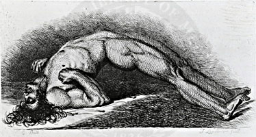
C Bell 1865: From NLM

Putnam


Isaac's Syndrome

Associated disorders
Clinical
Differential diagnosis
EMG
Laboratory


Familial Dyskinesia & Facial Myokymia 10

  Adenylate cyclase 5 (ADCY5) ; Chromosome 3q21.1; Dominant

Stiff-person (Stiffman) syndrome

Antibodies
Associated disorders
Clinical features
Laboratory
Treatment
Variants


Clinical features 7 Associated disorders Laboratory Disease mechanisms Antibodies: Stiff-person syndromes Therapy (Rarely completely effective) Stiffperson syndrome: Variants


Strychnine 11


Hyperekplexia (Startle disease)

  Glycine receptor, α-1 subunit (strychnine binding) ; Chromosome 5q33.1; Dominant or Recessive

Muscle Fibers & Nerves: Activity Patterns

Muscle fibers
Axon firing patterns
Complex repetitive discharges
Cramp
Endplate Spikes
Fasciculations
Fibrillations
Insertion activity
Miniature endplate potentials (MEPPs)
Positive Sharp Waves

Also see: Spontaneous activity
Neural Origin
Amyotrophic Lateral Sclerosis
Black widow spider
Blepharospasm
Complex repetitive discharges
Cramp
Dystonia variants
Fasciculations
Geniospasm
Isaac's Syndrome
Motor neuropathies & neuron disorders
Motor units
Muscle fiber activity
Myokymia
Neuromyotonia
Polyneuropathy
Startle syndrome (Hyperekplexia)
Stiff-person Syndrome
Tetanus
Tetany
Tremor
Whipple's


Motor Unit Potentials


Fibrillations


Positive Sharp Waves


Endplate Spikes


Miniature endplate potentials (MEPPs)


Motor Axons: Firing Patterns


Fasciculations


Insertional Activity


Complex repetitive discharges


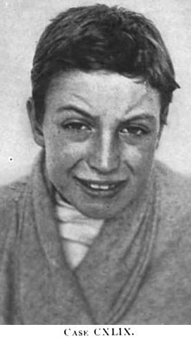
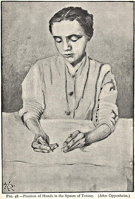
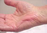
Tetany with carpopedal spasm
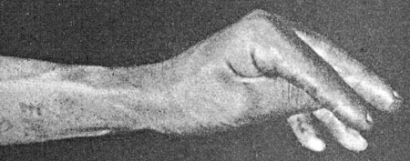
  • Pathophysiology
    • Abnormal Ca++ control of Na+ channels
    • Axonal hyperexcitability: Due to
      • Membrane depolarization
      • Reduced action potential threshold
    • Spontaneous repetitive discharges
      • Frequency: 5 to 15 Hz most common
      • Source: Arise in peripheral axons
  • Causes
    • Ion changes
    • Alkalosis: Respiratory
    • Normocalcemic tetany: Rare
      • Acquired: Associated with neoplasm
      • Hereditary: Dominant with epilepsy
  • Exacerbated by: Hyperventilation; Ischemia
  • EMG
    • Spontaneous axon discharges
      • High rates: Up to 300 Hz
    • Repetitive discharges: Doublets or Multiplets
    • Intense spasm: Maximal interference pattern
    • Normal doublets occur at < 10 Hz
  • Diagnosis: Ischemic forearm test
  • Treatment: Calcium; Magnesium; Phenytoin


Barker


Geniospasm (GSM1) (Trembling chin)

  Chromosome 9q13-q21; Dominant

Palmaris brevis spasm syndrome 2


From: M. Al-Lozi
Movie
From: R Bucelli
Palmaris brevis spasm

Superior Oblique Myokymia


Dopa responsive dystonia


Crisponi syndrome

  Cytokine Receptor-Like Factor 1 (CRLF1) ; Chromosome 19p13.11; Recessive

Whipple's


Muscle Cramp Syndromes: Autosomal Dominant

Familial cramp syndrome
Familial dwarfism with muscle spasms
Muscle cramp & neuropathy syndrome
HANAC
Cramps & myalgias

Muscle cramp & Neuropathy syndrome 1
  Autosomal Dominant
Familial Cramp Syndrome
  Autosomal Dominant
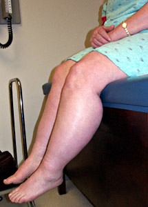
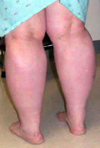
Hereditary Angiopathy, Nephropathy, Aneurysms & Muscle Cramps (HANAC) 35
  Collagen Type IV α1 (COL4A1) ; Chromosome 13q34; Dominant or de novo
  • Epidemiology: > 130 patients with HANAC
  • Genetics
    • Mutation locations: Located in glycine coding regions in exons 24 & 25
    • Mutation effects
      • Dominant negative
      • COL4A1 General: Reduced secretion; Intracellular accumulation
      • Intracellular accumulation
        • More with mutations closer to carboxy-terminus of triple-helical domain
      • Myopathy: More with mutations nearest amino terminus
    • Allelic disorders: Other COL4A1 mutations
      • Brain
        • Brain small vessel disease 1 (BSVD1), Dominant
          • ± Hemorrhage or Ocular disorders
          • Porencephaly I
        • Infantile Hemiparesis
        • Schizencephaly
        • Microangiopathy & Leukoencephalopathy, Pontine, Dominant (PADMAL)
      • Eye
        • Anterior segment dysgenesis
        • Optic nerve dysgenesis
        • Glaucoma, Secondary
        • Retina: Hemorrhages
        • Cataract: Congenital
    • Incomplete penetrance
    • Similar spectrum of disorders with COL4A2 mutations
  • Collagens
    • COL4A1: Roles in muscle, myelination, vessels
  • HANAC: Clinical
    • Nephropathy
      • Hematuria: Persistent; Microscopic or Gross
      • Cysts: Bilateral; Large or Small
    • Angiopathy
      • Affects both small vessels and large arteries
      • Causes
        • Leukoencephalopathy
          • Bilateral
          • Supratentorial (Fronto-Parietal)
        • Retinal arteriolar tortuosity
        • Intracranial aneurysms
          • Small
          • Multiple
          • Extra or intradural carotid siphon
          • Unruptured
        • Brain microbleeds
          • Basal ganglia
          • Supratentorial white matter
          • Infratentorial: Cerebellum, Pons
      • Raynaud phenomenon
      • Supraventricular arrhythmia
    • Cramps (15%)
      • Painful
      • Paroxysmal: Last seconds to minutes, occasionally hours
      • Triggers: Exercise; Alcohol
      • EMG: Electrically silent
    • Weakness
      • Proximal
      • Mild
  • Laboratory
    • Serum CK: High, 2x to 7x (20%)
    • Brain MRI: Leukoencephalopathy
    • EMG: Normal
    • Muscle biopsy: Patchy pathology (Gly773Arg mutation)
      • Fiber size: Marked variation
      • Endomysial fibrosis
      • Fat replacement: Focal
      • Fiber types: Normal
      • COX & SDH staining: Reduced in muscle fibers in myopathic areas
    • Peripheral nerve: Hypomyelination in mouse models
HANAC

FLAIR hyperintensities +
  Foci of prior hemorrhage

Multiple small hypointense lesions

Also see

Sporadic Cramp Syndromes


Cramp-Fasciculation syndromes

Pregnancy-associated cramps
Return to Neuromuscular home page
Return to Ion Channel Disorders
Return to Myopathy & NMJ Index

References
1. J Neurol Neurosurg Psychiatry 1999;67:116-119
2. J Neurol Neurosurg Psychiatry 1995:59:182-184
3. Neuromuscular Disorders 1999;9:604-607, J Neurol Neurosurg Psychiatry 2010;81:125-126, Neuromuscular Disord 2011; Online Feb
4. Neuromuscular Disorders 1997;7:241-249
5. Human Genetics May 2000
6. Neurology 2000;55:383-388
7. Neurology 2000;55:1531-1555, Movement Disord 2002;17:853-866
8. J Neurol Neurosurg Psychiatry 2000;69:110-113
9. Neurology 1999;52:1453-1459; Muscle Nerve 2001;24:340-344
10. Ann Neurol 2001;49:486-492, Neurology 2015; Online November
11. NEJM 2001;344:1232-1239
12. Science 2001;293:864-867
13. PNAS 2001;October On-Line
14. Muscle Nerve 2001;24:1542-1547
15. Neurology 2001;57:1849-1857
16. Muscle Nerve 2002;S11:S55–S58, Brain 2002;125:1887-1895
17. Muscle Nerve 2002;S11:S103–S107
18. Brain 2002;125:2392=2407
19. Ann Neurol 2002;51:361–368
20. Neurol India 2002;50:398-407
21. Ann Neurol 2003; Online March
22. Muscle Nerve 2003;27: 449–455
23. Am J Med Genet 2003;118A:52–54
24. BMC Neurology, Neuromuscular Disorders 2002;13:347–351
25. Muscle Nerve 2002;26:702-707
26. Am J Hum Genet 2004; February
27. Arch Neurol 2001;58:225-230
28. Brain 2004; Online July
29. Arch Neurol 2004;61:1938-1942
30. Muscle Nerve 2005; Online February
31. J Neurol Neurosurg Psychiatry. 2005;76:999-1001
32. J Neurol 2006; Online March
33. Infection 2006;34:35-38
34. Muscle Nerve 2007 Apr 2
35. N Engl J Med 2007;357:2687-2695, Curr Opin Neurol 2011;24:63-68, Am J Hum Genet 2019;104:847-860
36. Muscle Nerve 2007;36:400-403, Rheumatology (Oxford) 2023 Feb 7
37. Neurology 2008;70:353-359
38. Muscle Nerve 2007;36:349-356
39. Hum Mutat 2008;29:E100-E102
40. Muscle Nerve 2010 March
41. Brain 2010; Online July, Neurology 2012;79:1136–1144
42. Arch Neurol 2011;68:1180-1184
43. Brain 2011; Online November
44. Clin Exp Neurol 1979;16:49-56
45. Neuromuscular Disorders 2012;22:944–954
46. Neurology 2012;79:1–7
47. Neurology: Neuroimmunology & Neuroinflammation 2015; Online July
48. JNNP 2017l; Online Jan
49. Muscle Nerve 2017 Jan 7, J Neurol Sci 2023;445:120540
50. Neurology 2018; Online Jan
51. Neurol Neuroimmunol Neuroinflamm 2018;5:e438
52. Muscle Nerve 2020 Jan 16
53. Brain 2020;143:452-466
54. Pflugers Arch 2020 May 2
55. J Neurol Neurosurg Psychiatry 2020 Jul 10
56. J Neurol 2020 Dec 2
57. Muscle Nerve 2021 Mar 6
58. Curr Opin Neurol 2021 Jul 20
59. Ann Neurol 2021 Aug 9
60. Brain 2022;145: 607-620
61. JAMA Neurol 2022 Jun 13
62. Am J Hum Genet 2022 Aug 6
63. Neurol Genet 2023;9:e200073
64. Front Neurol 2022;13:830707
65. Brain 2022;145:607-620

2/29/2024