Neuromuscular

Home, Search, Index, Links, Pathology, Molecules, Syndromes,
Muscle, NMJ, Nerve, Spinal, Ataxia, Antibody & Biopsy, Patient Info

BOTULISM 4

Clinical features
Clostridium botulinum
Diagnosis
Mechanisms of action
Pathology
Prevention
Prognosis
Structure
Types
Clinical Syndromes
  Food-borne
  Iatrogenic: Generalized
  GI colonization
  Infantile
  Inhalation
  Wound

Justinus Kerner
1st Described Botulism

Emile Van Ermengem
Isolated
Clostridium botulinum

Clostridium
botulinum

Causative organisms
Botulinum Toxin: Proteins & Transport
  • General: Botulinum toxins
    • Production
      • Only when the spores germinate
      • Germination circumstances
        • Anaerobic conditions
        • Low acidity (pH > 4.5)
        • Low salt & sugar content,
        • Temperatures
          • 37°F–99°F (3°C–37°C)
          • Varies with serotype
      • Most strains produce single serotype
    • Molecular
      • Zinc-endopeptidase protein
      • Produced as
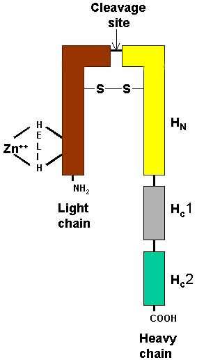
        • Single chain protein (protoxin)
        • Molecular weight ~150 kDa
      • Types share common domain organization
      • Processed into mature toxins by cleavage into 2 subunits
        • 50 kDa light chain (LC)
        • 100 kDa heavy chain (HC)
        • Subunits remain connected through a conserved disulfide bond
        • Disulfide bond is not present in BoNT/Wo
      • Released from bacteria as part of noncovalent multimeric complex
    • Auxiliary proteins
      • Hemagglutinins (HA)
      • Nontoxin, nonhemagglutinin (NTNH)
      • Complex with botulinum
        • May protect toxin at low pH in GI tract
        • Dissociates spontaneously at physiologic pH
        • Some components necessary for toxicity
      • No direct role of auxiliary proteins in toxin-induced Ach blockade
  • Neurotoxic types of botulinum toxin
    • A , A2 , B , C1 , D , E , F , G
    • Type A: Most severe disease & frequent need for ventialtory support
    • Type B: Milder disease
    • Types C & D: Not reported in recent outbreaks
    • Type D: May not be absorbed from human GI tract
    • Type E: Foods of aquatic origin; Varied disease severity; GI symptoms common
    • Type F: Rapid progression & earlier rocovery
  • Tetanus toxin: Sequence homology 30% to 40%
  • Cleavage of Botulinum protoxins: Into 2 chains
    • Heavy (100 kDa) chain
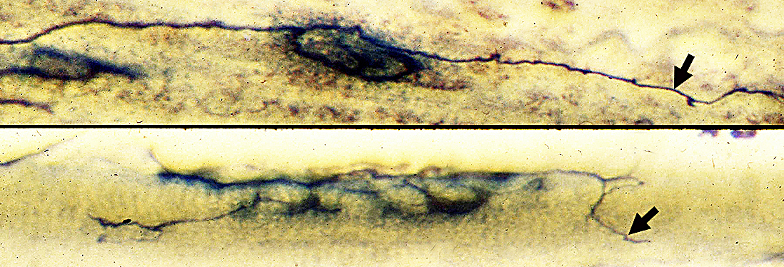
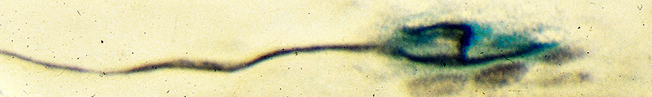
      • C-terminal region (HC) of Heavy (H) chain: Binds to surface of target nerve cells
      • N-terminal region (HN) of H chain: Translocates L chain across membranes
    • Light (L) chain (50 kDa)
      • Light chains have a tetrahedral zinc binding motif: Contains
        • Consensus HELIH amino acid sequence
        • 2 histidines, glutamate & water molecule
        • Structure resembling thermolysin-like endoproteases
      • Contain toxic activity
        • Zinc-dependent endopeptidase
        • Cleave proteins forming synaptic vesicle docking & fusion complex
      • 3-D Geometry: Catalytic sites buried deeply within protein
    • Heavy & Light subunits linked by disulfide bond
    • Double chain product is active form: Inhibits cholinergic transmission
    • Protease producing cleavage
      • Often contained by producing organism
      • May also occur in GI tract
  • Toxin passes from GI tract to vasculature: Mechanisms
    • Transport cell: Absorptive enterocyte
    • Binding to the apical surface of epithelial cells
    • Receptor-mediated endocytosis
    • Transcytosis through cells
    • Delivery to the basolateral surface of cells
    • Ability of toxin to cross cells depends on
      • Toxin type
        • Able to cross cells: Botulinum type A & B
        • Not able to cross cells: Botulinum type C & Tetanus
      • Heavy chain of botulinum toxin: C-terminus
  • Toxin passes out of vasculature to presynaptic regions
    • ? Mechanism
    • Does not cross blood-brain barrier
  • Other absorption routes
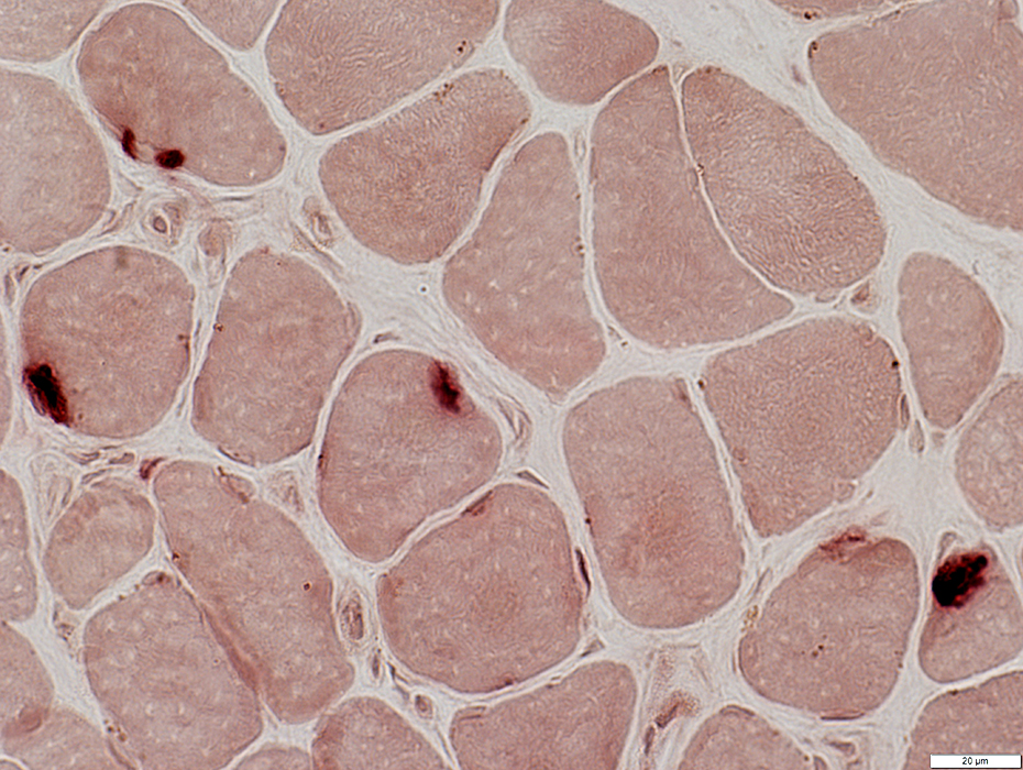
    • Botulinum toxin may be absorbed from respiratory system
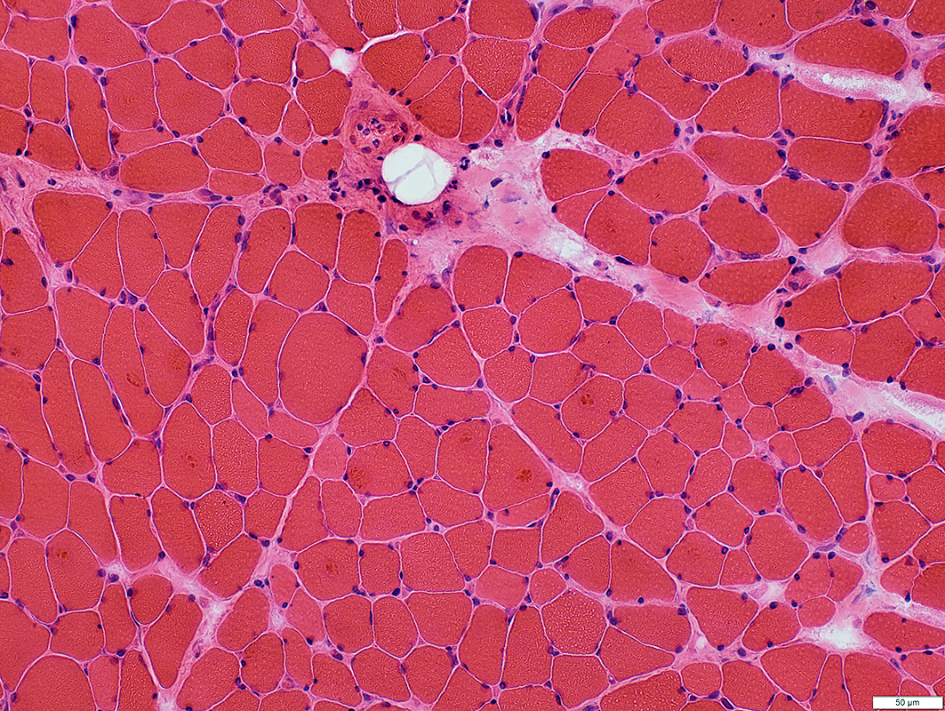
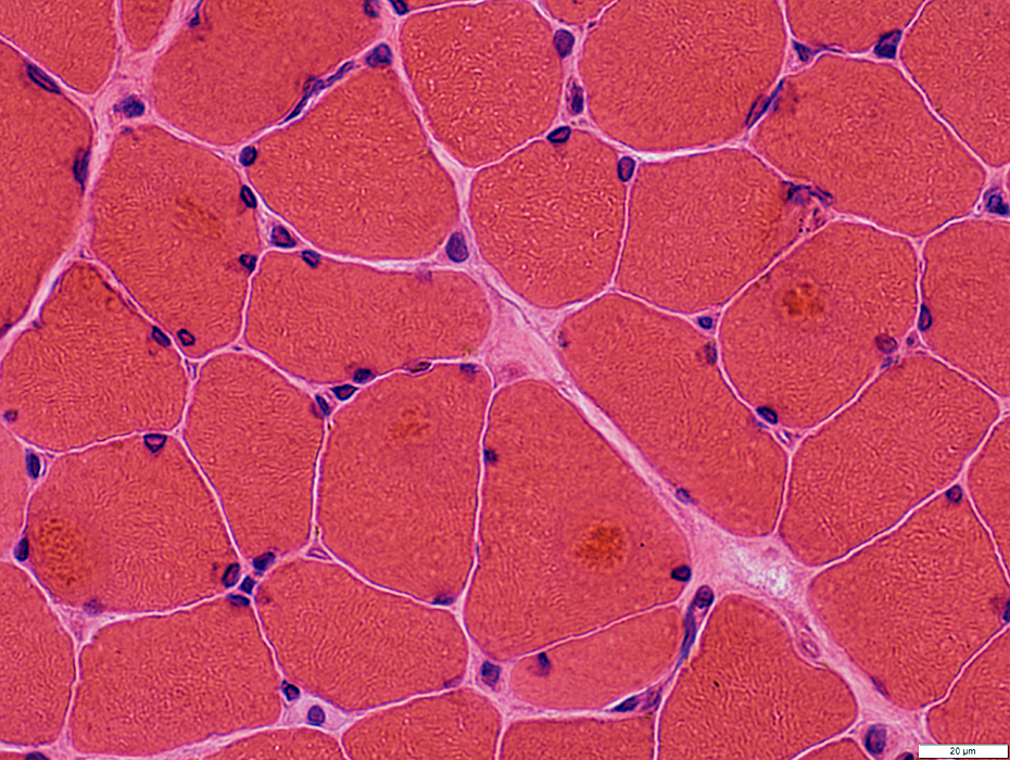
  • General action: Blocks exocytosis
    • Most potent action: Blockade of cholinergic terminals
    • Other blockade of exocytosis: At higher concentrations of toxin
      • Other nerve terminals: Norepinephrine, Serotonin
      • Non-neural cells: If membrane receptors present
Botulinum Toxin
Type B

Botulinum toxin: Mechanism of action at presynaptic nerve terminals 2
  • Overall action: Blocks neurotransmitter release at peripheral cholinergic nerve terminals
    • Neuromuscular junction
    • Autonomic nerve terminals: Sympathetic & Parasympathetic
  • Endplate electrophysiology
    • MEPP frequency: Reduced
    • EPP quantal content: Reduced
  • 4 STEPS in development of presynaptic blockade at NMJs
    1. Binding to receptors on unmyelinated presynaptic membrane
      • Mediated by: Carboxy (C) terminal of Botulinum toxin heavy chain
        • In native state heavy chain is associated with light chain
      • Receptors: Double receptor model
        • First receptors: Presynaptic membrane
          • Receptors: Gangliosides with > one neuraminic (sialic) acid, e.g. GT1b
          • Type of binding
            • Lock & Key
            • Little or no change in conformation of bound botulinum neurotoxin
            • Low affinity
          • Role: Bring toxin into proximity with second receptor
        • Second receptors: In lumen of synaptic vesicles
          • Synaptotagmin 1 : BoNT/B, BoNT/G & mosaic toxin BoNT/DC
          • SV2C : BoNT/A, BoNT/E, BoNT/F, TeNT
      • Susceptibility of cholinergic & non-cholinergic neurons to specific Botulinum & Tetanus toxins 13
        • May be related to ability of toxins to bind to receptors on presynaptic terminals
    2. Uptake of toxin into nerve terminals by endocytosis
      • Bound toxin is internalized by endocytosis
        • Botulinum toxicity is inhibited by Botulinum anti-toxin only in 1st 30 minutes after toxin exposure
      • L-chain is translocated across presynaptic membrane through a channel
      • Translocation is mediated by (N) terminal of botulinum toxin heavy chain (HN)
        • Heavy chain then forms channel releasing light chain into terminal cytoplasm
        • Translocation domain: Kinked α-helices in HN
        • Low pH in endosome necessary
          • ? Brings amphiphilic/transmembrane segments of HN together to form transporter channel
          • ? Causes conformation change in N-terminus of heavy chain
    3. Translocation across endosome membrane
      • General: Movement of catalytically active domain from lumen of endosome to cytosol
      • pH-induced
        • Induces conformational changes in botulinum that expose hydrophobic domains
        • Inhibited by drugs that neutralize endosomal pH (Chloroquine)
      • Insertion of toxin into endosomal membrane
        • Mediated by N-terminus of heavy chain
        • Heavy & light chains become separated
        • Heavy chain: Has dual functions
          • Channel & chaperone, for light chain
          • Associated with cleft formation in endosomal membrane
        • Light chain
          • Transported from endosome to cytosol
          • In cytosol: More neutral pH restores light chain structure
    4. Inhibition of transmitter exocytosis from presynaptic terminal
      • Action of toxin: Zinc dependent proteolysis of protein components of exocytosis (SNARE complex)
        • Enzyme class: Endopeptidase
        • Toxin actions antagonized by metalloproteinase inhibitors
          • Captopril; Phosphoamidon
        • Inhibition produced by: Light chains of Botulinum & Tetanus toxins
        • Protein substrates can only be cleaved before assembly of SNARE complex
        • Endopeptidase activity
          • Mainly seen with reduction of disulfide bond between 2 botulinum toxin chains
      • Molecular targets: Botulinum subtypes & Tetanus toxin
        • General
          • Location of substrates: Nerve terminal cytosol
          • Specific substrates
          • Mechanism of action: Proteolysis of substrates
        • Vesicle associated membrane protein (VAMP; Synaptobrevin)
          • Present in synaptic transmitter-containing vesicles
          • Site cleaved is extravesicle loop of VAMP
            • Botulinum type B: Gln76-Phe77
            • Tetanus: Gln76-Phe77
            • Botulinum type D: Lys59-Leu60
            • Botulinum type F: Gln58-lys59
        • Synaptosomal-associated protein (SNAP-25 )
          • Attached to syntaxin & presynaptic membrane
          • Cleaved near C-terminus by
            • Botulinum type A: Gln197-Arg198
            • Botulinum type E: Arg180-Ile181
        • Syntaxin 1
          • Presynaptic membrane protein associated with
            • Ca++ channels & SNAP-25
          • Cleaved near C-terminus by
            • Botulinum type C1
            • Only when in membrane
      • Termination of action
        • Mechanism unknown
        • Type A toxin with longer action than type B

Clostridium
toxins
Target proteins
cleaved
Botulinum
    A
    B
    C
    D
    E
    F
    G
    Wo
Tetanus

SNAP-25
VAMP (Synaptobrevin)
SNAP-25; Syntaxin
VAMP (Synaptobrevin)
SNAP-25
VAMP (Synaptobrevin)
SNAP-25; VAMP
VAMP (Synaptobrevin)

Botulinum toxin: Subtypes & Features
Botulism: Clinical features
Botulism: Diagnosis 15
Botulism: Clinical syndromes

Food-borne
GI colonization
Iatrogenic
Inhalation
Wound


Botulism: Prevention

Botulism: Pathology

Muscle fibers
NMJs
Terminal Axons
  Other disorders

Terminal axons


Botulinum toxin treatment: Terminal axons at Neuromuscular junctions (Rat)
Botulinum toxin induces: Enlargement of NMJs (middle) & Sprouting of nerve terminals (middle & top)

Normal NMJ
Terminal axon branches at NMJ (Surrounded by esterase stain (Blue))


Botulinum toxin: Muscle pathology

H&E stain
Muscle Fiber Sizes
  Varied
  All fibers are small
  Aggregates in some muscle fibers are targets

H&E stain


NADH stain
Botulinum toxin: Targets, NADH stain
  Rounded pale regions in muscle fibers
  Usually surrounded by dark rim

NADH stain


VvG stain
Botulinum toxin: Targets, VvG stain
  Rounded dark regions in muscle fibers
  Usually surrounded by irregular cytoplasm

VvG stain

ATPase pH 9.4 stain
Botulinum toxin: Fiber types
  Atrophy: All muscle fibers
  Type 2 fibers (Dark): Smaller than type 1
  Targets: More in larger, type 1 muscle fibers

ATPase pH 9.4 stain


ATPase pH 4.3 stain
Botulinum toxin: Fiber types
  Few small type 2 C fibers


Esterase stain
Botulinum toxin: Neuromuscular Junctions
  Varied morphology
    Dark-stained
    Several patches on one fiber
    Small & Pale

Esterase stain

Botulinum Toxin, Type B

Return to Neuromuscular Home Page
Return to Neurotoxins

References
1. Muscle Nerve 2000;23:1137-1144
2. Nature Structural Biology 2000;7:617-619, Toxicon 2020;175:36-43
3. JAMA 2001;285:1059-1070
4. Annu Rev Pharmacol Toxicol 2004;44:167–193
5. Neurology 2005;64:2029–2032
6. Emerg Infect Dis 2009;15:69-71
7. J Zoo Wildl Med 2012;43:388-390
8. Case Rep Neurol 2012;4:113-115, Neuromuscul Disord 2012 Jun 22
9. Neurology 2005;65:1694-700, Emerg Infect Dis 2012;18:102-104
10. Clin Microbiol Infect 2015; Oct 20
11. Toxicology 201;335:79-84
12. MMWR Morb Mortal Wkly Rep 2019 ;67(5152):1415-1418
13. Toxicon 2020;178:20-32
14. Nat Commun 2019;10:2869
15. MMWR Recomm Rep 2021;70:1-30

2/23/2022