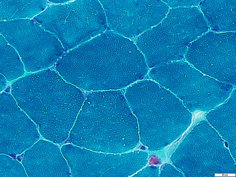
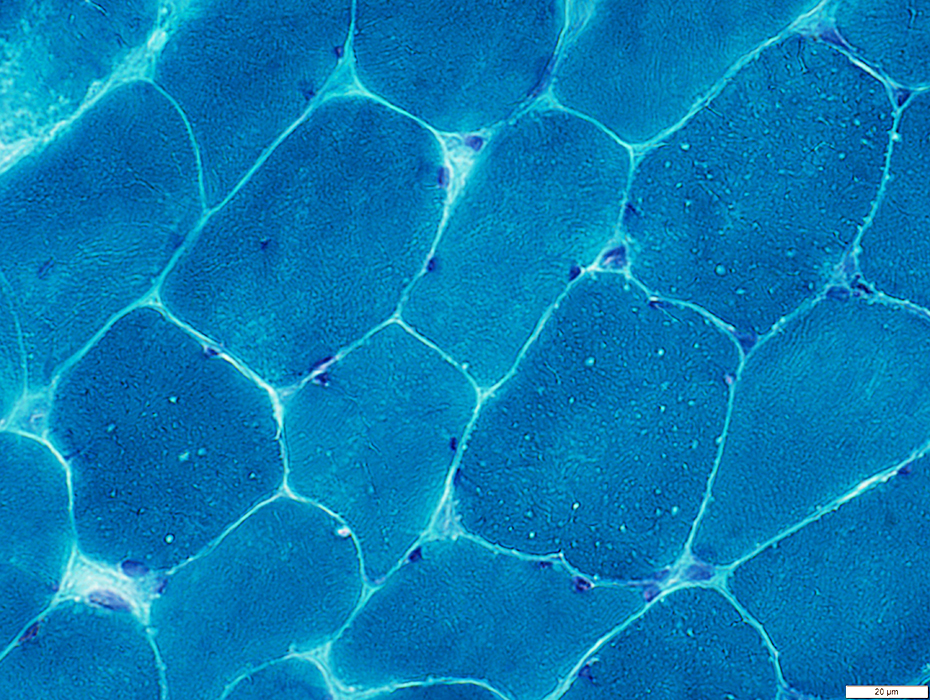
Myotonia Congenita
CLCN-1: Gly230Glu Mutation, Dominant inheritance (Thomsen Syndrome)
52 year old patient
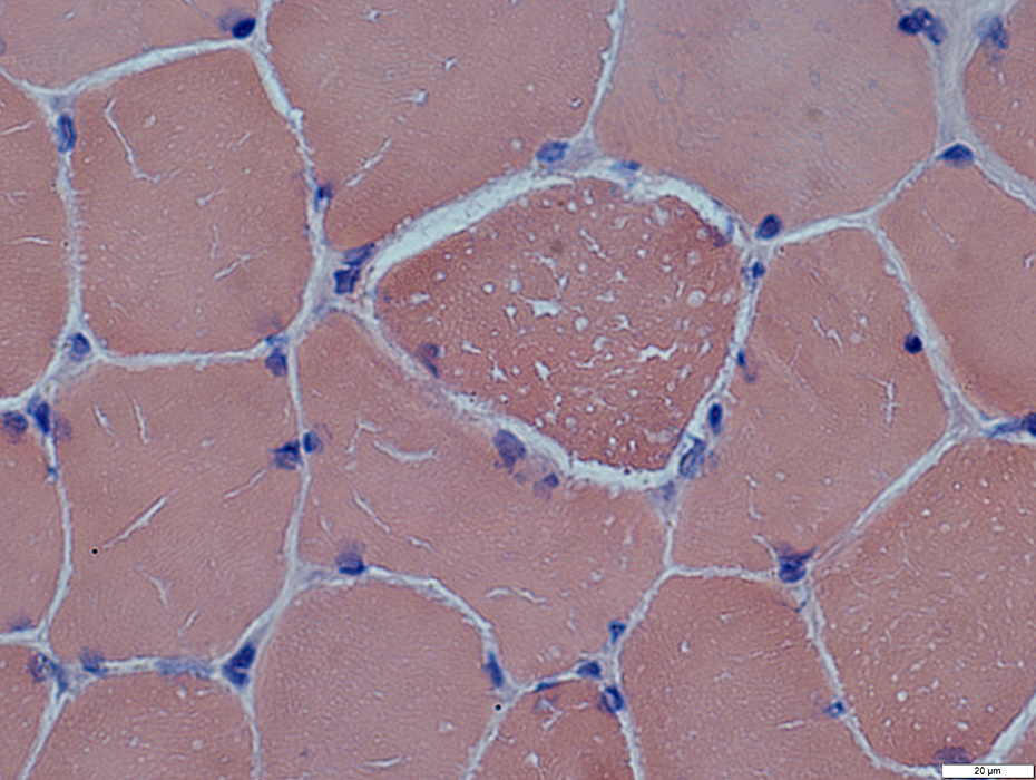
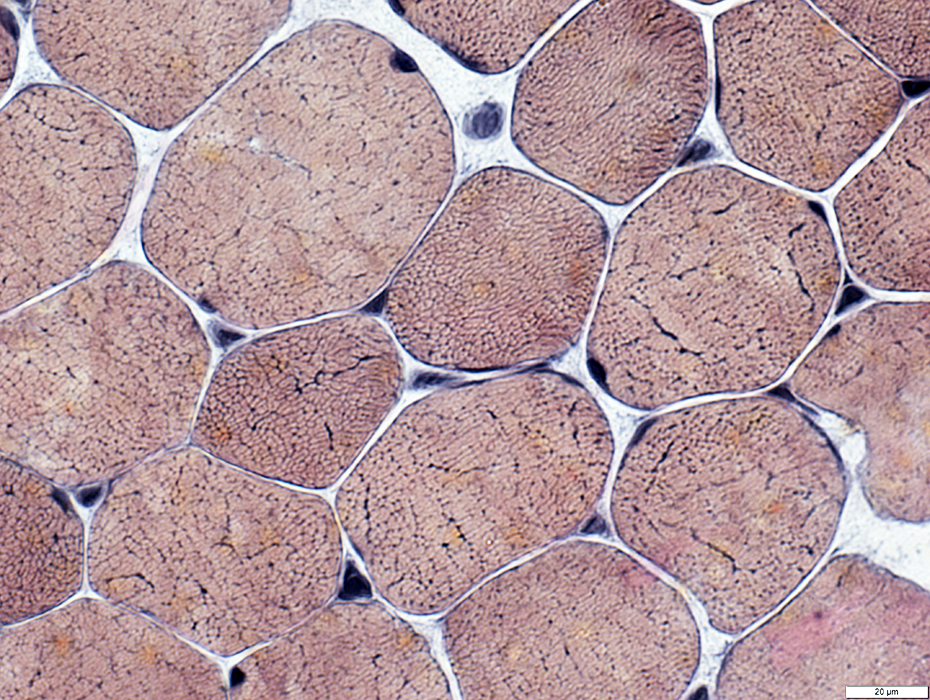
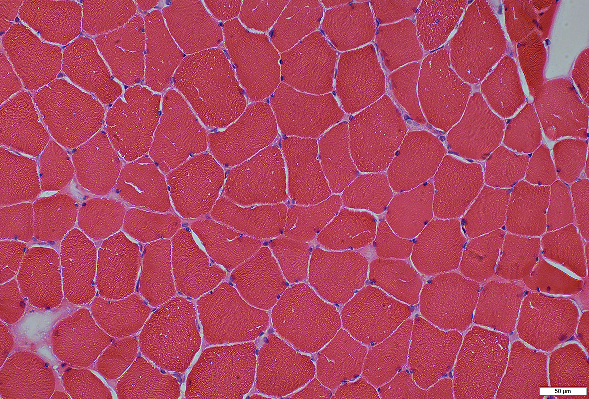 H&E stain |
Fiber Sizes: Mildly varied
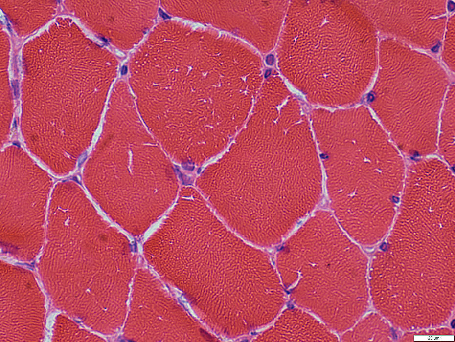 H&E stain |
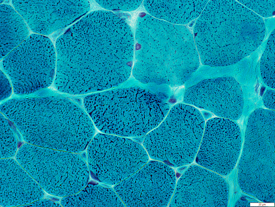
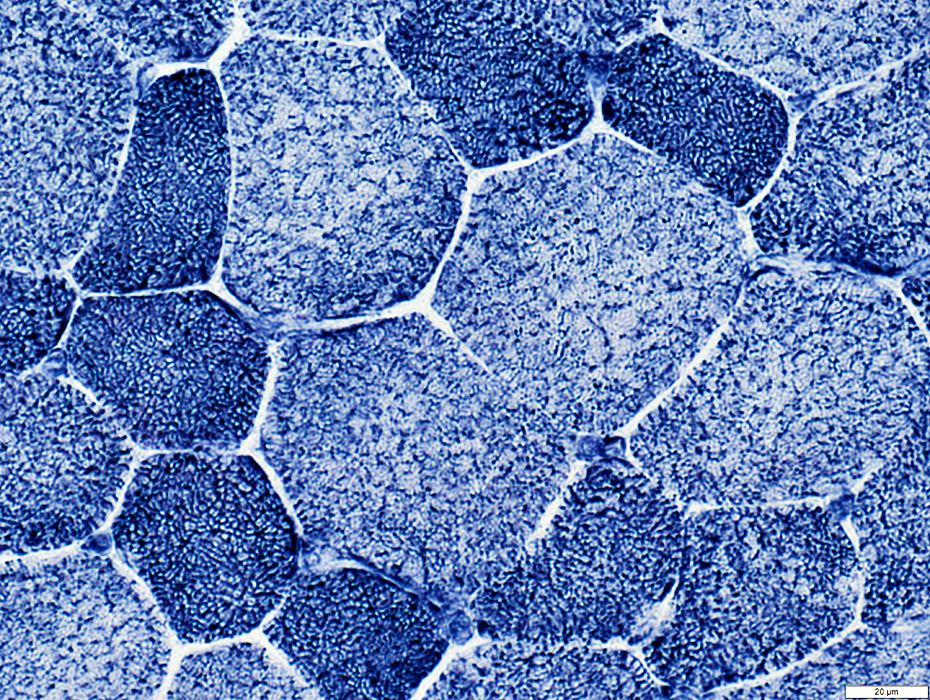
 Gomori trichrome stain |
Small, Irrregular-shaped or Rounded, Clear regions scatttered in fiber cytoplasm
 Gomori trichrome stain |
 Congo red stain |
Small, Irrregular-shaped or Rounded, Clear regions scatttered in fiber cytoplasm
 VvG stain |
A few fibers have dark-stained punctate regions in cytoplasm
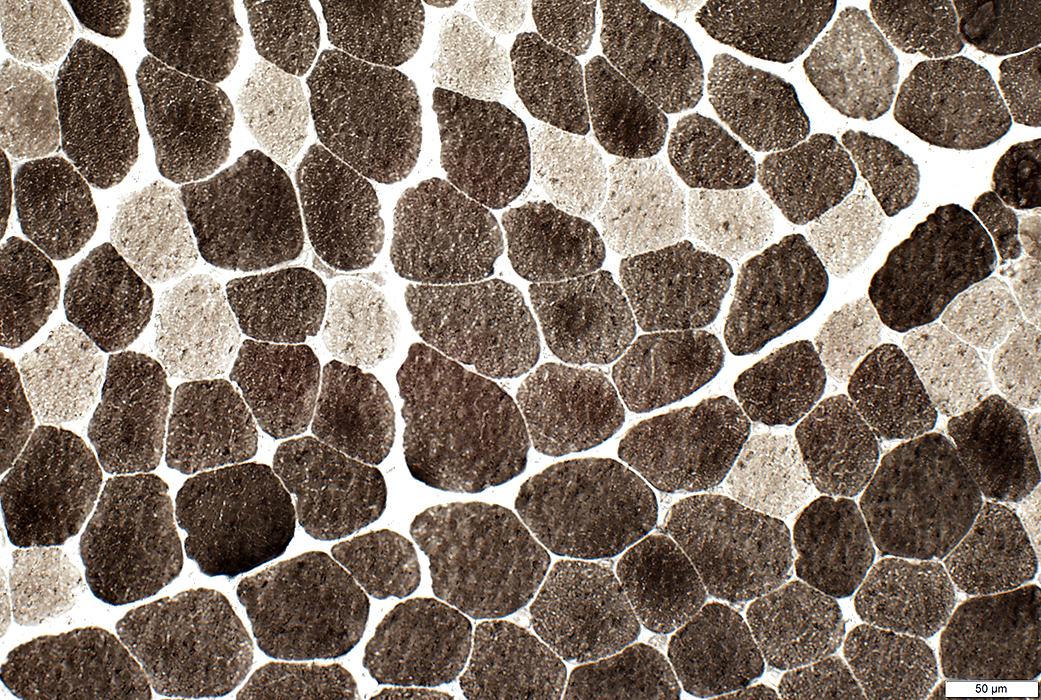
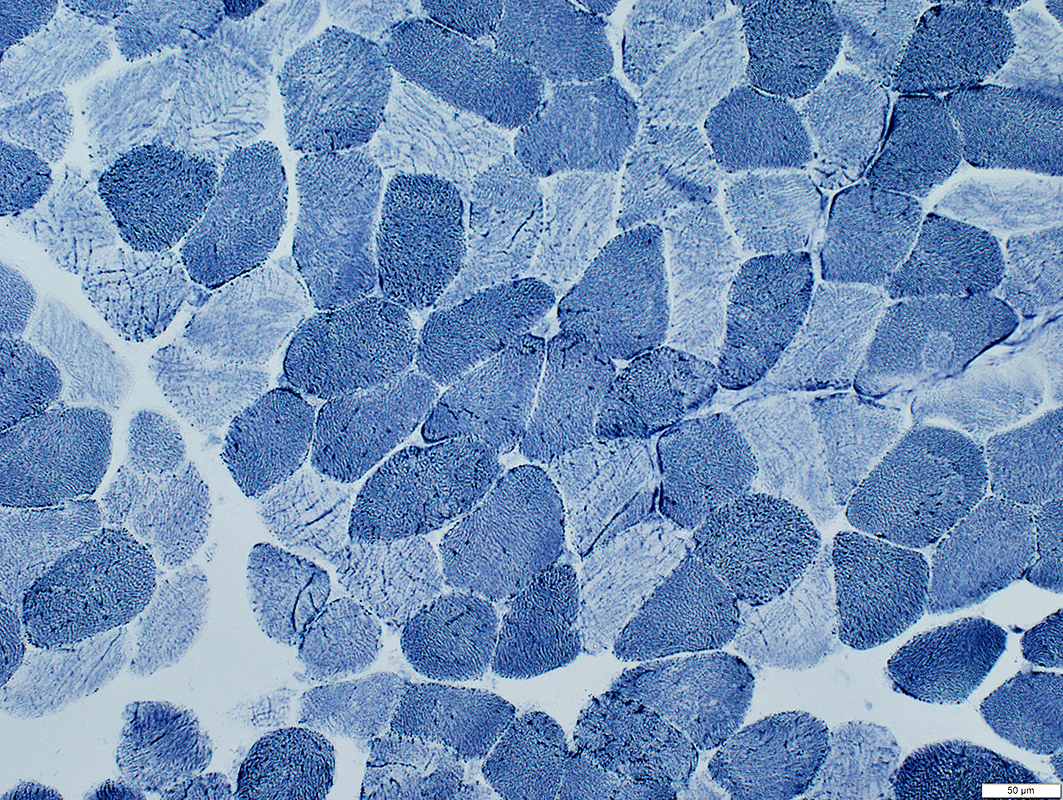 NADH stain |
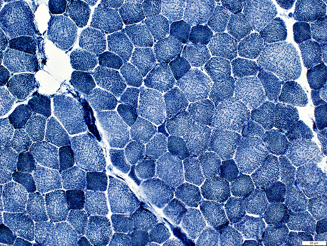
Darker (Type 1) fibers: Punctate or Irregular linear staining
Lighter (Type 2) fibers: Linearization of internal architecture
 NADH stain |
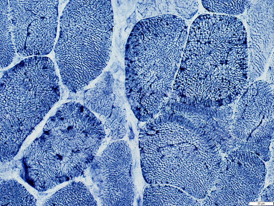 NADH stain |
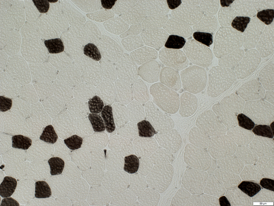
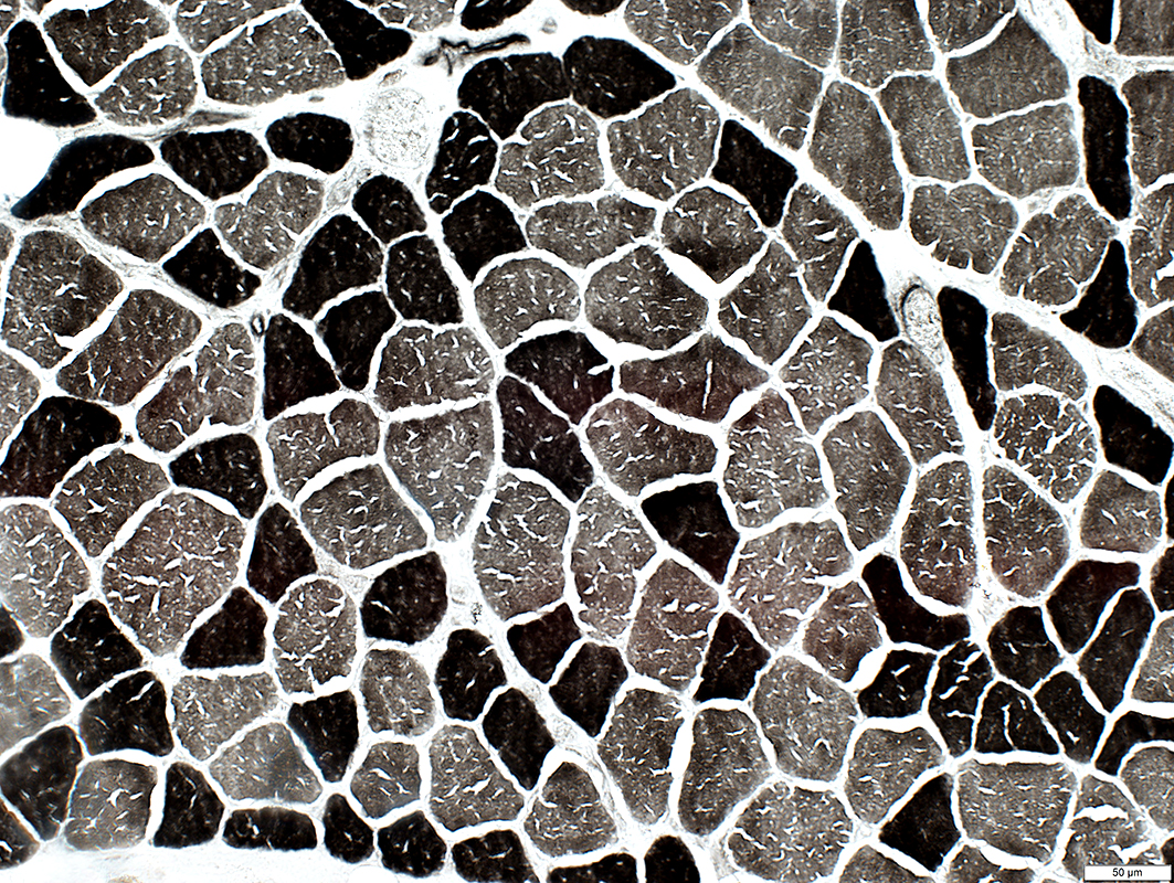 ATPase pH 9.4 stain |
Irrregular "cracks": More prominent in type I fibers
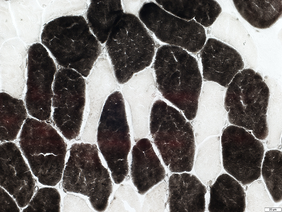 ATPase pH 4.3 stain |
No 2C fibers
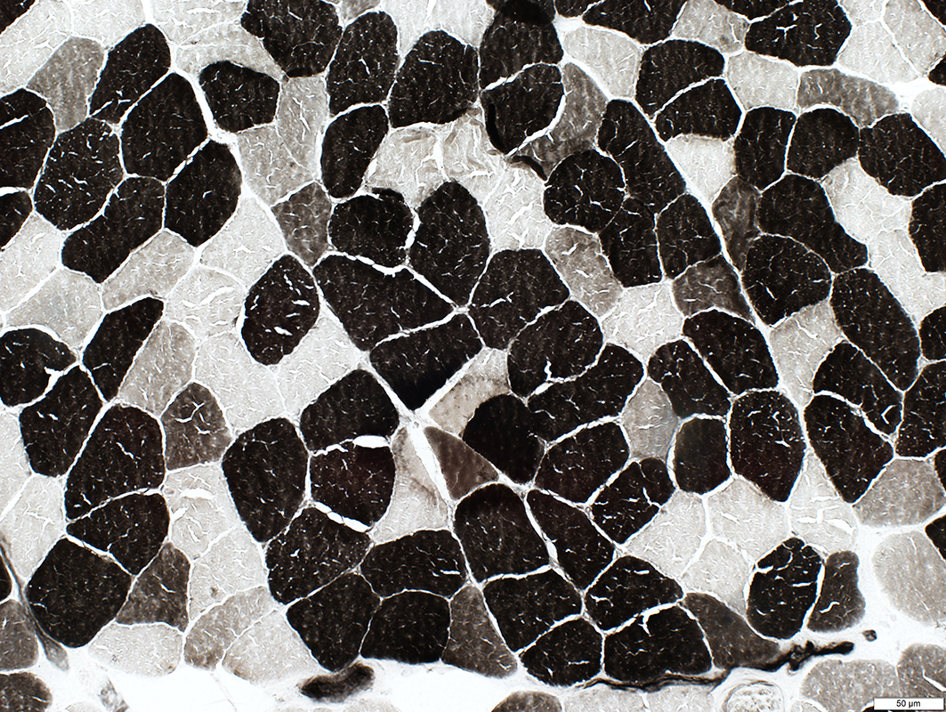 ATPase pH 4.6 stain |
Many 2B fibers fibers
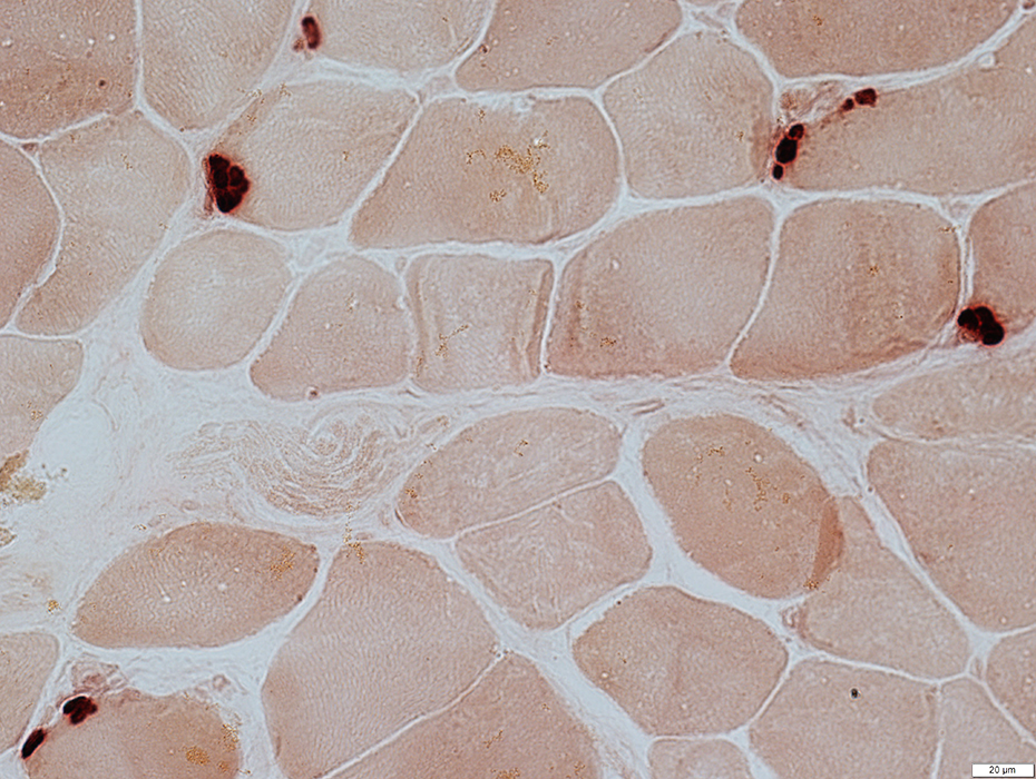 Esterase stain |
Several patches of esterase staining
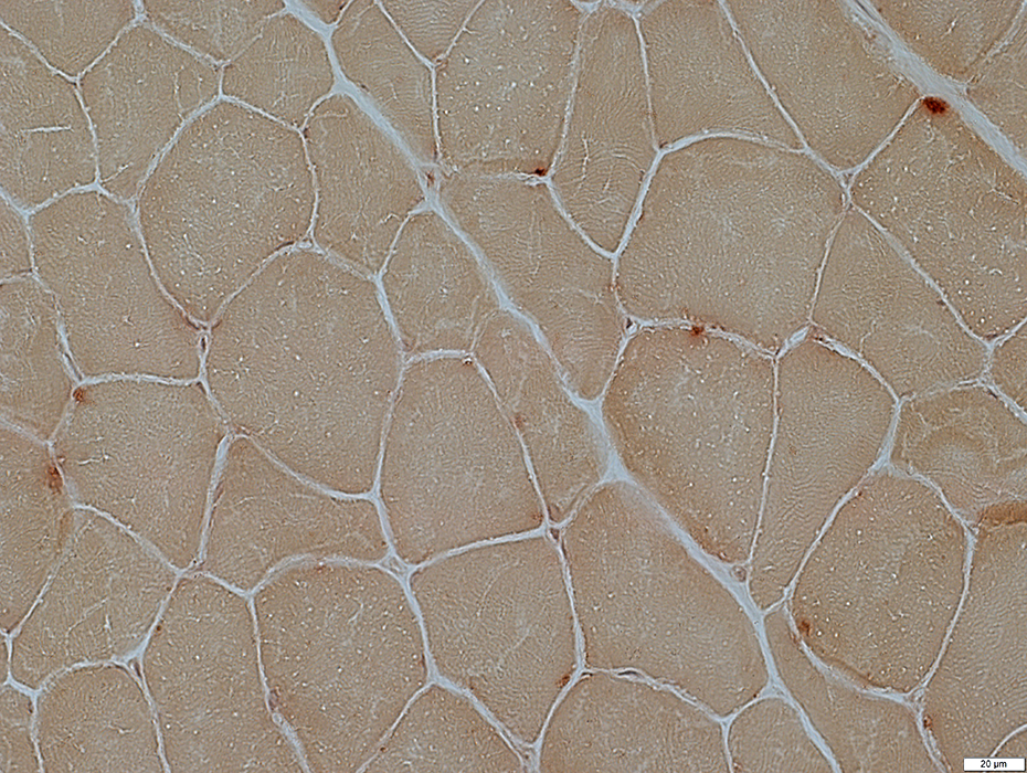 Acid phospbhatasestain |
No lipofuscin
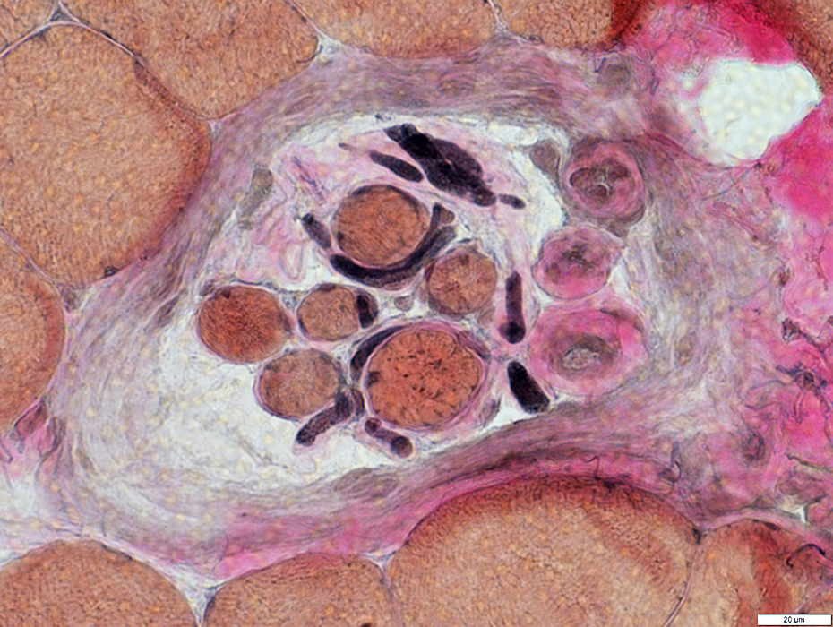 VvG stain |
Unusual feature: Small, thick-walled vessels inside spindle
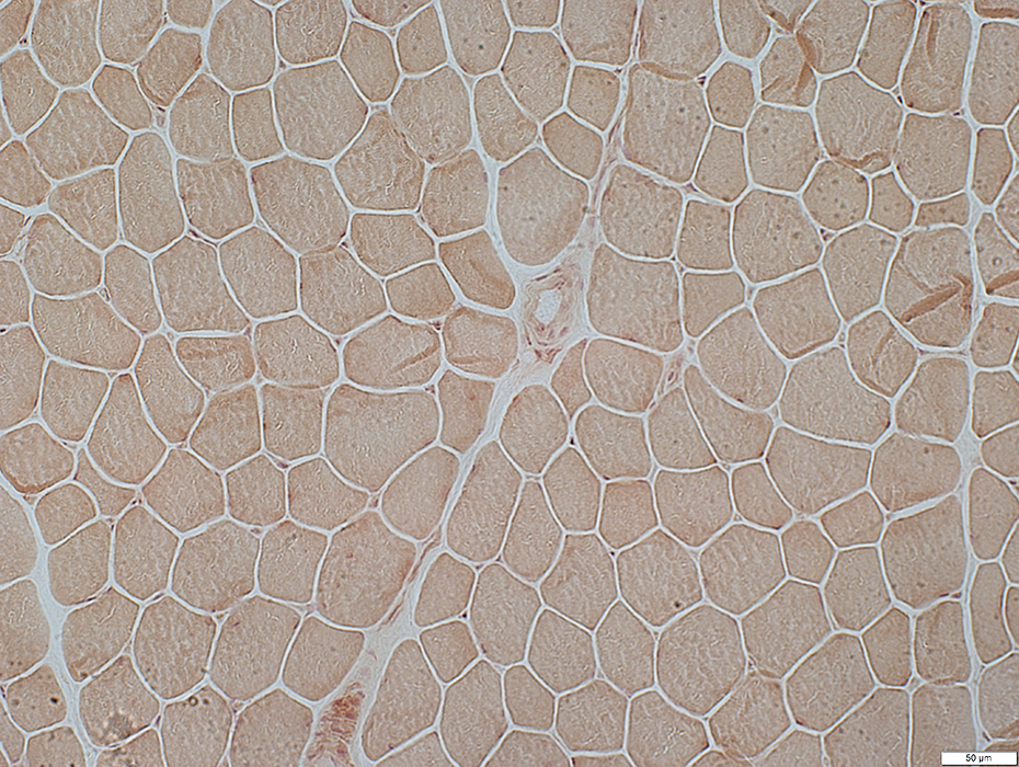
Myotonia Congenita
CLCN-1: Gly230Glu Mutation, Dominant inheritance (Thomsen Syndrome)
15 year old patient
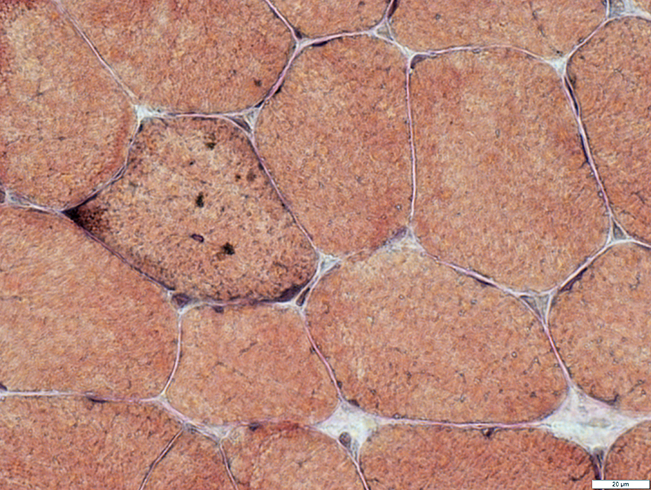
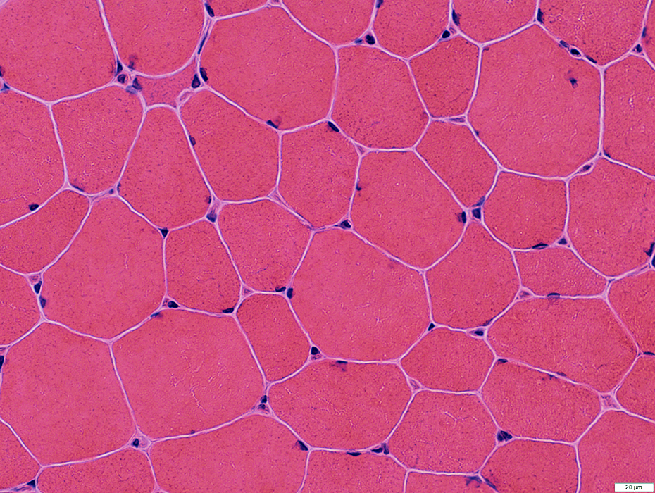 H&E stain |
Varied size
Few small angular fibers
 Gomori trichrome stain |
Internal architecture: Prominently stained
 VvG stain |
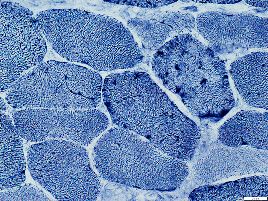
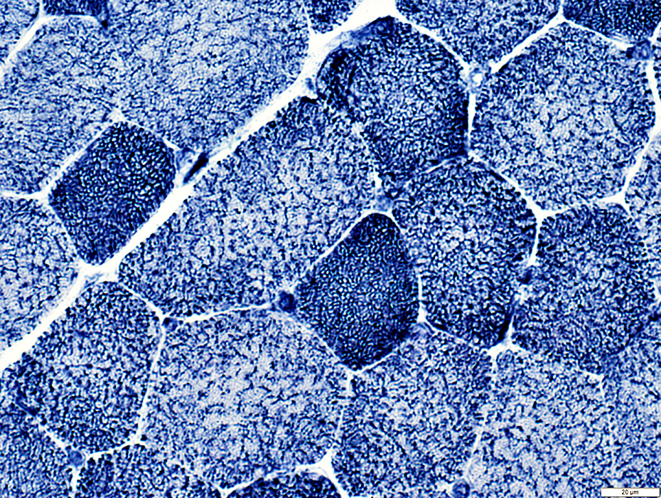
 NADH stain |
Type 1 fibers: Dark & Continuous (Immature)
Type 2 fibers: Punctate or Irregular
 NADH stain |
 NADH stain |
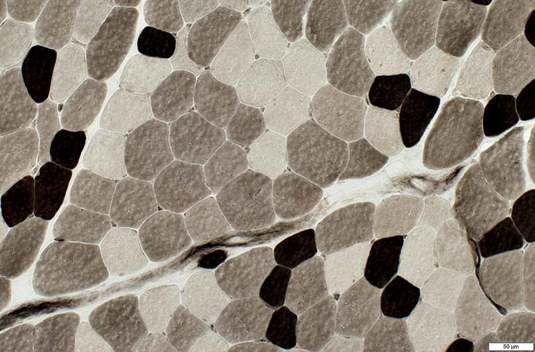
 ATPase pH 9.4 stain |
Type 2 fibers: Predominance; Mildly larger than type 1
 ATPase pH 4.3 stain |
 ATPase pH 4.6 stain |
Many Type 2B fibers: Larger than other fiber types
 Acid phosphatase stain |
No lipofuscin
Return to Neuromuscular Home Page
5/10/2020