Neuromuscular

Home, Search, Index, Links, Pathology, Molecules, Syndromes,
Muscle, NMJ, Nerve, Spinal, Ataxia, Antibody & Biopsy, Patient Info

NERVE: PROXIMAL MOTOR

Brachial neuropathies
  Acquired
  Hereditary
  Radiation vs Neoplastic
Demyelinating neuropathies
Diabetic amyotrophy
Lumbosacral plexopathies
  Types
  Radiation vs Neoplastic
Motor neuropathy, Progressive
Myopathy
Rule out
Spinal muscular atrophy
Thoracic outlet syndromes


Putnam 1909
Wasting of Shoulder muscles


BRACHIAL NEUROPATHIES

Brachial Plexus Structure
  Upper extremity innervation
  Cervical radiculopathy

Hereditary disorders
  HNPP
  HNA: Acute attacks
  HNA2: Chronic, Undulating
  HNA3: Acute attacks

Acquired disorders
  Diagnoses by anatomy
    Supraclavicular
      Lower plexus
      Upper plexus
    Infraclavicular
    Root avulsion
  Types
    Amyotrophic Diplegia
    Burner syndrome
    Cervical polyradiculopathy
    Gunshot wounds
    Heroin
    Infection
      West Nile
    Neonatal (Obstetric)
    Neoplastic
    Neuralgic amyotrophy
    Postoperative
    Radiation
    Rucksack paralysis
    Thoracic outlet syndromes
    Traction


Brachial Plexus: Structure 8

  • Length: Mean = 15.3 cm
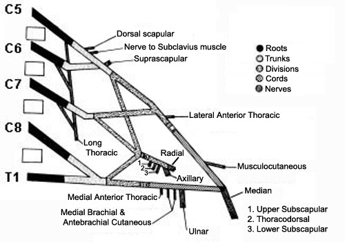
  • Regions
    • Roots
      • Number
        • Usual: 5; C5 to T1
        • Variants
          • C4 & T2 may contribute axons to plexus
          • No effect on lesion location
      • Most proximal areas of plexus
      • Root branches to brachial plexus: Anterior primary rami
        • Located deep in neck: Between anterior & middle scalene muscles
        • Nerves directly from anterior primary rami
          • Scalene muscles (C5 - C8)
          • Longus coli (C5 - C8)
          • Long thoracic nerve to serratus anterior (C5 - C7)
          • Phrenic nerve, inferior contribution (C5)
          • Dorsal scapular to rhomboids & Levator scapulae (C5)
          • No somatic axons originate from T1 anterior primary rami
      • Most motor axons: C5 & C6
      • Most sensory axons: C7
    • Trunks
      • Number: 3; Upper, Middle, & Lower
      • Location
        • Posterior cervical triangle
        • Behind clavicle & sternocleidomastoid muscle
      • Lower trunk: Adjacent to subclavian artery & apex of lung
    • Divisions
      • Number: 6; 3 Anterior & 3 Posterior
      • Location: All retroclavicular
      • Functions
        • Anterior divisions: Tend to supply flexor muscles
        • Posterior divisions: Tend to supply extensor muscles
    • Cords
      • Number: 3; Lateral, Posterior & Medial
      • Location: Near axillary artery & lymph nodes; Below pectoralis minor
    • Terminal nerves
      • Location of origin: Distal axilla
  • Anatomical classification of lesions
    • Supraclavicular
      • Anatomy: Root & Trunk lesions
      • Frequency: More common
      • More frequently due to closed traction
        • Produces long lesions
      • More severe
      • Myelogram: Root avulsion
        • Deformed dural pouches
        • Poor root sleeve filling
        • Cord edema or atrophy
        • Most reliable for C8 & T1 lesions
      • Outcome
        • Generally worse
        • Upper lesions: Best prognosis
          • More commonly demyelinating
          • Closer to innervated muscles
          • Surgically accessible
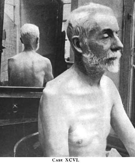
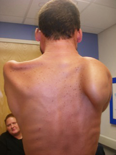
      • Specific syndromes
    • Retroclavicular
      • Anatomy: Divisions
      • Usually associated with upper or lower plexus lesions
      • Isolated lesions: Uncommon
    • Infraclavicular
      • Anatomy: Cords & Terminal nerves
      • No regional differences in incidence, severity, prognosis
      • Associated lesions: Humeral fracture; Glenohumeral dislocation
  • Electrophysiology of brachial plexus lesions
    • Useful for differentiating pre- & post-ganglionic lesions
    • SNAPs: Useful for localization of root lesions
    • CMAPs: Useful for determining
      • Demyelination
      • Severity of lesions (before reinnervation)
   
Muscle Innervation: Brachial Plexus
Trunks
Upper trunk (C5-C6)
  Supraspinatus
  Infraspinatus
  Biceps
  Deltoid
  Teres minor
  Triceps
  Pronator teres
  Flexor carpi radialis
  Brachioradialis
  Extensor carpi radialis
  Brachialis

Middle trunk (C7)
  Pronator teres
  Flexor carpi radialis
  Triceps
  Anconeus
  Extensor carpi radialis
  Extensor digitorum communis

Lower trunk (C8-T1)
  Abductor pollicis brevis
  Flexor pollicis longus
  Pronator quadratus
  Extensor indicis proprius
  Extensor pollicis brevis
  Extensor carpi ulnaris
  First dorsal interosseous
  Abductor digiti minimi
  Adductor pollicis
  Flexor digitorum profundus
  Flexor carpi ulnaris
Cords
Lateral cord
General: Musculocutaneous & Median
  Biceps
  Brachialis
  Pronator teres
  Flexor carpi radialis

Medial cord
General: Median & Ulnar nerves
  Abductor pollicis brevis
  Opponens pollicis
  Flexor pollicis longus
  First dorsal interosseous
  Adductor pollicis
  Abductor digiti minimi
  Flexor carpi ulnaris
  Flexor digitorum profundus

Posterior cord
General: Axillary & Radial nerves
  Latissimus dorsi
  Deltoid
  Teres minor
  Triceps
  Anconeus
  Brachioradialis
  Extensor carpi radialis
  Extensor digitorum communis
  Extensor pollicis brevis
  Extensor carpi ulnaris
  Extensor indicis proprius

ACQUIRED BRACHIAL PLEXOPATHIES
Differential diagnosis by anatomy

Supraclavicular Infraclavicular plexus Total plexus Root avulsion: Tearing of ventral or spinal roots from spinal cord

ACQUIRED BRACHIAL PLEXOPATHIES: Types

Burner syndrome
Gunshot wounds
Heroin
Medial brachial fascial compartment syndrome
Neonatal (Obstetric; Erb's)
Neoplastic
  Primary
  Secondary
Neuralgic amyotrophy
Pancoast
Postoperative
Radiation
Rucksack paralysis
Thoracic outlet syndromes
Traction
Trauma


General: Vulnerability to trauma or compression


Brachial plexus: Traction lesions


Postoperative paralysis


Post-Median Sternotomy Paralysis


Neuralgic
  Amyotrophy 10


Rucksack paralysis


"Burner" syndrome


Thoracic outlet syndromes


Gunshot wounds


Neonatal brachial plexopathy 7


Heroin plexopathies


Pancoast syndrome


Medial Brachial Fascial Compartment Syndrome


Primary Brachial Plexus Neoplasms


BRACHIAL PLEXOPATHY: RADIATION vs NEOPLASTIC
Neoplastic Radiation 12
  • Onset
    • Pain: Shoulder & hand
    • Mass
    • < 6 months after radiation
  • Clinical
  • Neoplasm
    • Lung: 3%; Smoking history
    • Metastases elsewhere
  • Imaging: Focal mass
  • Pathology: Neoplasm
    • 1°: Neurofibraoma
    • 2°: Lung; Breast
  • Onset
    • Years (Up to 2 decades) after radiation
    • Paresthesias: Median-innervated hand
  • Clinical features
    • Infraclavicular lesion
    • Weakness: Arm, Distal then Proximal
    • Fasciculations
    • Long duration (> 2 to 4 years)
    • Progression: Slow
    • Little pain
    • Treatment: ? Anticoagulation
  • Neoplasm
    • Breast
    • Head & Neck
  • Risk factors
    • Radiation
      • High dose: > 44 to 55 Gy
      • Posterior axillary
    • Axillary nodes: # Removed
    • Systemic: Smoking; Diabetes
  • Electrodiagnostic
    • EMG
    • NCV
      • Median sensory: Small
      • Conduction block across clavicle
      • Segmental slowing
  • Radiology
    • Fibrosis: Lung apex; Pericardium
    • Bone necrosis
  • Pathology
    • Vascular endothelium (usual)
    • Radiation induced fibrosarcoma
      • 4 to 41 yrs later

HEREDITARY BRACHIAL PLEXOPATHIES: Autosomal dominant

HNPP: Recurrent; Painless
HNA: Acute attacks; Pain
HNA2: Chronic, Undulating; Pain
HNA3: Acute attacks; Pain

HNA eyes: Hypotelorism


Neuropathy with liability to pressure palsies (HNPP)
  PMP-22 deletion: Chromosome 17p12; Dominant


Neuralgic Amyotrophy (HNA1)
  SEPT9 ; Chromosome 17q25; Dominant - distinct from HNPP


From: A Connolly


Chronic Neuralgic Amyotrophy (HNA2) 4
  Autosomal Dominant

Chronic Neuralgic Amyotrophy (HNA3) 6
  Autosomal Dominant: Not linked to 17q25

Lower Motor Neuron Syndrome: Proximal Arm Predominant 1, 2


LUMBOSACRAL PLEXOPATHIES

Circulatory
  Arterial injections
  Hemorrhage
  Ischemic
Diabetic Amyotrophy
Differential diagnosis:
  Cauda equina lesion
  Hip disease
  Episodic
Hereditary
  HNPP
  Neuralgic amyotrophy
Heroin
Immune
Infection
Neoplastic
Obstetric-Gynecologic
Radiation
Trauma

Lumbosacral Plexus

Psoas hematoma (arrow): CT scan


Lumbosacral plexopathy: Immune (Small vessel vasculopathy)

Lumbosacral plexopathy: Hemorrhage

Lumbosacral plexopathy: Intra-arterial injections

Lumbosacral plexopathy: Ischemic 3

Lumbosacral plexopathy: Trauma

Lumbosacral plexopathy: Obstetric-Gynecologic complications


LUMBOSACRAL PLEXOPATHY: Episodic
Cauda equina lesion Exacerbated walking downhill
No effect of bicycle exercise
Pain & Sensory loss: Distal
Ischemic plexopathy Exacerbated walking uphill
No effect of bicycle exercise
Pain & Sensory loss: Proximal & Distal
Peripheral Arterial:
  Occlusive disease
Distal pulses: Reduced
Pain: Distal
No progressive sensory-motor loss during exercise


LUMBOSACRAL PLEXOPATHY: RADIATION vs NEOPLASTIC
Neoplastic Radiation
  • Onset
    • Pain: Leg
    • Weakness: Proximal
  • Progression: Rapid
  • Weakness
    • Unilateral; Asymmetric
    • Proximal
      • Femoral lesions
  • Pain: Often severe
  • Mass: Palpable rectal
  • Cancer type
    • Colon; Prostate; Sarcoma
  • Imaging: Focal mass
  • Onset
    • Weakness: Distal legs
    • Latency: Months to Years
  • Clincal
    • Weakness: Distal; Asymmetric
    • Progression: Slow
    • Pain: Late; Not disabling
  • Cancer type: Gynecological; Testicular
  • Predisposing treatment
    • High radiation dose: > 55 Gy
    • Chemotherapy
    • NOT hormonal
  • Electrodiagnostic
    • EMG: Myokymia
    • NCV: Absent late reflexes
  • MRI
    • Fibrosis: Multifocal
    • Nerve: Enlargement; Enhancement


Return to Polyneuropathy Index

References
1. Ann Neurol 1990;27:316-326
2. Neurology 1999;53:1071-1076, J Neurol 2015 Dec 24
3. JNNP 1999;67:793-795
4. Brain 2000;123:718-723
5. Neuropediatrics 2000;31:328-332
6. J Neurol 2001;248:861-865
7. Arch Dis Child Fetal Neonatal Ed 2003;88:F185–F189
8. Muscle Nerve 2004;30:547–568
9. Nature Genetics 2005; Online September 25
10. Muscle Nerve 2017 Jan 3
11. Neurology 2017; Online August
12. PM R 2024;16:85-91

2/4/2024