Home
,
Search
,
Index
,
Links
,
Pathology
,
Molecules
,
Syndromes
,
Muscle
,
NMJ
,
Nerve
,
Spinal
,
Ataxia
,
Antibody & Biopsy
,
Patient Info
SPINAL ARACHNOIDITIS
Clinical syndrome
Pain: Low back & radiating down both legs
Weakness: One or multiple lumbar or sacral root distribution
Sensory loss: One or multiple lumbar or sacral root distribution
Causes
Spinal surgery: Especially multiple
Chemical
Oil based radiographic contrast agents
Spinal drugs: Anesthetics; Steroids; Amphotericin B; Methotrexate
Infections: Tuberculosis; Cryptococcosis; Syphilis; Viral
Trauma: Vertebral injuries; Disc herniation
Spinal subarachnoid hemorrhage
TOXIC MYELOPATHIES
Systemic
Organophosphates
:
TOCP
Source: Contaminants in cooking oil or flour; Alcohol substitute
Myelopathy & Polyneuropathy
Pathology: Distal axonal degeneration: CNS & PNS
Iodochlorhydroxyquinoline (Clioquinol)
Nitrous oxide
Substance abuse
Heroin
; ? Cocaine
Anterior spinal artery syndrome
Hexacarbons
: Occasional delayed myelopathy
Muzolimine
Use: Diuretic
Toxicity especially with renal insufficiency
Myelopathy
Polyneuropathy: Demyelination
1-Bromopropane
(n-propyl bromide)
Konzo
Lathyrism
Zinc
Direct
Angiographic contrast agents
Myelographic contrast agents
Acute onset spinal cord irritability: Often transient
Arachnoiditis
Chymopapain: Used to treat herniated discs
Spinal anesthesia: ? direct toxic vs. vascular
Chemotherapeutic agents: Methotrexate; Cytosine arabinoside
Often in patients treated with both systemic & intrathecal drugs
Onset: Acute (Hours) to Subacute
Risk increased
More frequent treatments;
High cumulative dose
Concurrent methotrexate & cytarabine
Spinal radiotherapy
Pathology: 2 types
Spinal white matter: Peripheral cord; Lateral & posterior funiculi
Grey matter lesions
Amphoterecin B
Focal myelopathy
Diffuse radiculopathy
Radiation Myelopathy & Neuropathy
5
General
Disease presence: Dose related (> 4,000 rads)
Disease onset: Weeks to Decades after treatment
Weakness pattern: Asymmetric; Patchy
EMG with nerve damage:
Myokymia
common
Clinical: 4 syndromes
Sensory symptoms: Transient
Onset: 2 to 37 weeks after treatment
Lhermitte sign: Shock-like sensation after neck flexion
Course: Resolution common after 2 to 36 weeks
Myelopathy: Chronic progressive
Onset: Mean 17 months after Rx; Range 3 months to Decades
Asymmetric;
Brown-Séquard syndrome
Course: Progression over months
Treatment: ? Anticoagulation
Pathology: Vasculopathy; White matter > Grey
Transverse myelopathy: Acute
Amyotrophy +, Local
Lesion Locations
Spinal cord: Myelopathy
Radiculopathy
Plexopathy
Brachial
Lumbosacral
Neuropathy: Phrenic; Sciatic
Syndromes: Vary with onset timing
Early: < 6 Months after treatment
Pain: Common
MRI: T2 hyperintensity; Nerve large; Contrast +
Nerve pathology: Axon loss, patchy; Inflammation,Perivascular, Epineurial; Perineurium thick; Hemosiderin
Treatment: May improve incompletely with prednisone
May have monophasic course with improvement but not to normal
Chronic: Months to Decades after treatment
Pain: Some patients; Less severe
Progression: Slow
Nerve pathology: Axon loss; Epineurial vessel fibrosis
ELECTRICAL INJURY
Clinical
Lesion location: Most often cervical
Intensity
< 1,000 volts: Predominantly anterior horn cell damage
> 1,000 volts, or with burns near spinal cord: Lateral & posterior column damage
Course
Acute: Associated encephalopathy
Progressive for few months or static
Pathology
Perivascular & petechial hemorrhages
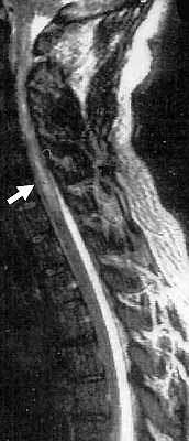
Heroin myelopathy
Increased T2 signal (Arrow)
in cervical spinal cord
HEROIN TOXICITY
Myelopathy
Neuropathy
Rhabdomyolysis
General
Drug route: Intravenous; Intranasal
Myelopathy
1
Onset
Acute, within hours after drug administration, after awakening from sleep
Often related to single dose after period of abstinence
Spinal cord syndrome
Weakness: Paraplegia or Quadriplegia
Tendon reflexes: Reduced or absent
Urinary retention
Prominent recovery may occur over weeks to months
Laboratory
CSF: Usually normal, Occasional pleocytosis or increased protein
MRI: Transverse myelitis-like findings
T2 imaging: Cord swelling; Enhancement, patchy, over several levels
Gadolinium enhancement: Acute
Mechanism of disease: ? Hypersensitivity
Treatment: ? Corticosteroids; Plasma Exchange
Rhabdomyolysis
Several mechanisms of
drug-toxin related rhabdomyolysis
Direct muscle toxicity
Adulterants (Quinine)
Compression: ? Most common for heroin
Inactivity for long period: Prolonged compression
Compartment syndromes
Leg: Gluteal
Associated with sciatic or lumbo-sacral plexus palsy
Arm: Associated with brachial plexus, especially upper, lesions
Ischemic necrosis due to direct arterial injection: Common mechanism for tamazepam
Exacerbating factors: Dehydration; Poor nutrition
Heroin-associated rhabdomyolysis may occur in association with acute myelopathy
Neuropathy
3
Local involvement: Compressive neuropathy
Non-compressive neuropathy
Onset
Time: 3 to 36 hours after heroin administration
Progression: Acute
Patient age: 22 to 42 years
Clinical syndromes
Plexopathy
Onset: Pain; Allodynia
Weakness: Distribution
Lumbosacral or Brachial
Proximal
Single extremity
Asymmetric
Course
Persistent proximal weakness
Pain resolution over months
Sensory-Motor Neuropathy
Onset: Acute
Clinical
Symmetric
Distal
Legs > Arms
Course: Mild or no improvement
Electrophysiology: Axon loss
Nerve pathology: Axon loss
Associated syndrome:
Rhabdomyolysis
Iodochlorhydroxyquinoline (Clioquinol) Myelopathy
Source: Used as anti-diarrheal drug
Epidemiology: Predominantly in Japan
Clinical features
Myelopathy
Dose: Oral intake of 2 g/day for >3 weeks
Onset: Numbness & pain in legs
Clinical
Sensation loss: Especially vibration
Hyperesthesia
Leg weakness & stiffness (50%)
± Optic neuropathy: Especially children
Course: Rapid onset; Slow recovery
1-Bromopropane (n-propyl bromide)
4
Source: Solvent
Substitute for ozone-depleting chloro-fluorocarbons
Vapor & immersion degreasing operations
Aerosol-applied adhesives.
Dry cleaning: Substitute for perchloroethylene (tetrachloroethylene)
Epidemiology: US; Aerosol exposure
Clinical: Myelopathy+
Onset: After exposure of 3 months to years; Ages 16 to 46 years
Systemic: Headache; Dizziness
Myelopathy
Spasticity: Legs
Gait disorder
Weakness: Legs
Reflexes
Tendon: Increased in legs
Plantar: Extensor
Sensory: Panmodal distal loss; Paresthesias & Pain
CNS (50%): Reduced memory; Altered mood; Cognitive change
Course: Partial improvement after exposure discontinued
Laboratory
Serum Chloride & Bromide: High
Nerve conduction testing: Normal or Borderline slow
Konzo
2
Epidemiology
Poor rural communities in Africa: Zaire, Mozambique, Tanzania & Central African Republic
Epidemics: Occur during dry season, especially in drought years
High risk: Women of child-bearing age; Children 3-13 years old
Lower risk: Men; Breast-fed children
Familial clustering
Etiology
Probable
cyanide
toxicity
Source: Cassava plant (Manihot esculenta esculenta)
Consumption of food processed from starchy roots
Roots have high cyanide levels: Patients exposed with poor removal of toxin during processing
Higher risk: Protein-deficient diet with low intake of sulphur amino acids
Sulphur substrates needed for conversion (detoxification) of cyanide to thiocyanate
Onset
Sudden: Less than 1 day
Leg weakness
Occasional: Low back pain; Paresthesias
Clinical
Spasticity: Paraparesis or Tetraparesis
Legs > Arms
Symmetric
Hip adductor spasms
Hands: Reduced fine movements
Tendon reflexes: Brisk
Plantar responses: Extensor
Strength: May be normal in spastic extremities
Course
Partial improvement over months
Then permanent & Non-progressive gait disability
Occasional patients with 2nd exacerbation
Contractures
Other transient features: May persist in severe cases
Eye: Blurred vision; Nystagmus
Speech difficulties: Spasticity
Normal: Mental status; Hearing; Sensation; Bladder, Bowel & Sexual functions
Laboratory
Somatosensory evoked potentials: Prolonged cortical responses & central sensory delay
Return to
Spinal disorders
Return to
Neuromuscular Home Page
References
1. Neurology 2000;55:316-317
2. Disability Rehabilitation 2001;23:731-736, Clinical Neurophysiology 2002;113:10–15
3. Journal of Peripheral Nervous System 2006;11:304–309
4.
Clin Toxicol (Phila) 2007;45:270-276
5.
Neurology 2023;101:e1455-e1460
2/4/2024