HEMOSIDERIN
- Stain: Prussian blue
- Description
- Brown, insoluble, granular pigment
- Contain: Iron
- Location: Extracellular; Within macrophages
- Disorders: Hemosiderin deposits in muscle or nerve occur in
- Hemosiderosis
- Hemochromatosis
- Other systemic iron overload
- Local hemorrhage
- Trauma
- Vasculitis & Vasculopathy
- Tissue Locations
- Endomysium: Near capillaries
- Epineurium: Near vessels
- Perimysium: Near vessels
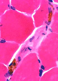
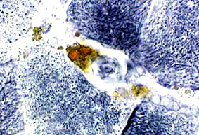
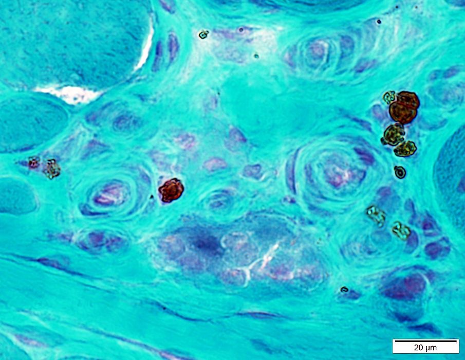
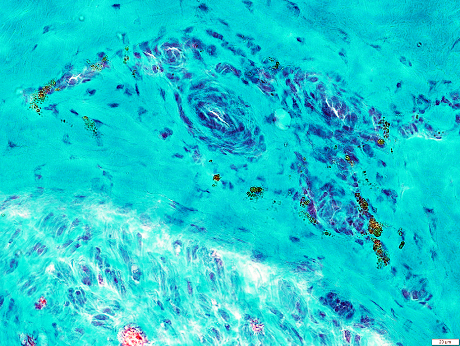
Hemosiderin deposits: Endomysial
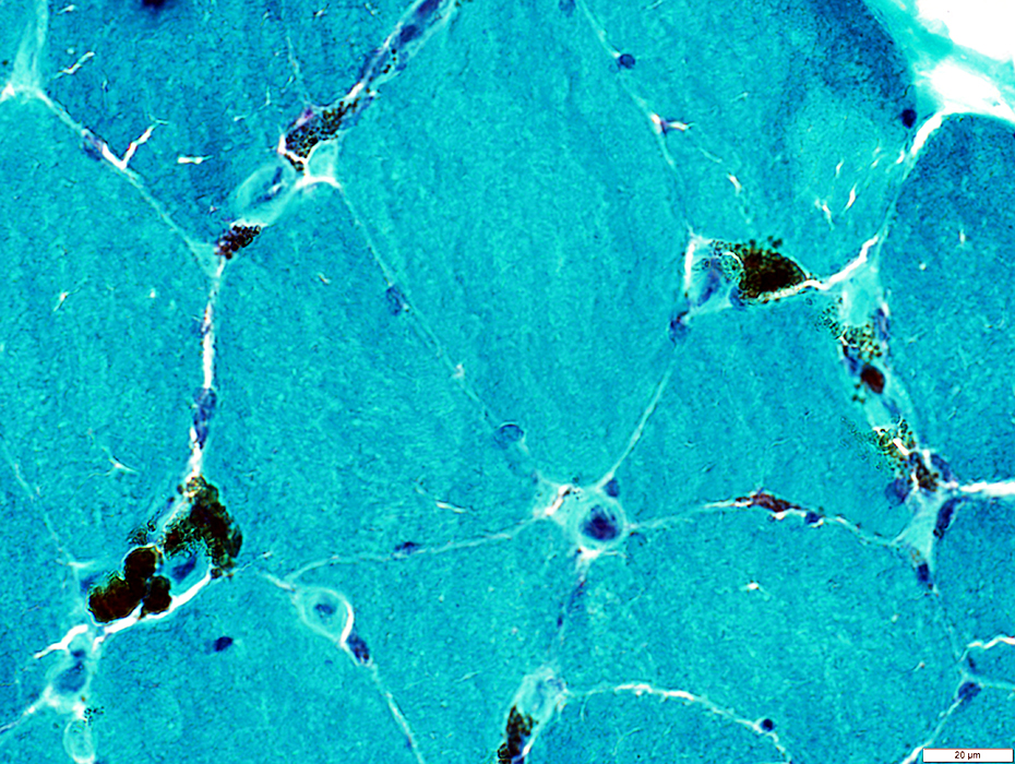 Gomori trichrome stain |
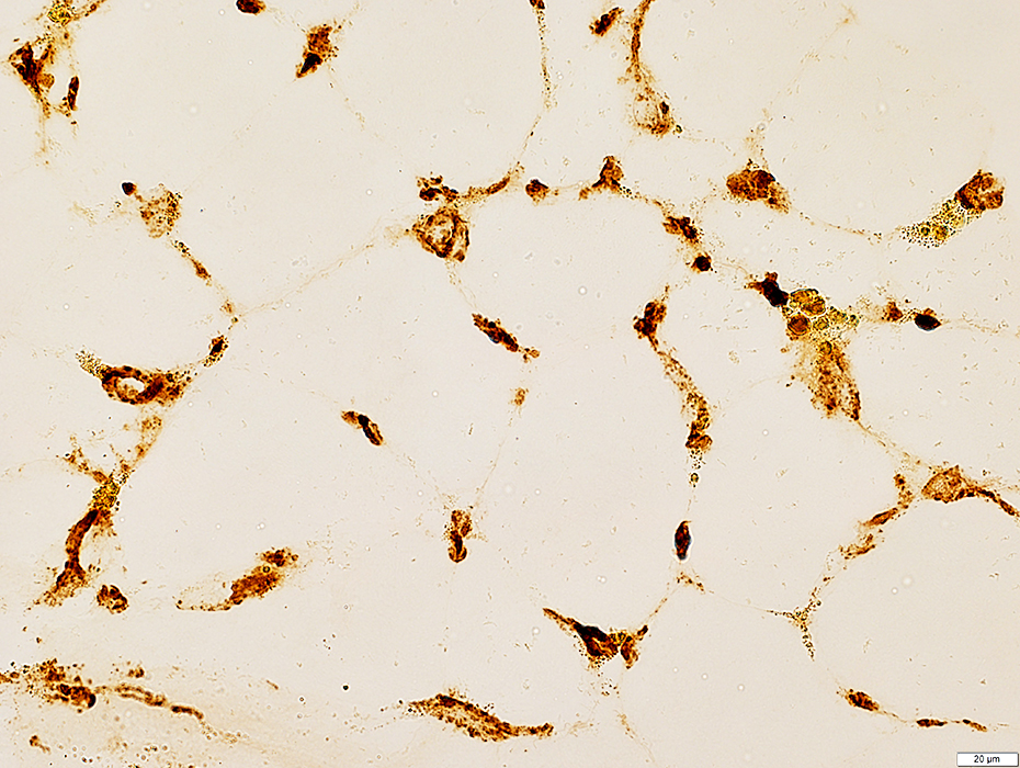
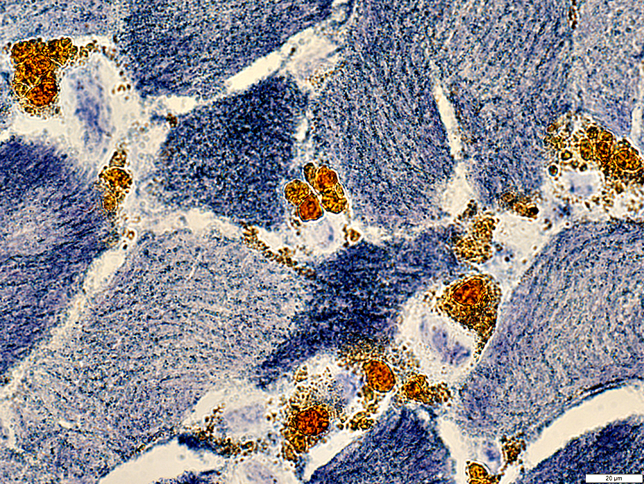 NADH stain |
 NADH stain |
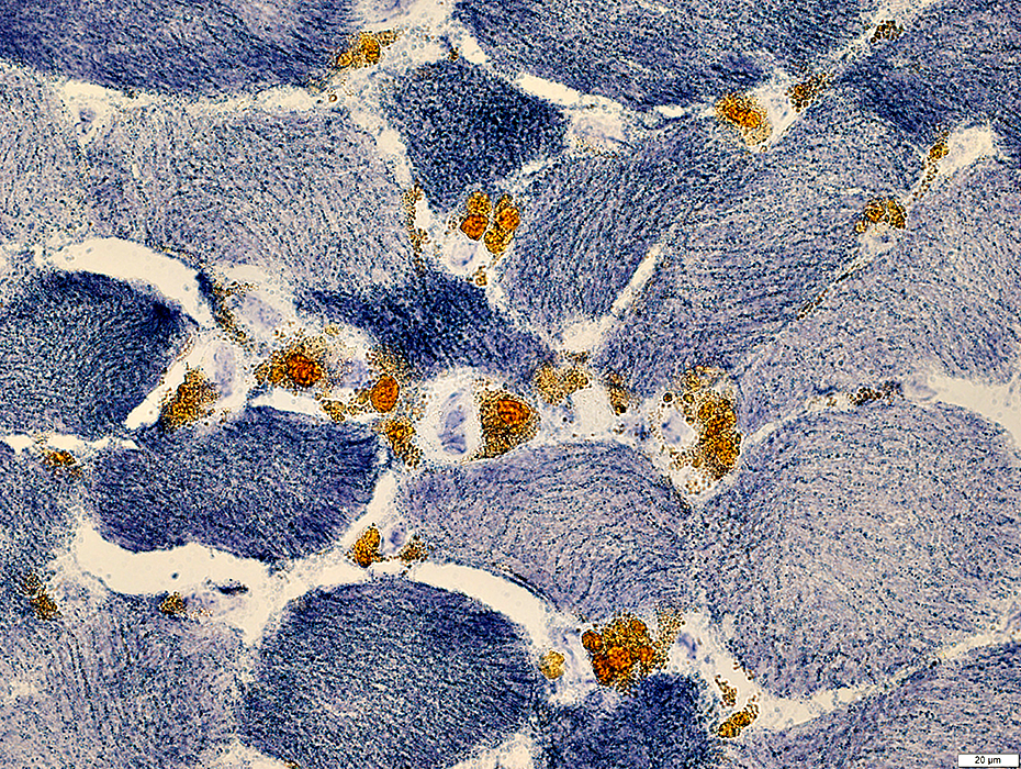
 Congo red stain |
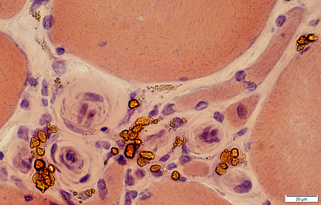
 H&E stain |
Around endomysial capillaries (Enlarged, with thick walls)
May also be present in perimysium in vasculitis
|
  NADH stain |
Outside vessel walls
Within a cluster of large capillaries
 Gomori trichrome stain |
 C5b-9 stain |
C5b-9 deposiits
Large size
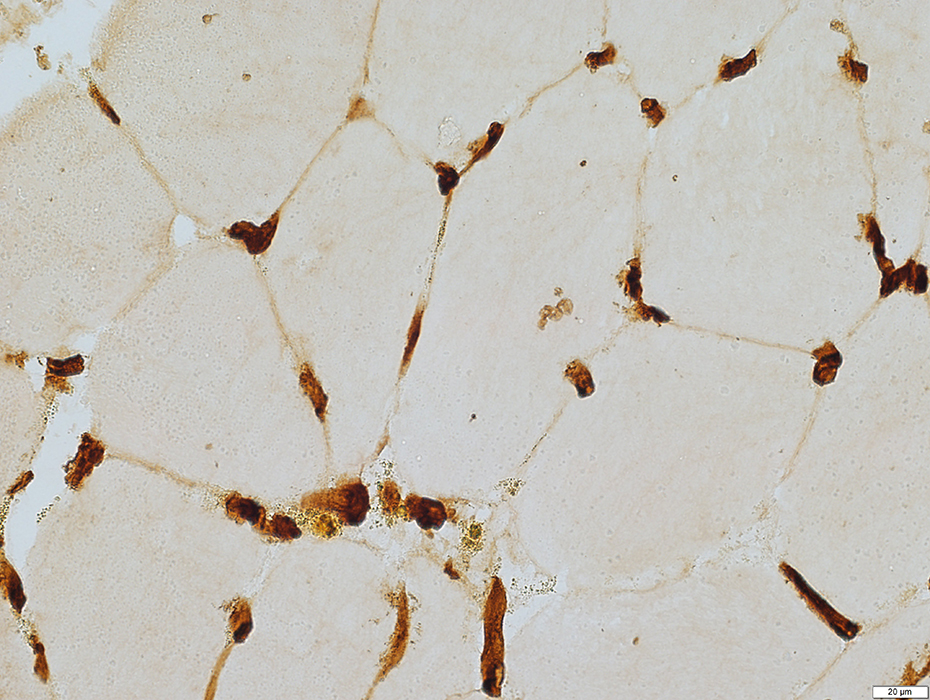 Ulex stain |
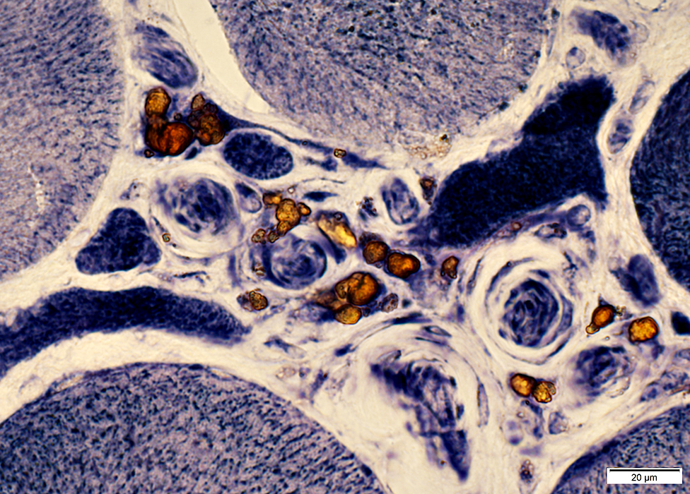
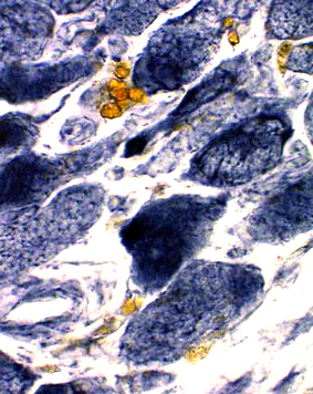
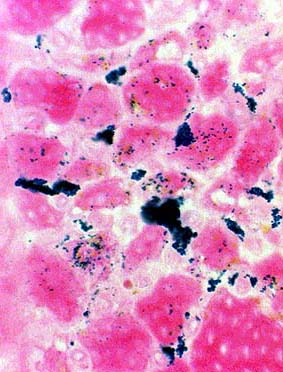
Hemosiderin deposits: Epineurial
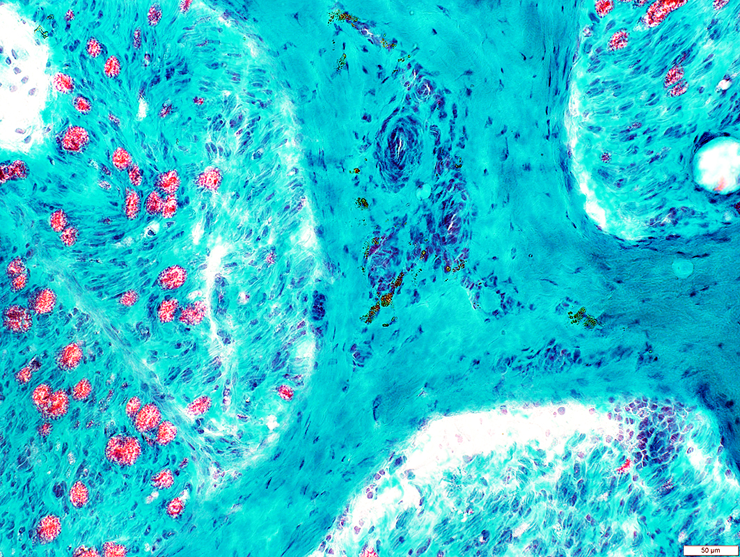 Gomori trichrome stain |
 Gomori trichrome stain |
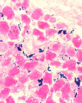 Prussian blue stain |
|
| Prussian blue stain: Iron in muscle with endomysial hemosiderin depositis | |
Return to Neuromuscular Home Page
6/1/2023