MYOTONIC DYSTROPHY
|
DM1 DM2 |
Myotonic Dystrophy 1 (DM1)
|
Chronic Late Mild |
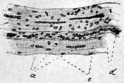 Rossolimo "De la myotonie atrophique" 1902 |
DM1: Adult, Chronic changes
|
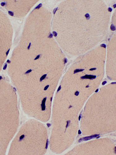
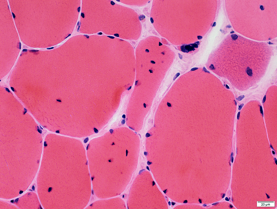
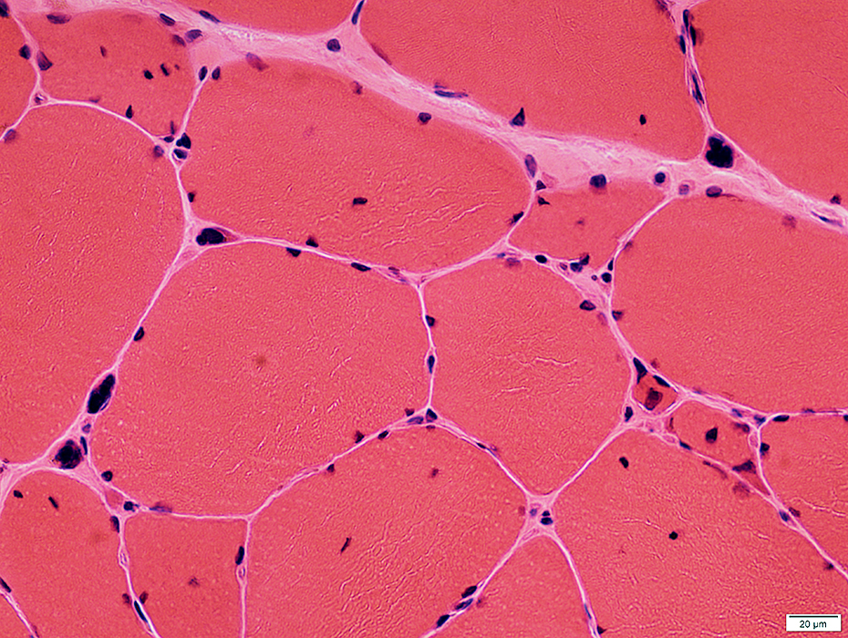
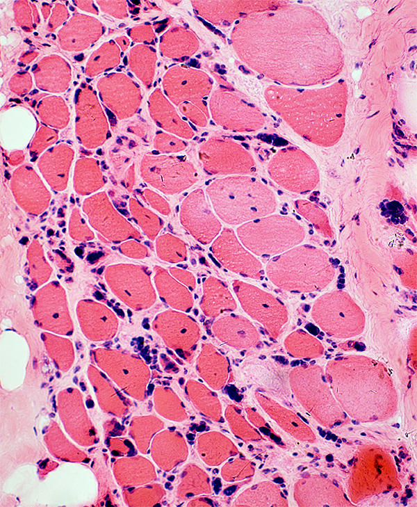
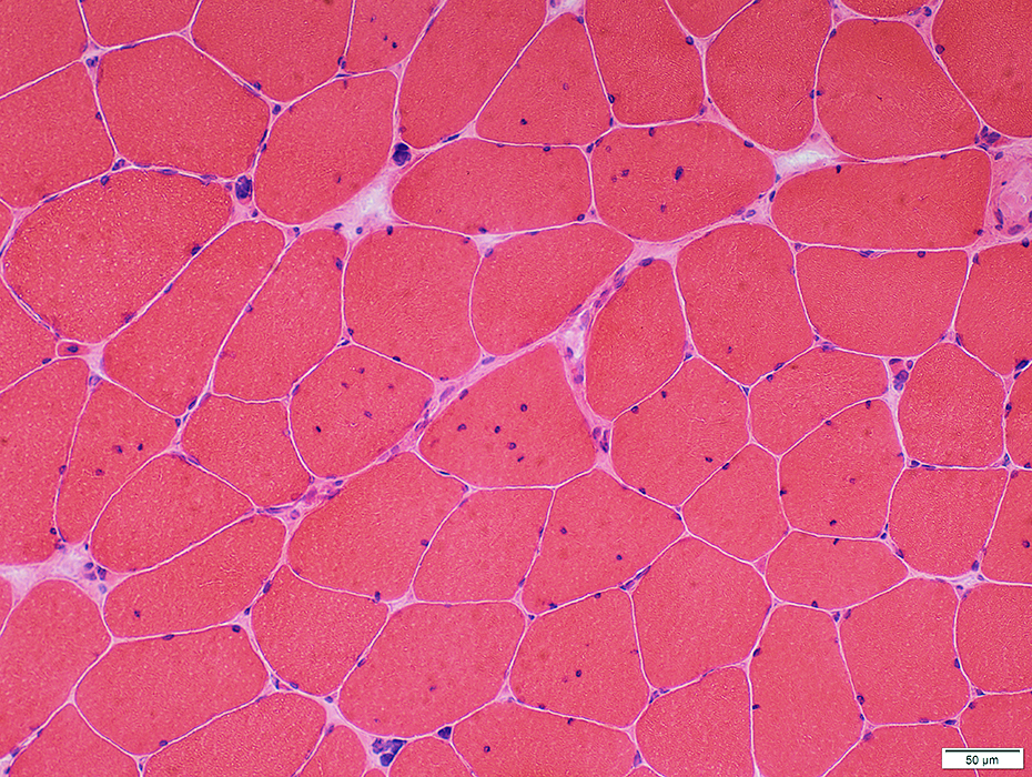
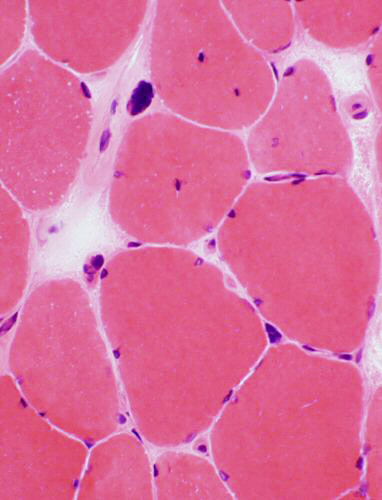
Muscle fibers Size: Variation Myonuclei Numbers: Increased Shapes: Irregular Internal Present in many muscle fibers Several in individual fibers Often clumped & in longitudinal chains Pyknotic clumps Acid phosphatase + granules Replacement by Fat 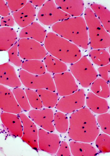 H&E stain |
Perimysium: Connective tissue replaced by fat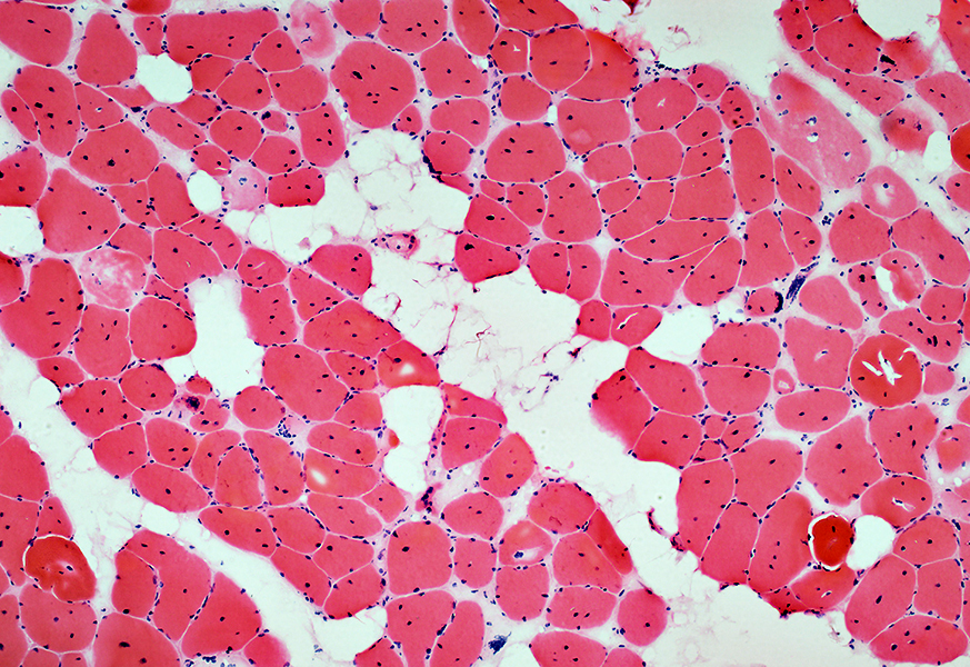 H&E stain |
 Congo red stain |
 Congo red stain |
 H&E stain |
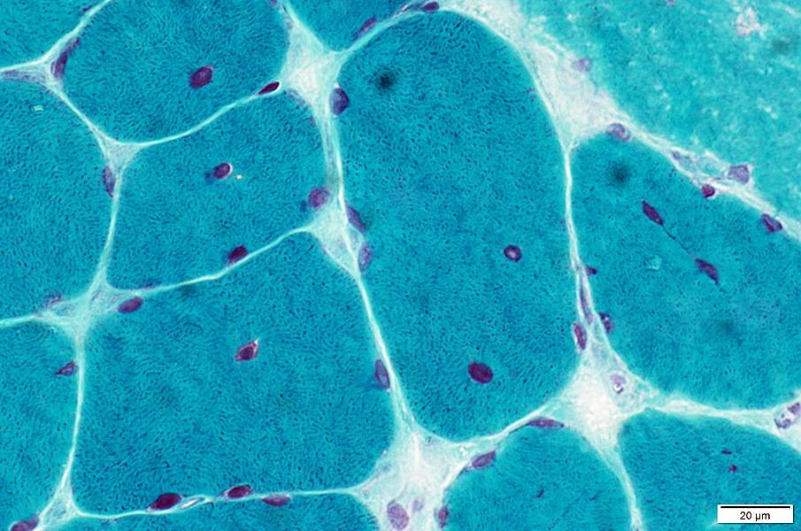
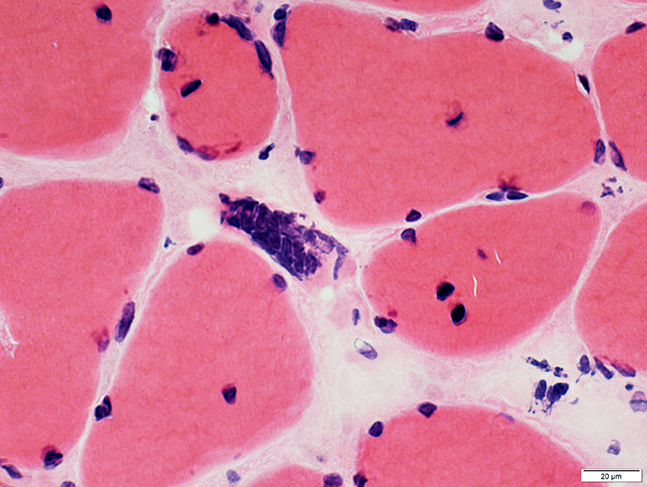
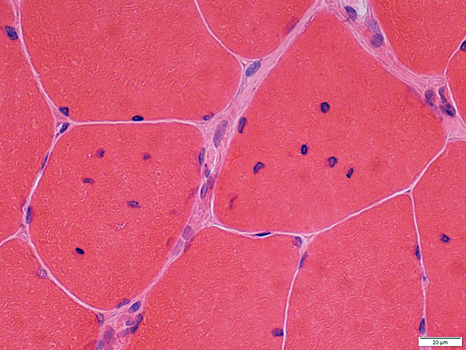
Internal nuclei: Many; Irregular shapes; Often in longitudinal chains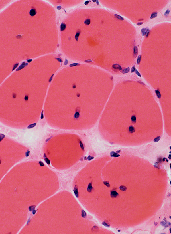 H&E stain |
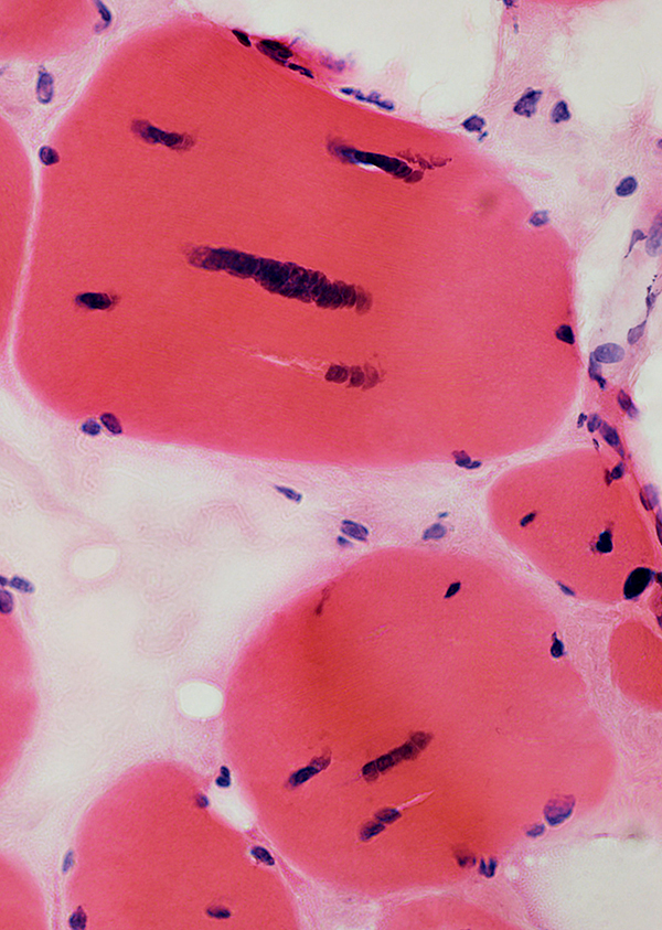 H&E stain |
 Gomori trichrome stain Internal nuclei Nuclei may be mildly large & clustered |
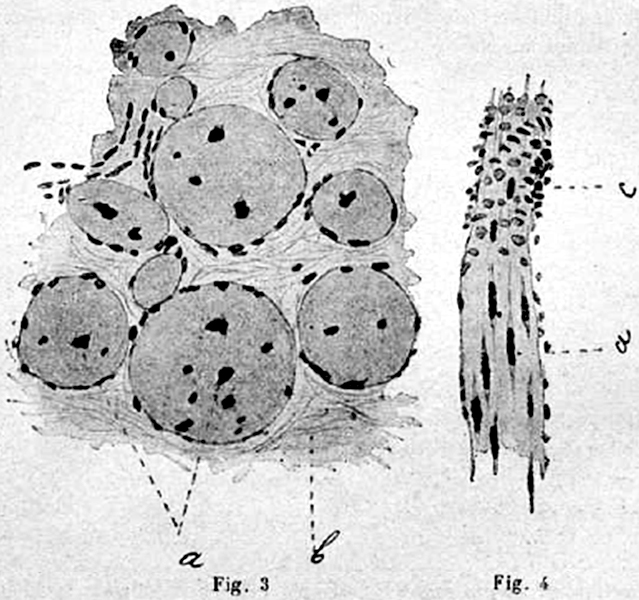 Rossolimo "De la myotonie atrophique" 1902 |
 Gomori trichrome stain |
|
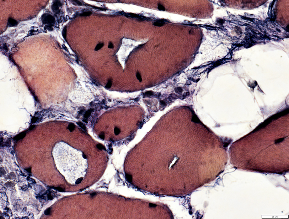
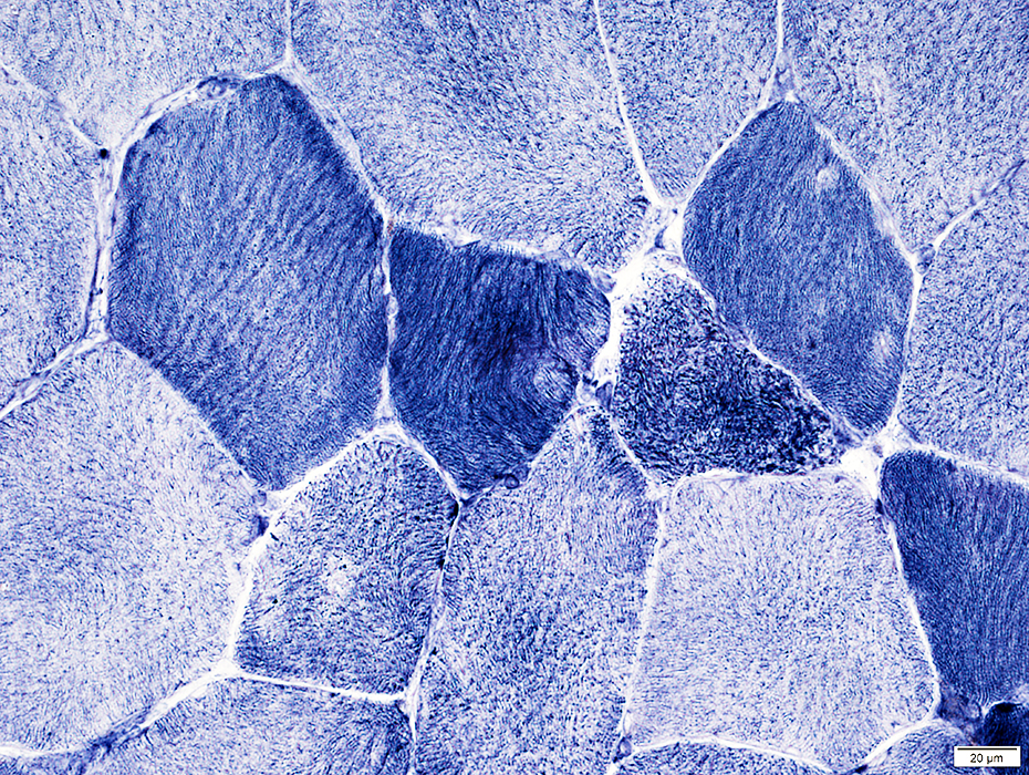
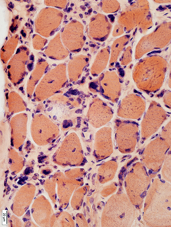
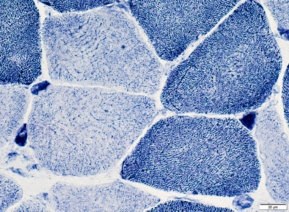
Internal architecture of muscle fibers: Disordered Aggregates Clear regions 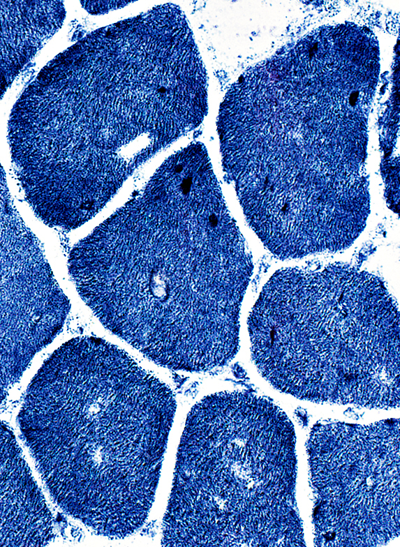 NADH stain |
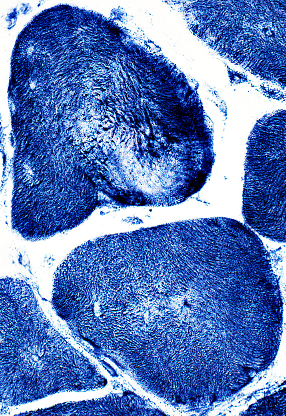 NADH stain |
Aggregates in muscle fiber cytoplasm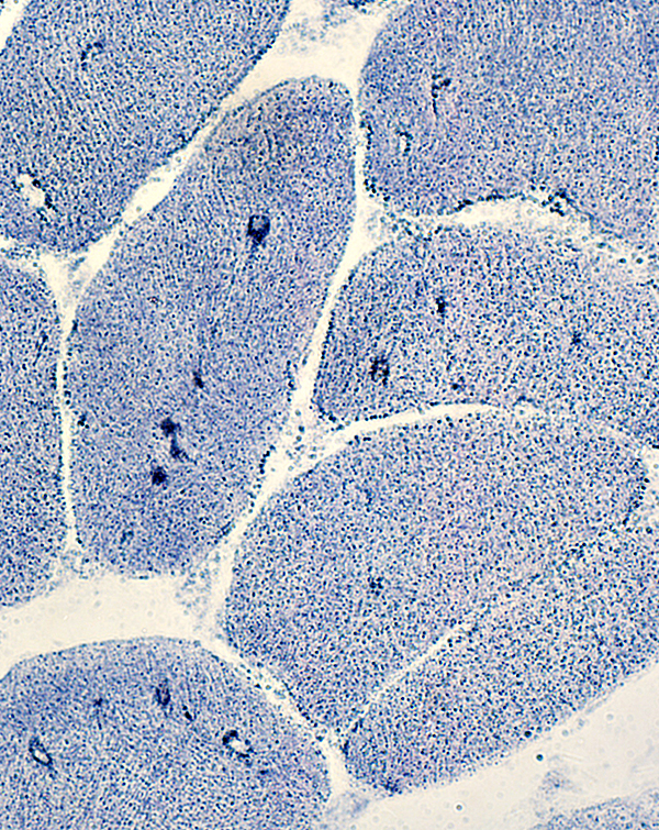 AMPDA stain |
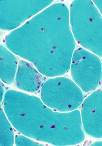
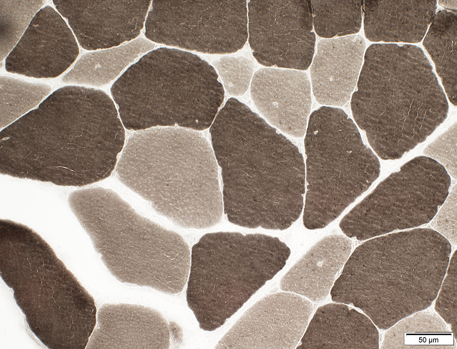
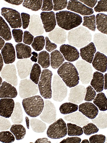
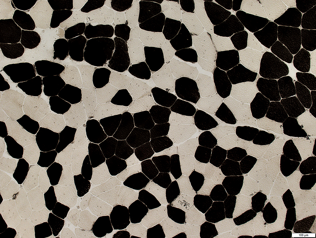
Fiber type abnormality: All fibers are type 1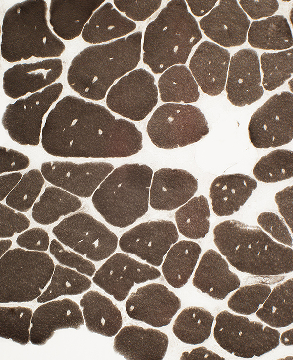 ATPase pH 9.4 stain |
|
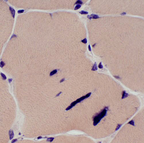
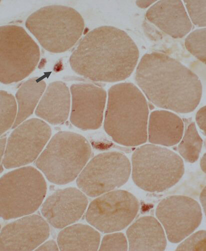
Acid phosphatase positive granules in muscle fibers
|
|
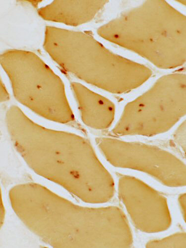 Acid phosphatase stain |
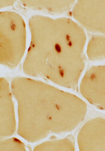 Acid phosphatase stain |
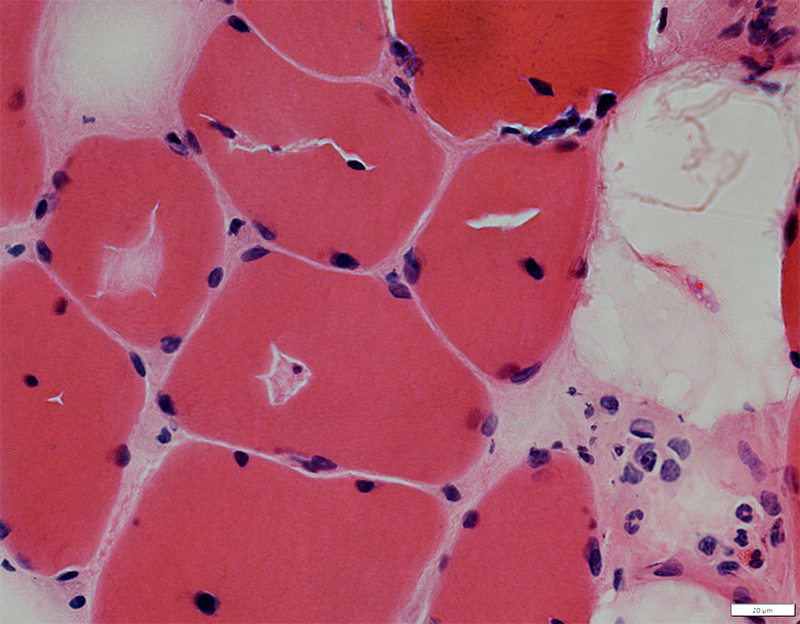
H&E stain |
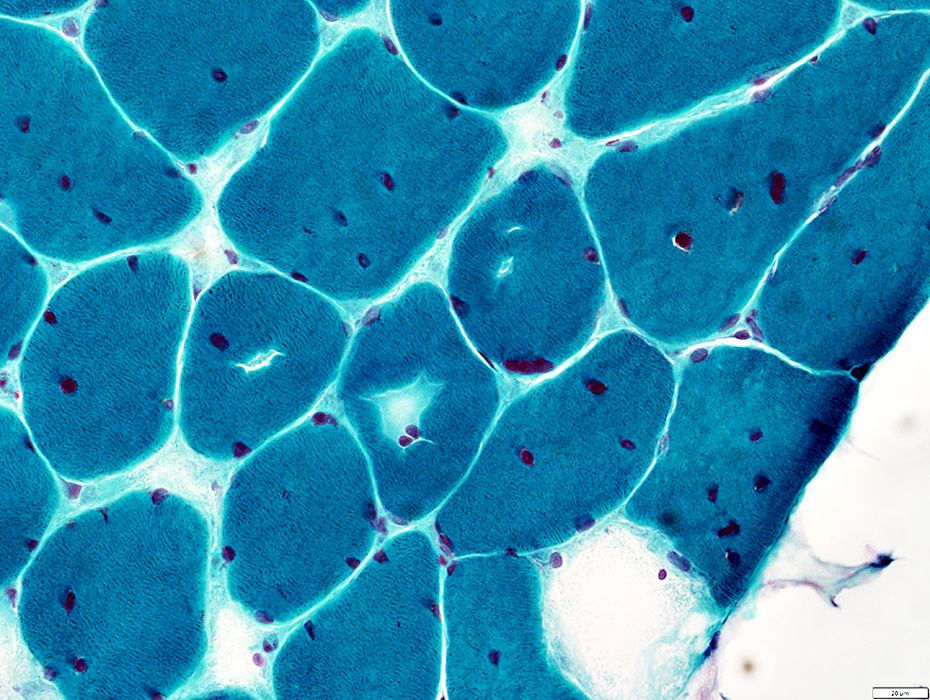 Gomori trichrome stain |
 VvG stain |
 H&E stain |
 Congo red stain |
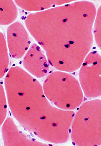
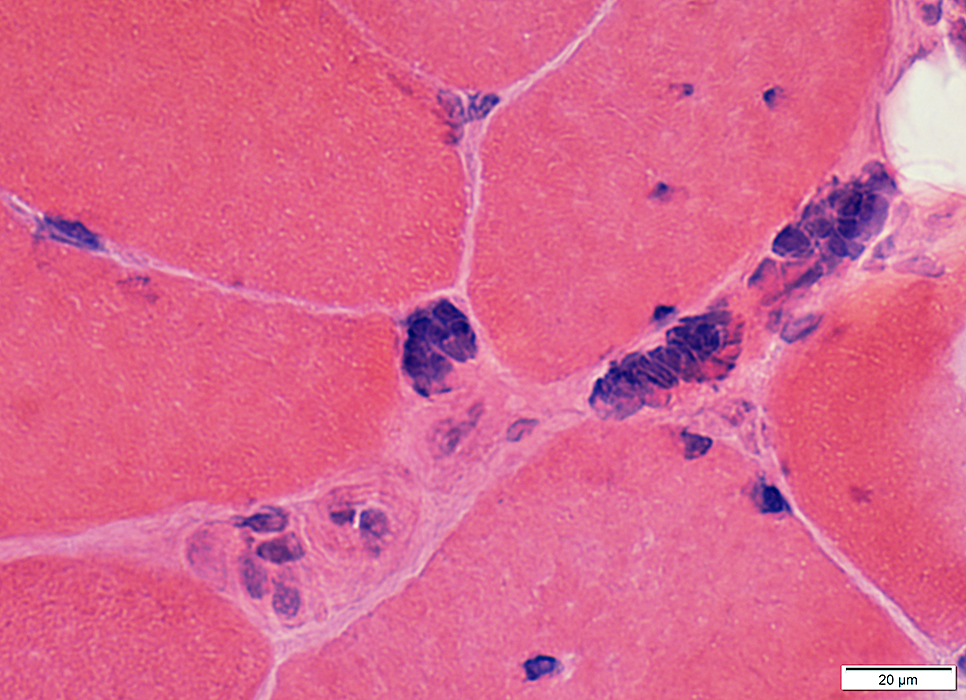
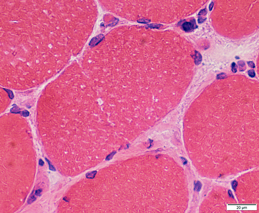
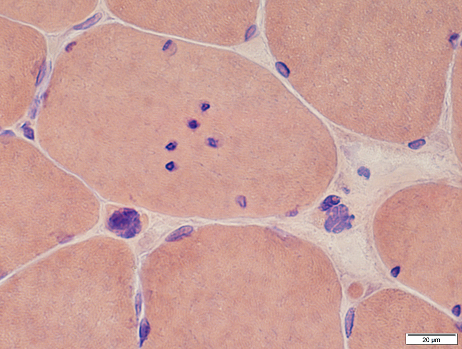
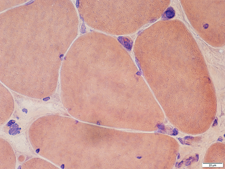
Myotonic dystrophy (DM1): Mild & Earlier changes
 H&E stain |
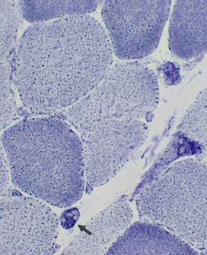
Pyknotic nuclear clumps
 H&E stain |
 H&E stain |
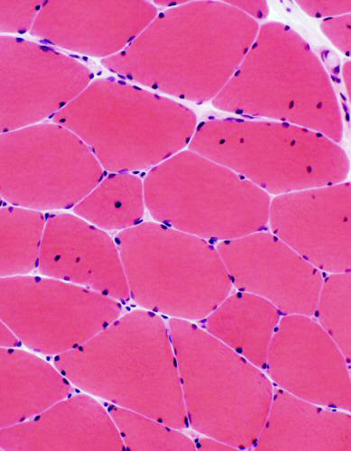 H&E stain Fiber sizes: Internal nuclei: In some fibers |
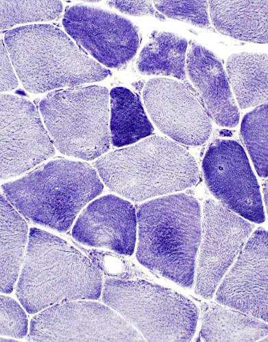 NADH stain Type I (darker) fibers: Smaller Internal architecture: Disordered |
 NADH stain |
Internal nuclei: In type 1 & 2 muscle fibers
 ATPase pH 9.4 stain |
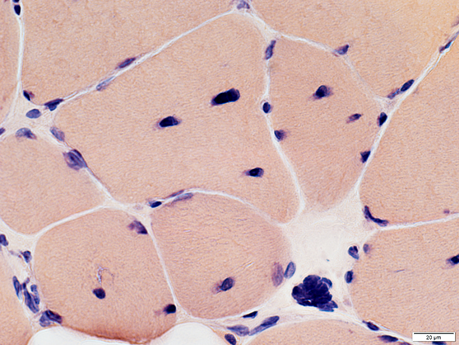
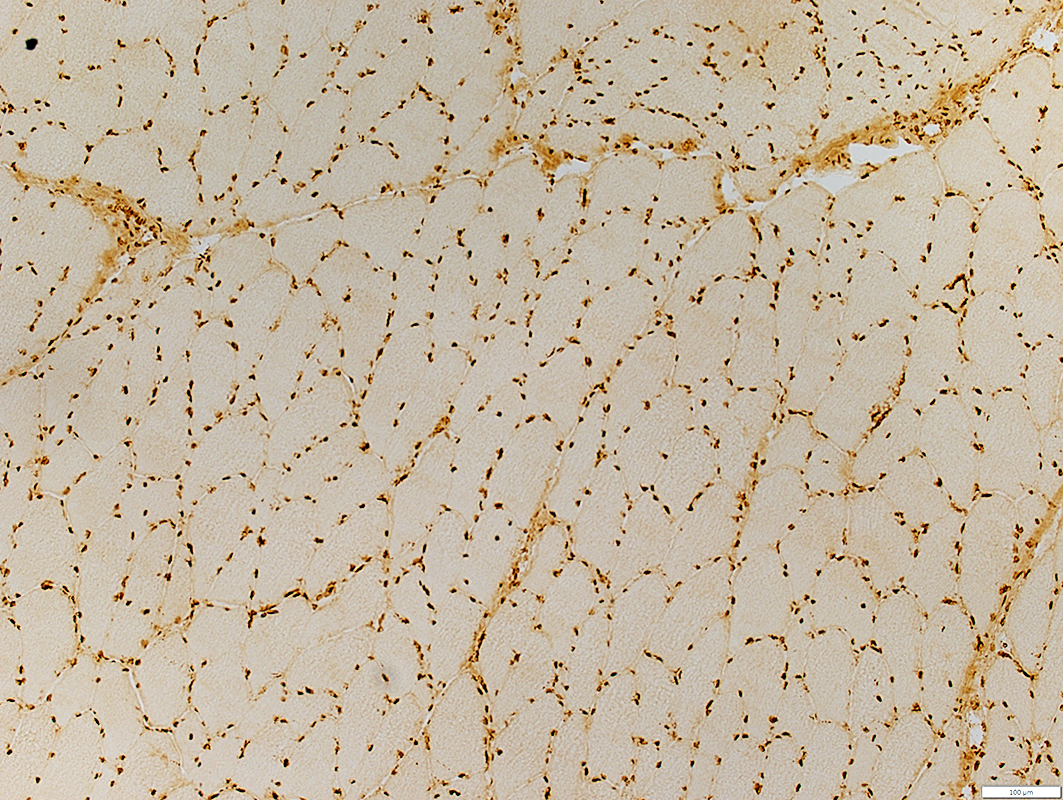 Emerin stain |
Increased numbers around periphery of muscle fibers
Irregular shapes (Below)
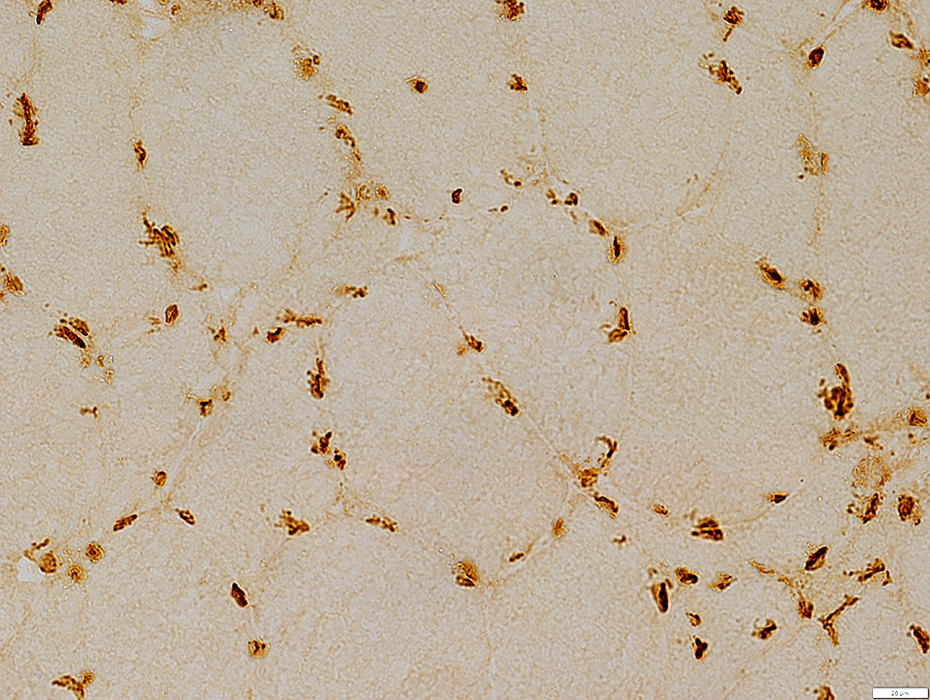 Emerin stain |
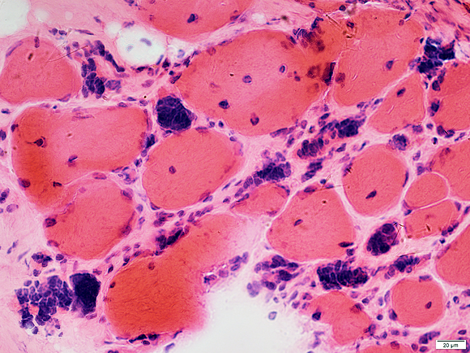
Myotonic dystrophy (DM1): Late changes
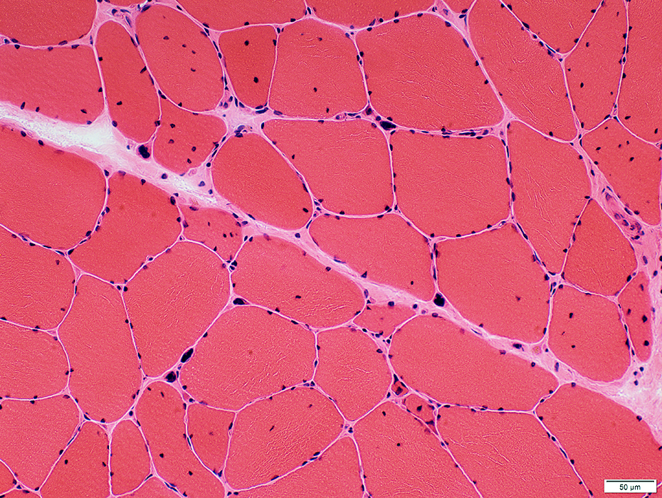
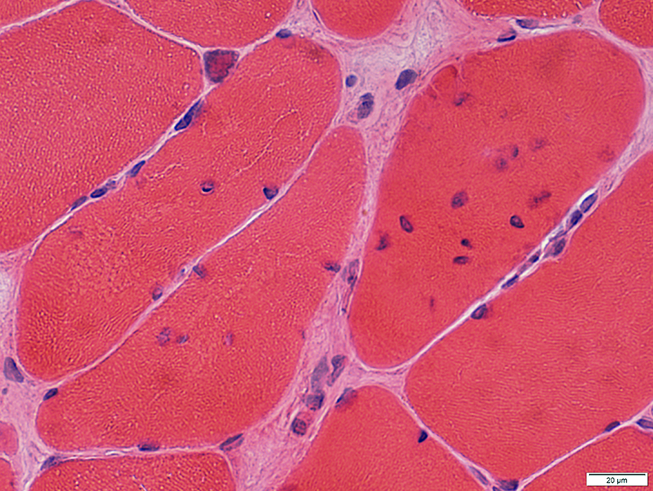
 H&E stain Muscle fibers Size: Atrophy & Some hypertrophy Pyknotic nuclear clumps Nuclei: Large & Irregular shapes Endomysial connective tissue: Moderately increased |
 Congo red stain |
 VvG stain |
 H&E stain |
Large
May be present in connective tissue with few intact muscle fibers
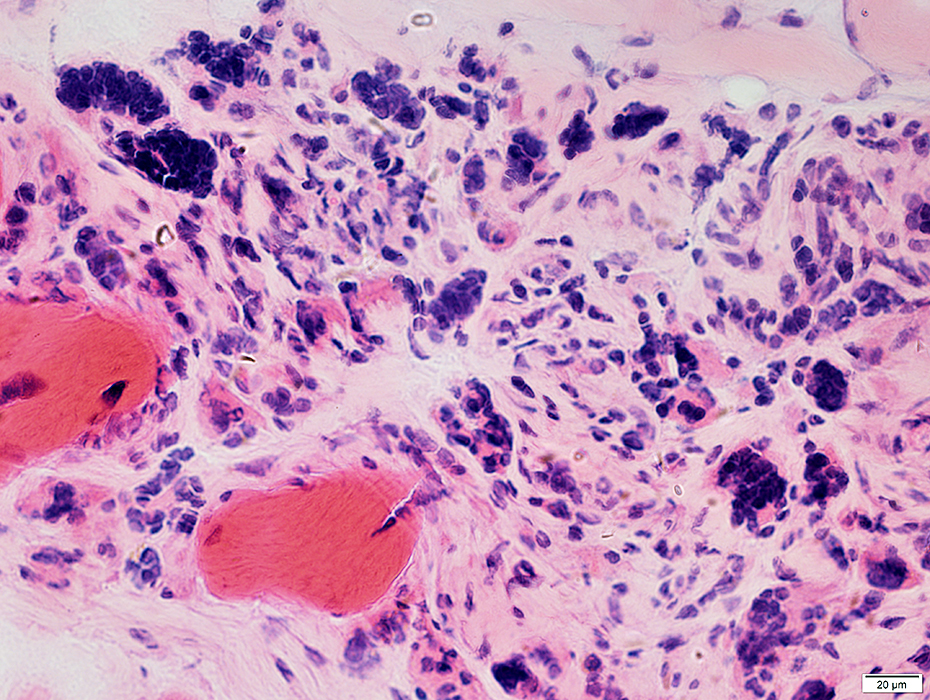 H&E stain |
Myotonic dystrophy, Type 2 (DM2)
PathologyNuclei: Internal; More in type 2 fibers
Pyknotic nuclear clumps
Replacement of muscle by fat
 H & E stain |
 H & E stain |
 H & E stain |
 Congo red stain |
 H & E stain Internal nuclei: Multiple in individual fibers |
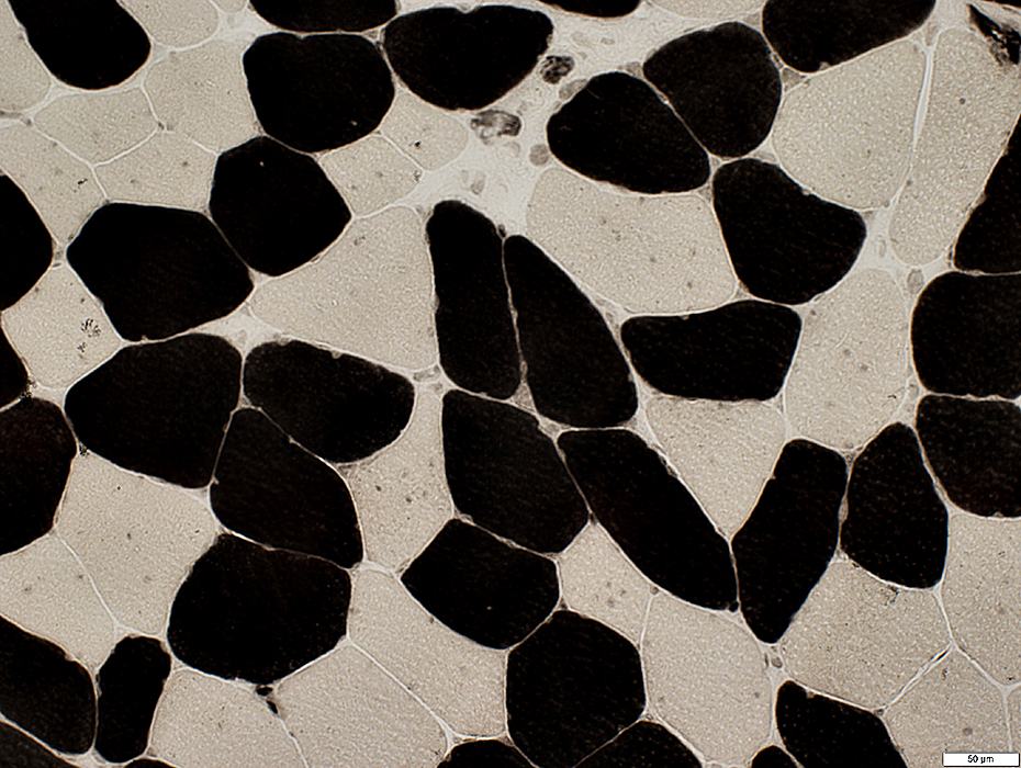
 ATPase pH 9.4 Internal nuclei (clear regions) Most commonly in type 2 (dark) muscle fibers |
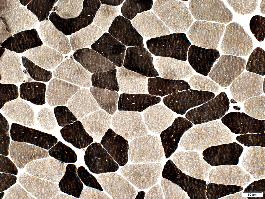 ATPase pH 9.4 |
Most common in type 2 (dark) muscle fibers
Fiber Sizes
Type 2 (Pale): Mildly smaller than Type I
DM2: Fiber types
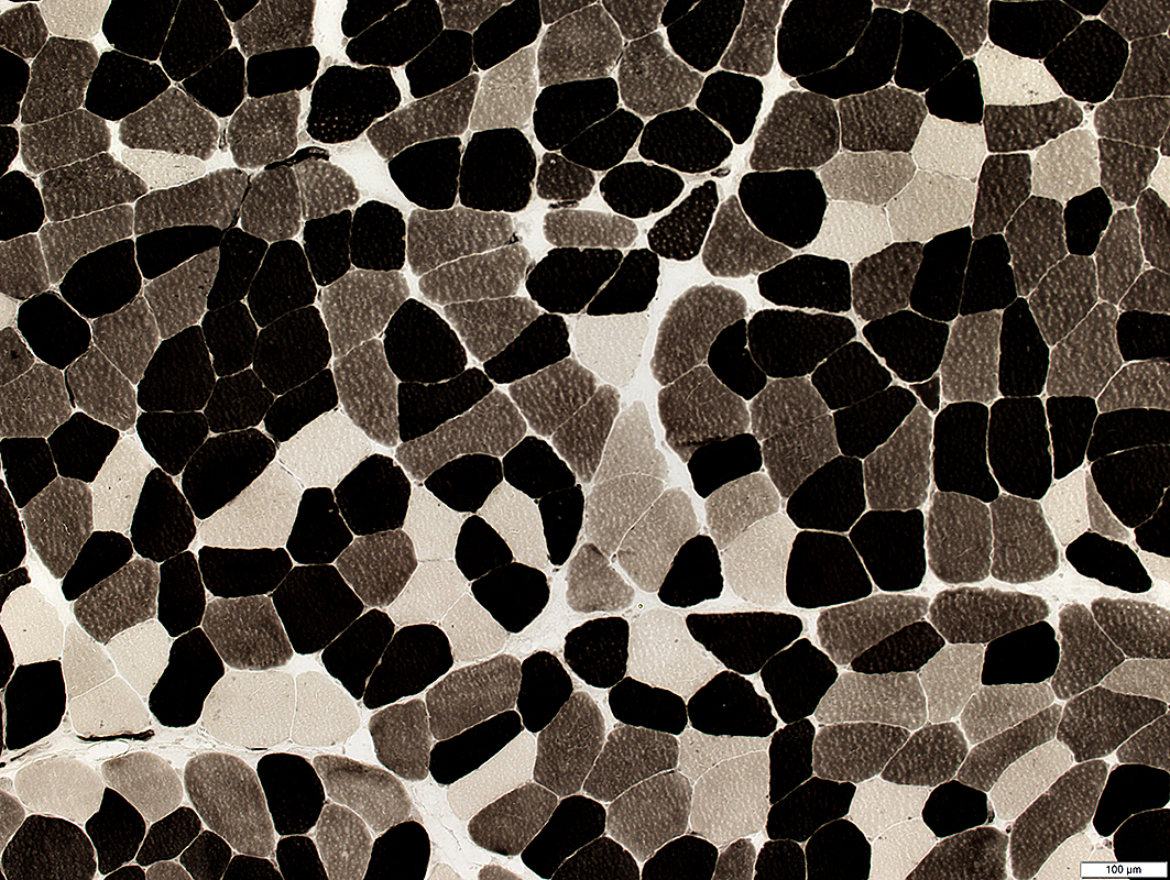 ATPase pH 4.6 |
2B fibers (Intermediate stain): Abnormally varied colors
ATPase pH 4.6 |
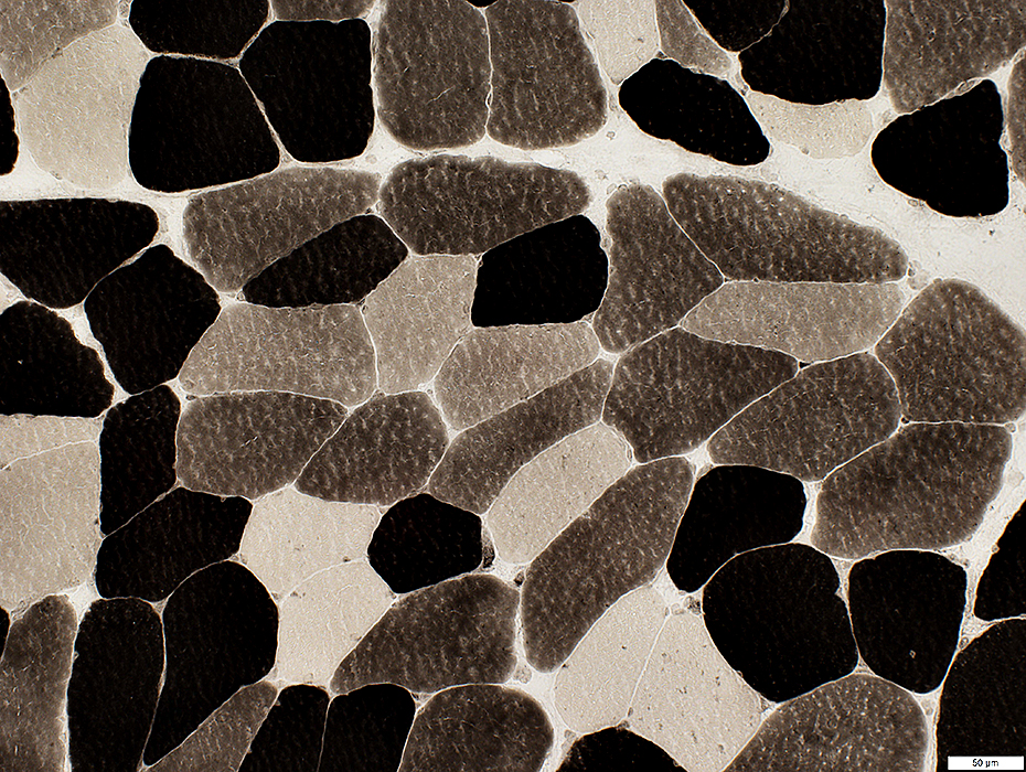
 ATPase pH 4.3 |
Normal staining for 2 fiber types type 1 (Dark) & type 2 (Light)
Type I fibers mildly larger than type 2
Few or No type 2C fibers (Intermediate color)
 ATPase pH 4.3 |
|
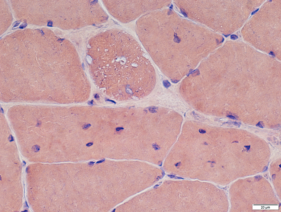
LYSOSOMAL Structures Cytoplasmic granules: Acid Phosphatase positive granules: Scattered in cytoplasm 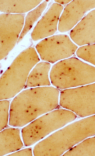 Acid Phosphatase |
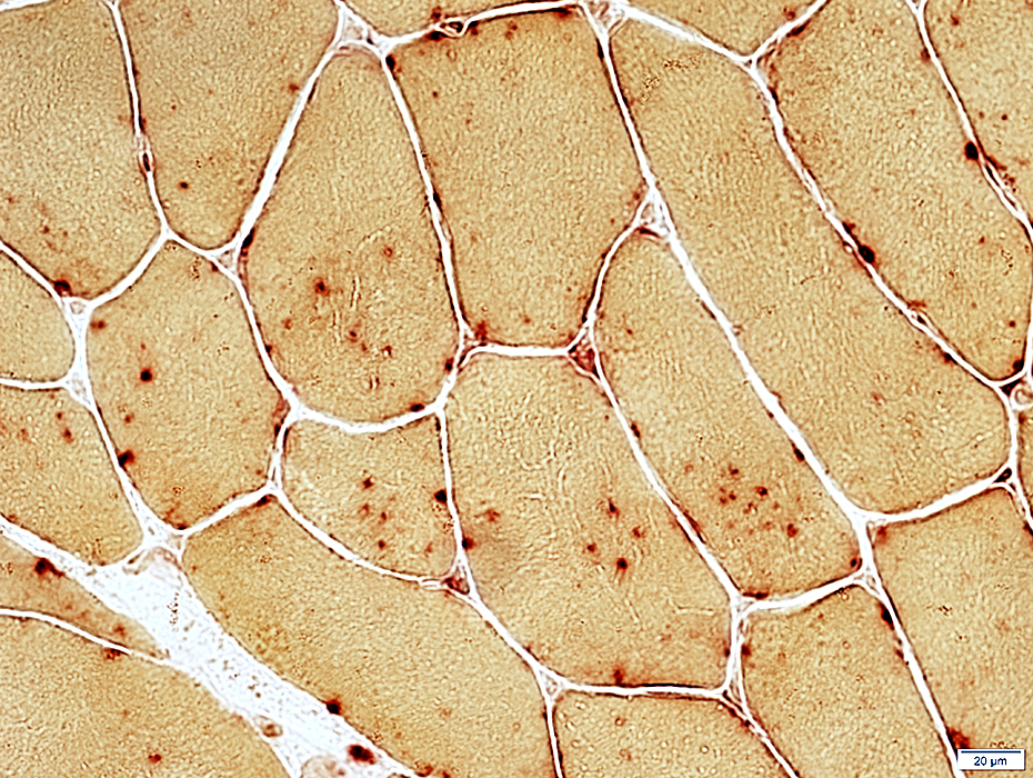 Acid Phosphatase |
Pyknotic nuclear clumps
 H & E stain |
 H & E stain |
 Congo red stain |
 Congo red stain |
 H & E stain |
 NADH stain |
 Esterase |
|
Pyknotic nuclear clumps: Dark-stained
|
||
 NADH stain |
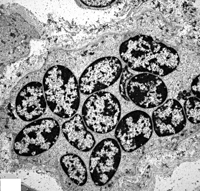 From: Cory Toth MD Pyknotic nuclear clump: Ultrastructure
|
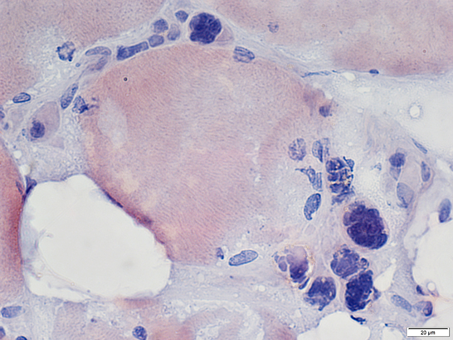
Myotonic dystrophy, Type 2 (DM2): Late
Pyknotic nuclear clumps: Large
Muscle fibers: Largest are hypertrophied
 Congo red stain |
Pyknotic nuclear clumps: Nuclei stained for emerin
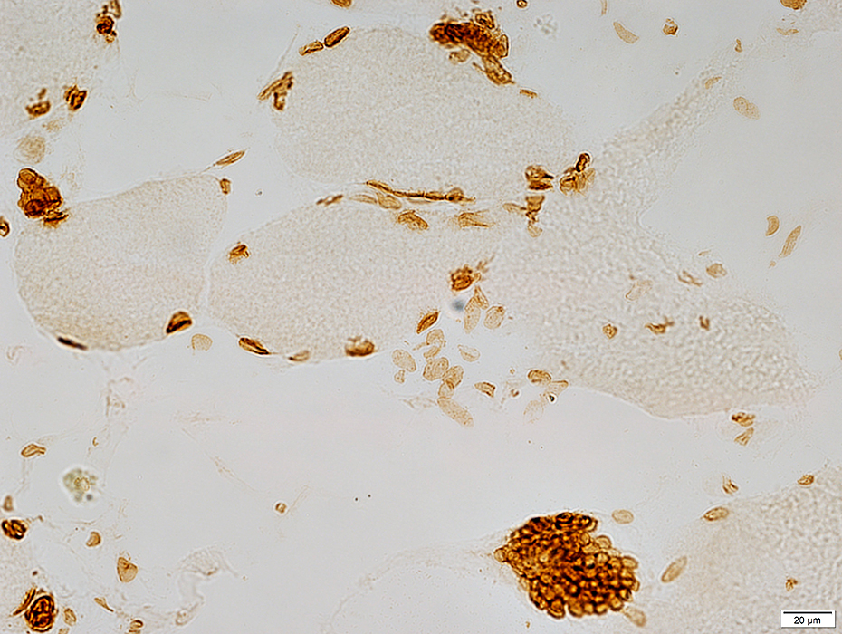 Emerin stain |
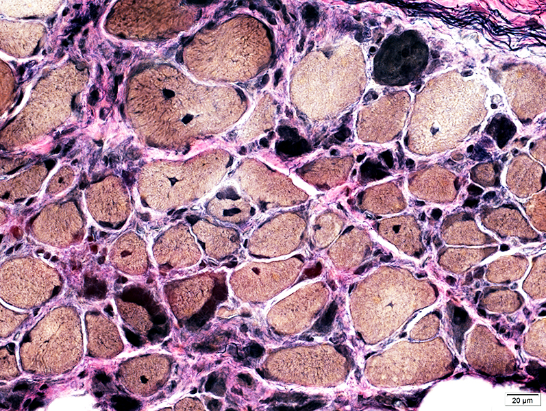
Muscle fibers & Perimysium: Replaced by fat
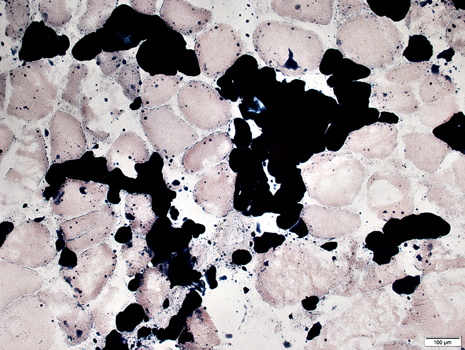 Sudan Black stain |
Return to Myotonic dystrophy
Return to DM2
Return to Neuromuscular syndromes
Return to Neuromuscular home page
6/2/2016