TYPE II MUSCLE FIBER ATROPHY
|
Clinical features Pathology Features Childhood Early Elongated fibers 2B fiber atrophy Moderately severe Atrophy: General |
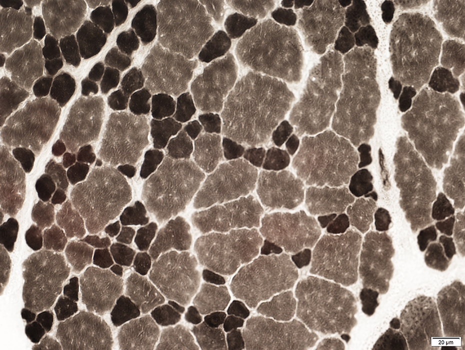
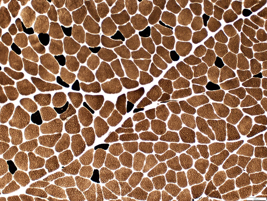 ATPase pH 9.4 stain |
Type II atrophy: Pathology
- Type II (Dark at pH 9.4) Muscle fibers
- Small: Reduced cross-sectional area\
- Shape
- Adult: Angular or Elongated
- Childhood: Polygonal or Round
- Early changes
- Disuse & weight loss atrophy: Small fibers are angular
- Congenital: Small fibers are polygonal or round
- Type I (Lighter at pH 9.4) Muscle fibers
- Larger than type II
- Often atrophic compared to normal type 1 fibers
- NADH stain: Small fibers usually pale
- Nuclei
- Number: Unchanged
- Myonuclear domain size: Reduced
- Recovery of fiber size after Disuse atrophy with exercise 1
- Mitochondrial oxidative enzyme activity reduction
3
- Complexes II & II+III > I
Type II Muscle Fiber Atrophy: Clinical associations
- Weakness: Proximal > Distal; Symmetric
- Muscle size: Wasting > Weakness
- Associated disorders
- Weight loss: > 15%
- Disuse
- Aging
- Systemic disease
- Childhood
- Congenital hypotonia
- Myasthenia gravis
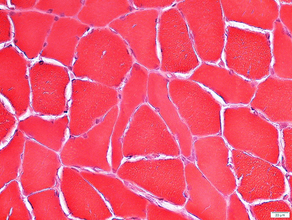
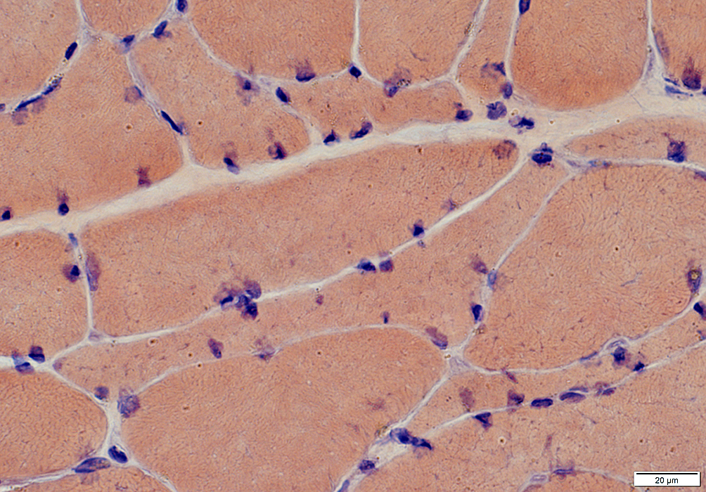
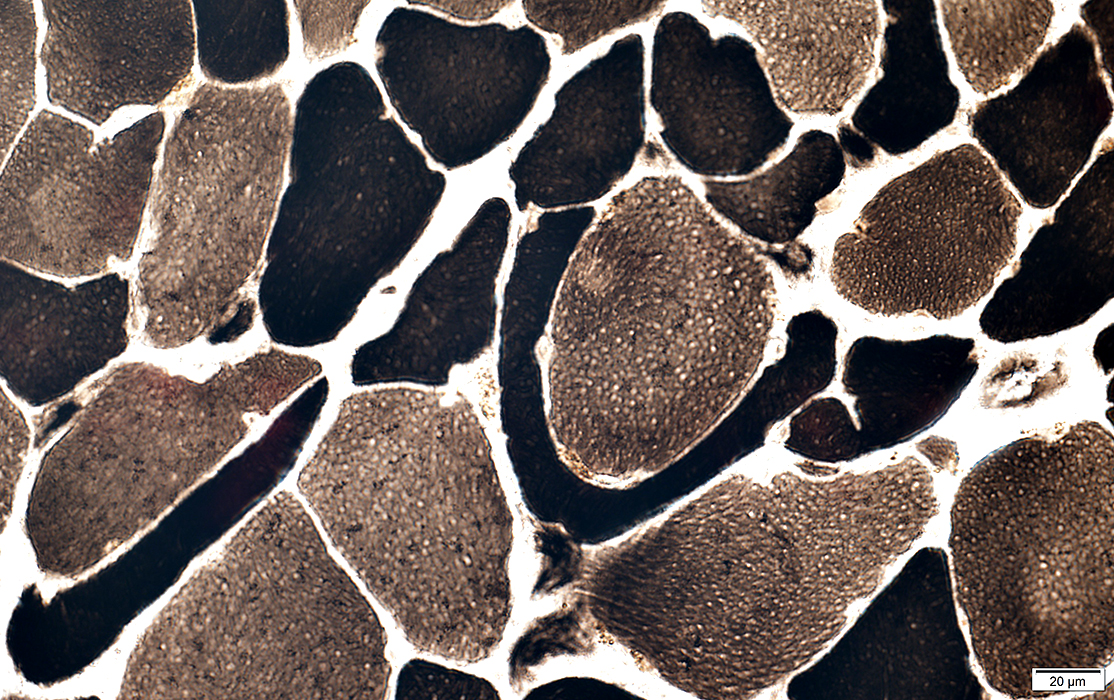
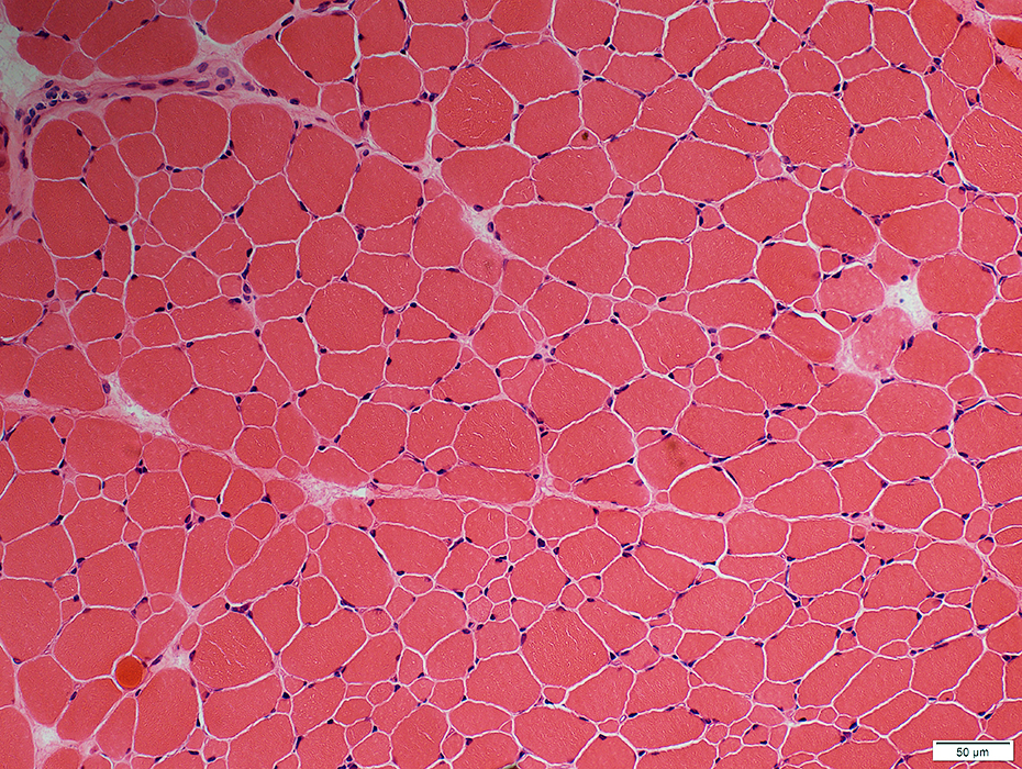
Type 2 Muscle Fiber Atrophy: Moderately Severe
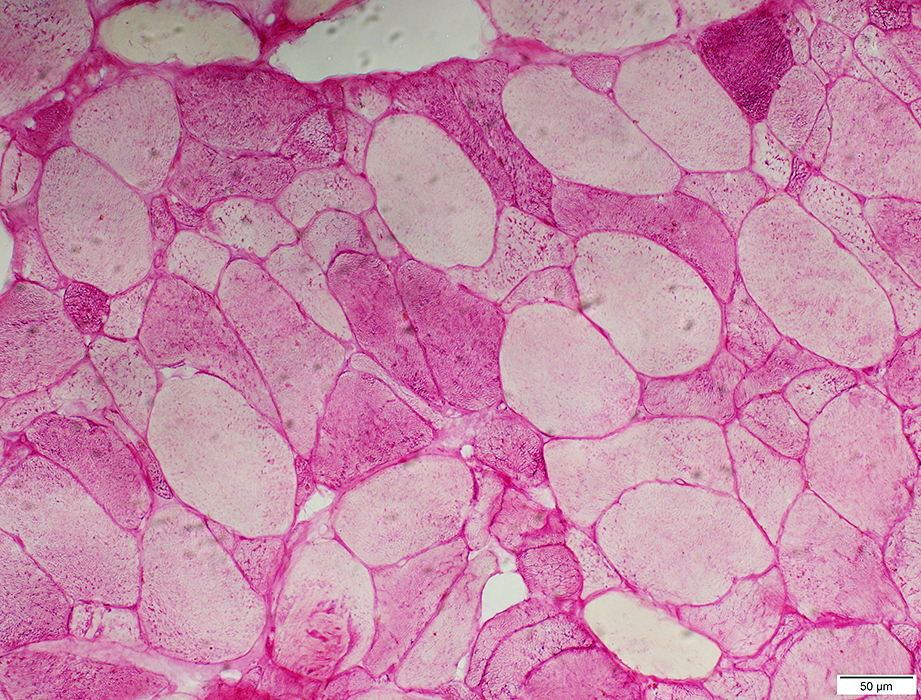
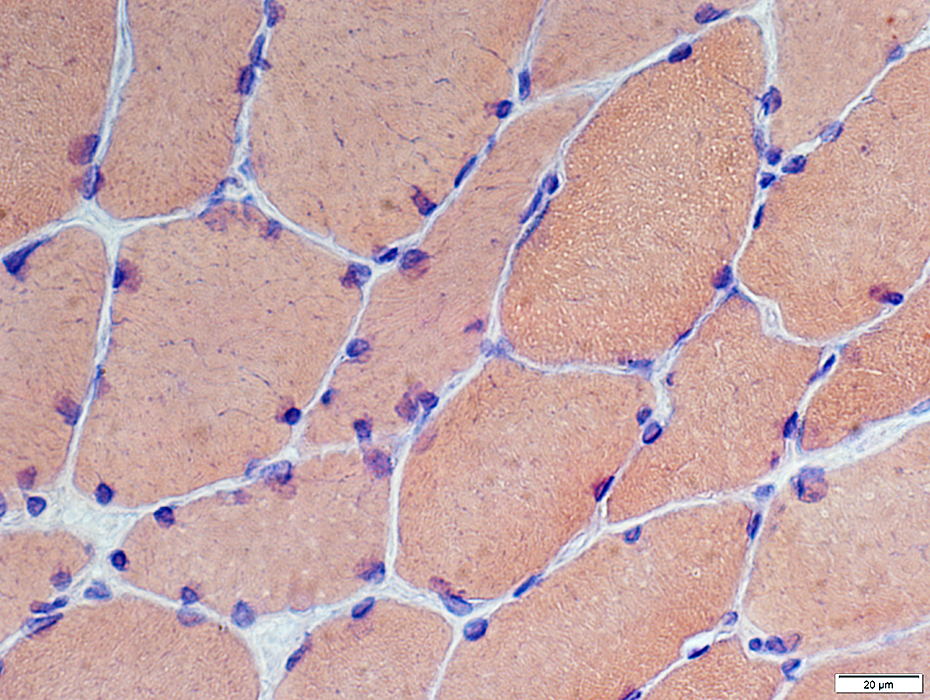
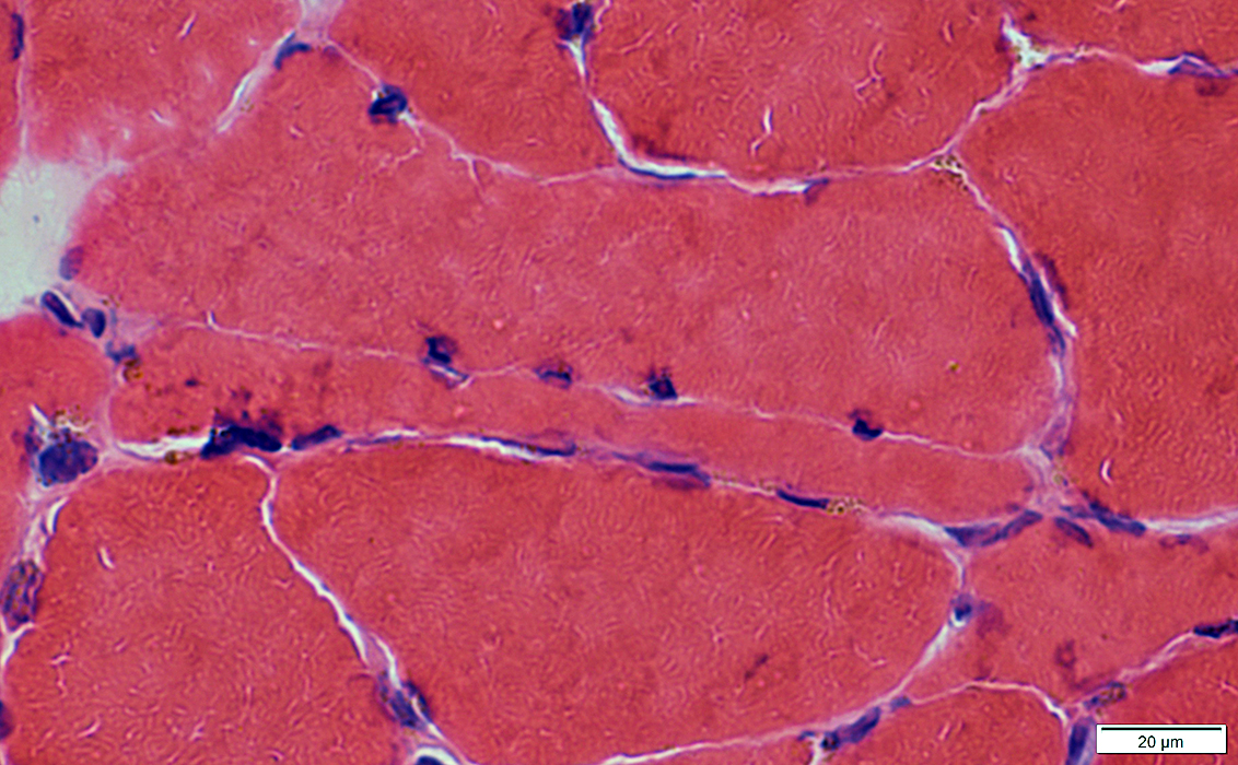
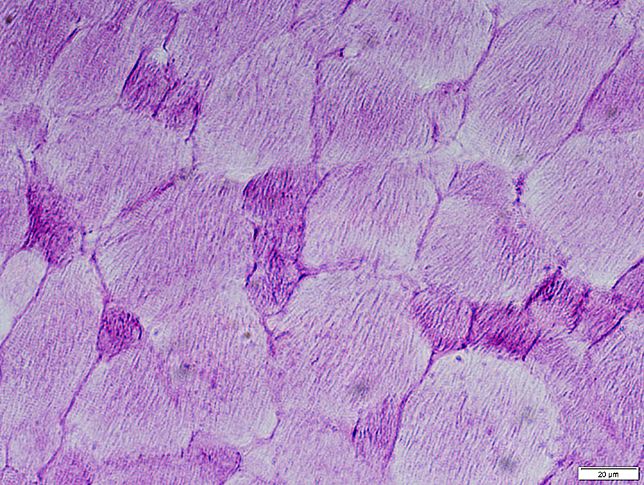
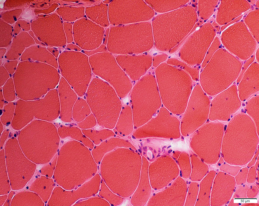 H&E stain |
Bimodal distribution
Type 1 (Larger) muscle fibers are normal or somewhat small
Small muscle fibers
Shape: Often angular; Some are elongated
Distribution: May appear clustered
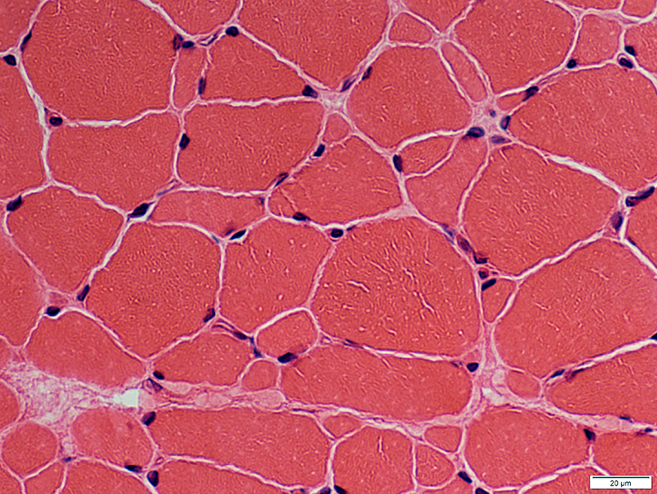
 H&E stain |
 VvG stain |
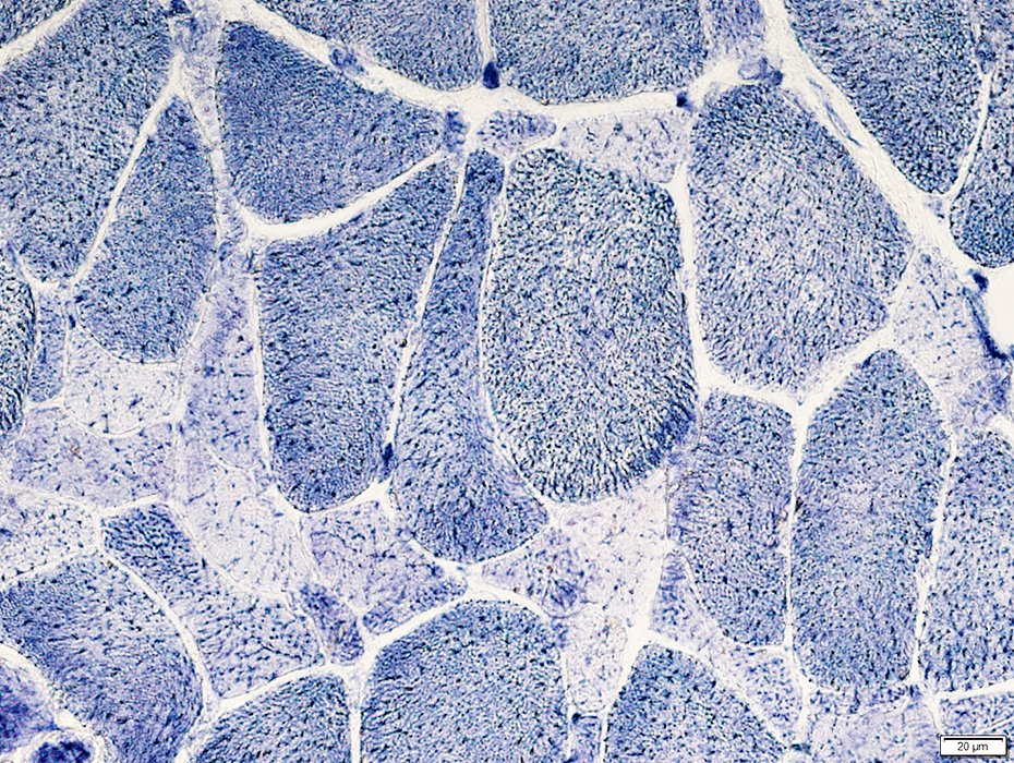
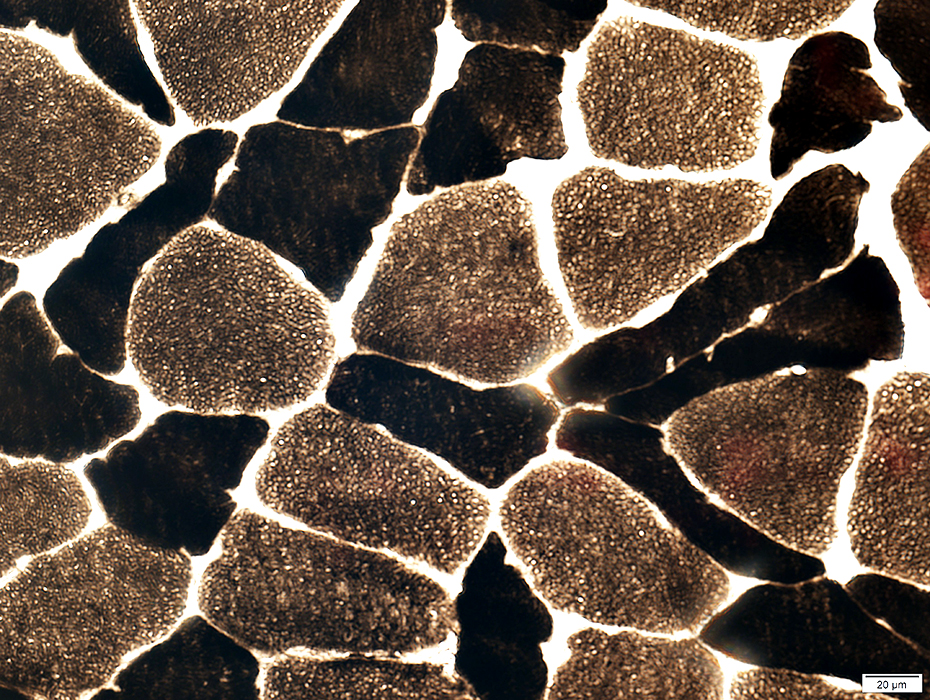
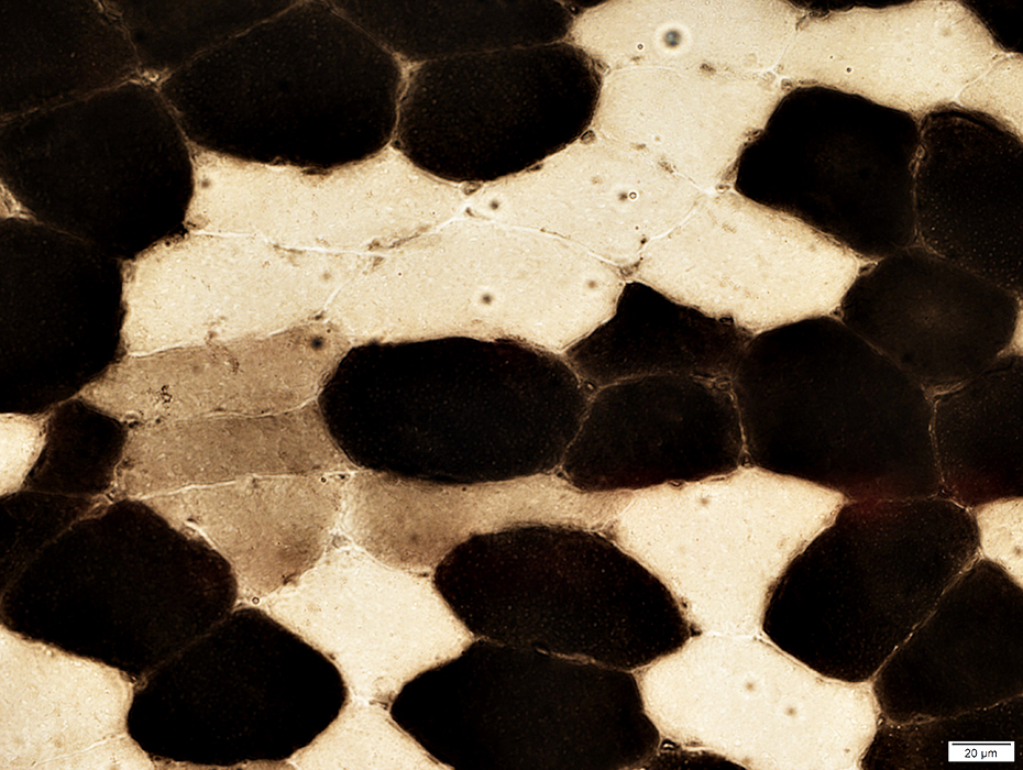
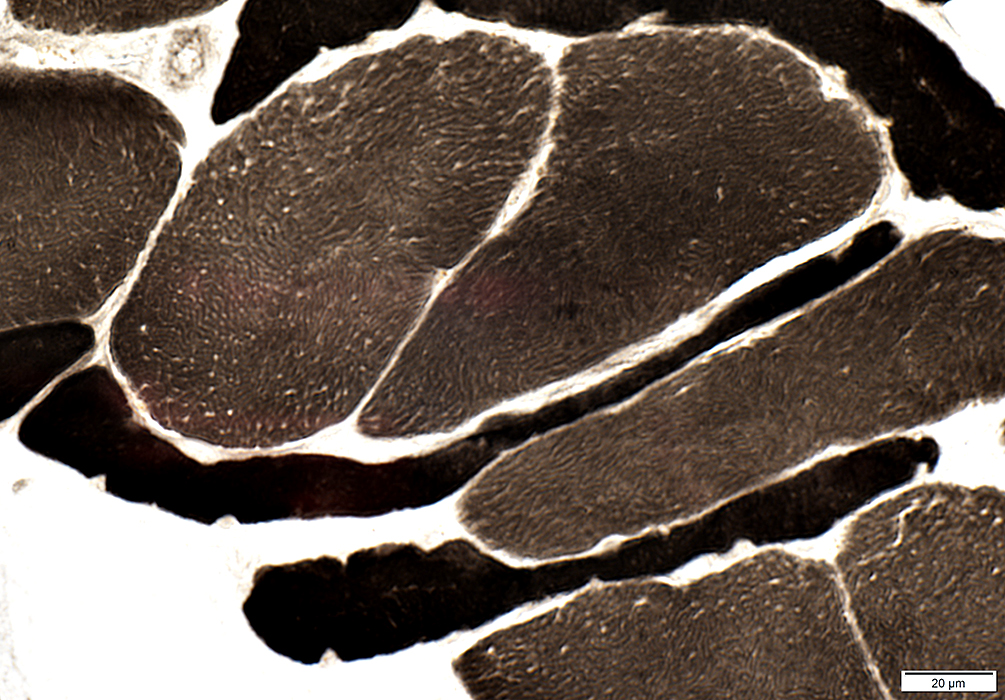
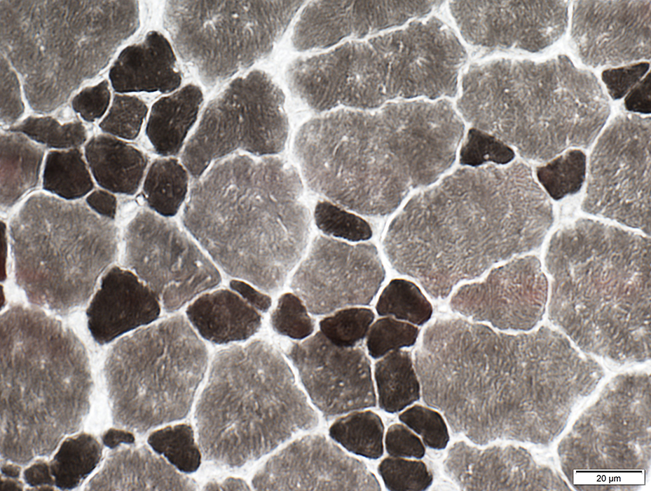
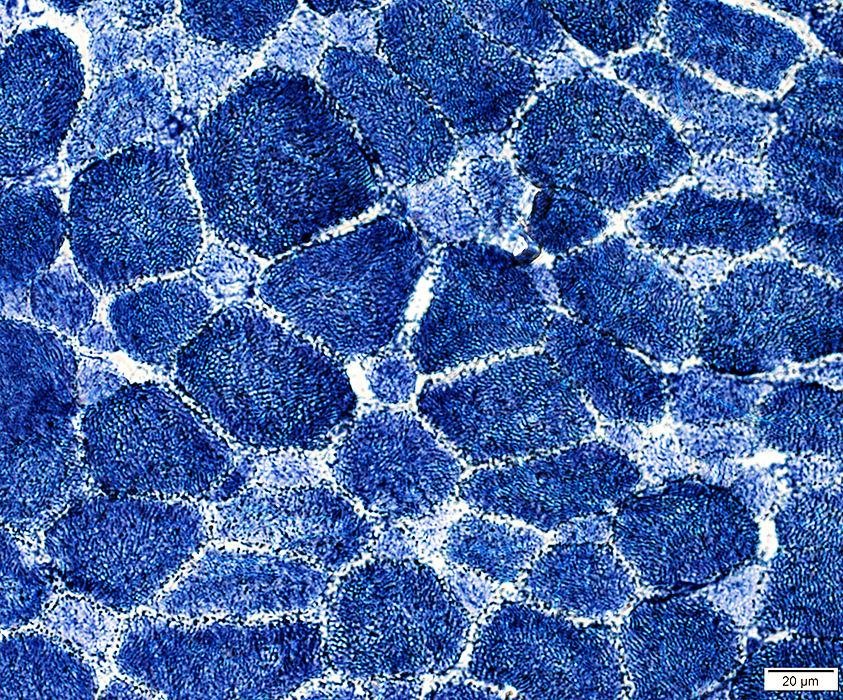
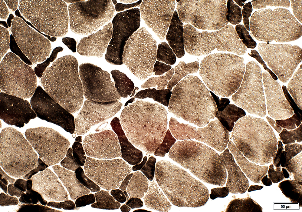 ATPase pH 9.4 stain |
Usually Type 2 (Dark stained at ATPase pH 9.4)
Shape: Often angular; Some are narrow and elongated
Distribution: May appear clustered
 ATPase pH 9.4 stain |
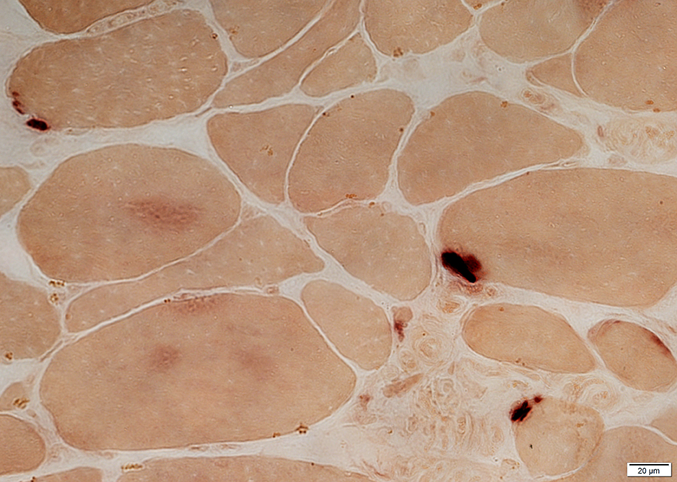
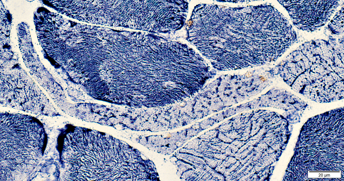
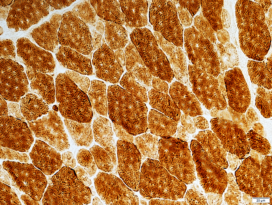
 ATPase pH 4.3 stain |
Pale stained at ATPase pH 4.6 & 4.3
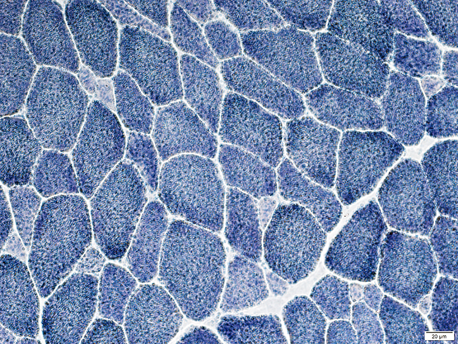
 ATPase pH 4.6 stain |
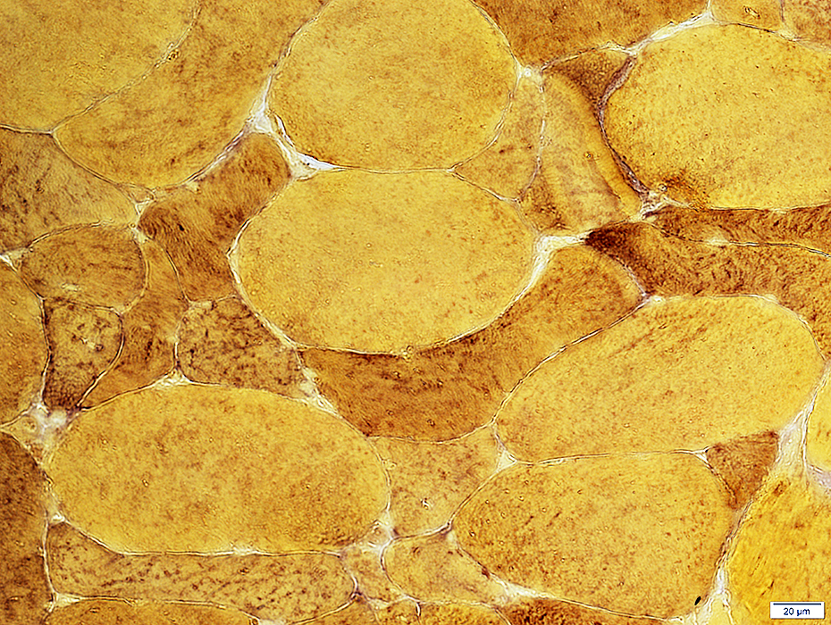
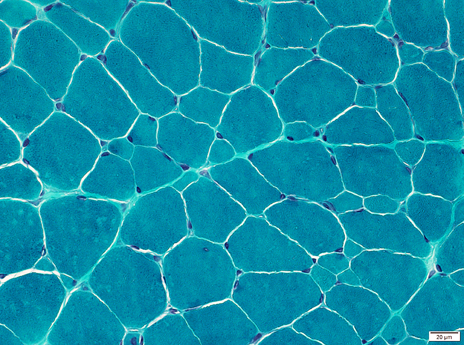
 NADH stain |
NADH: Pale
COX: Pale
Esterase: Not dark
 Esterase stain |
 PAS stain |
PAS: Darker
Phosphorylase: Darker
 Phosphorylase stain |
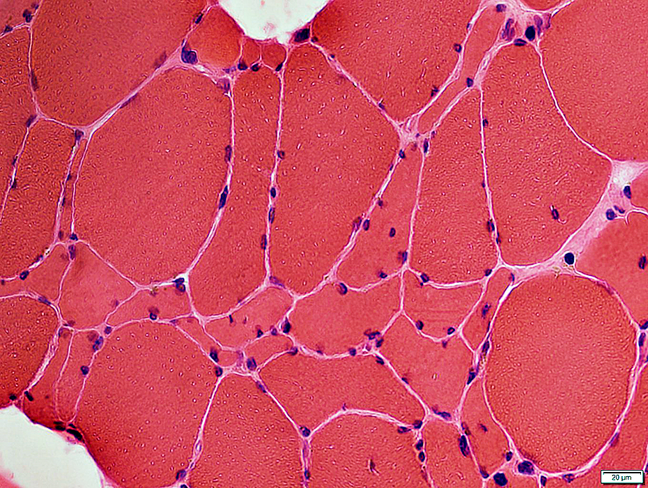
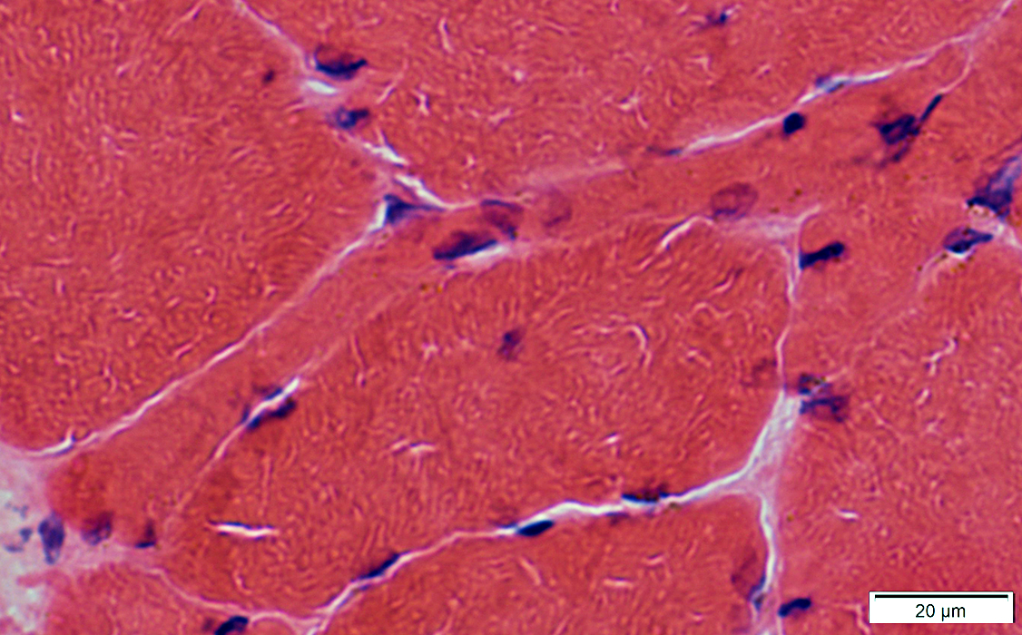
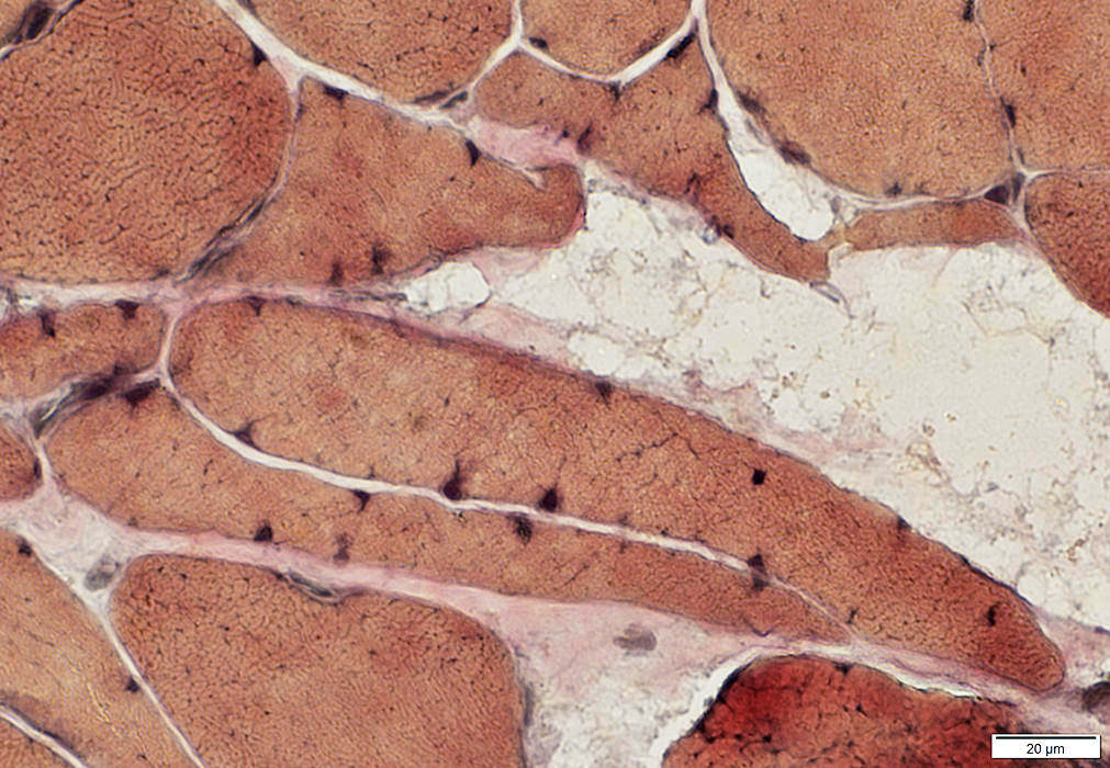
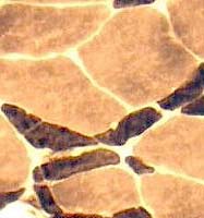
Type 2 Atrophy: Early
 H&E stain |
Elongated
Narrow
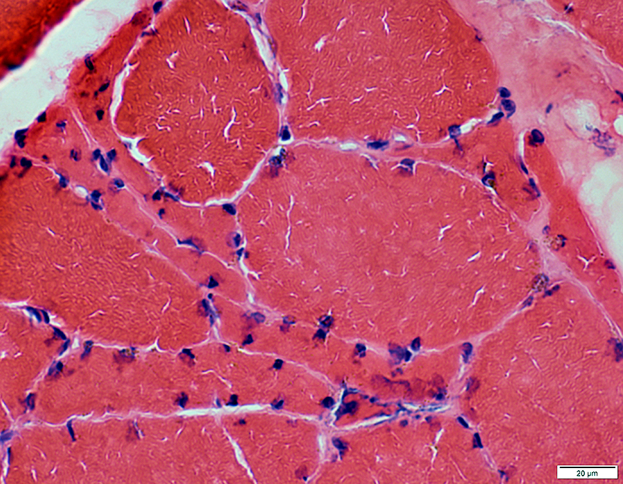 H&E stain |
 Gomori trichrome stain |
Elongated
Narrow
 Congo red stain |
 Congo red stain |
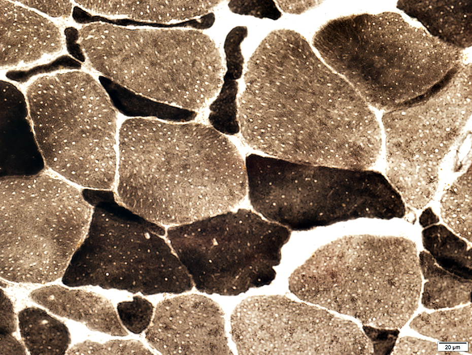
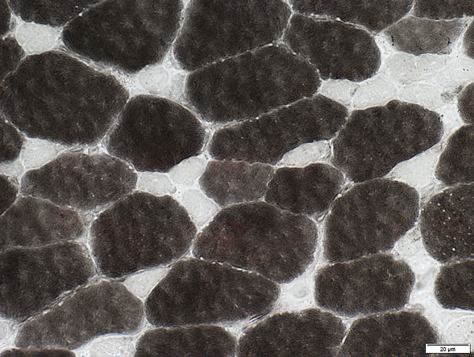
 ATPase pH 9.4 stain |
Shapes
Elongated & Narrow
Intermediate-sized & Polygonal
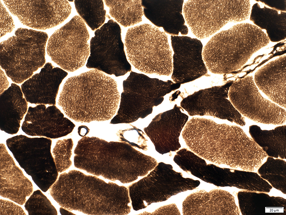 ATPase pH 9.4 stain |
 ATPase pH 4.3 stain |
Small & Angular
Pale stained with ATPase pH 4.3
Smallest muscle fibers may be 2B (Intermediate stain with ATPase pH 4.6)
 ATPase pH 4.6 stain |
 ATPase pH 4.6 stain |
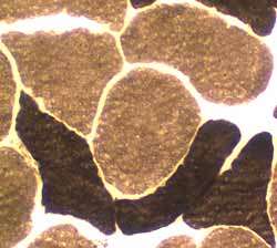
 H&E stain |
Elongated
Narrow
 H&E stain |
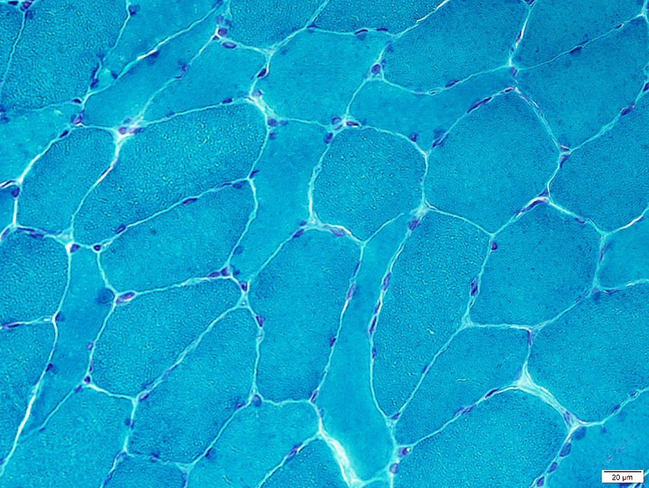
 VvG stain |
 Congo red stain |
 NADH stain |
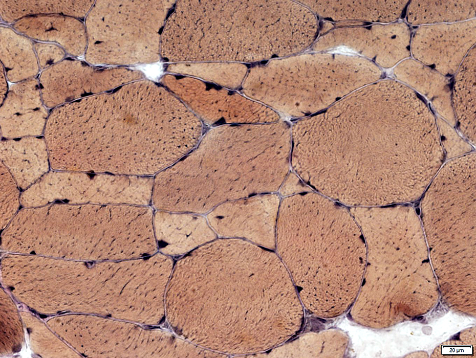
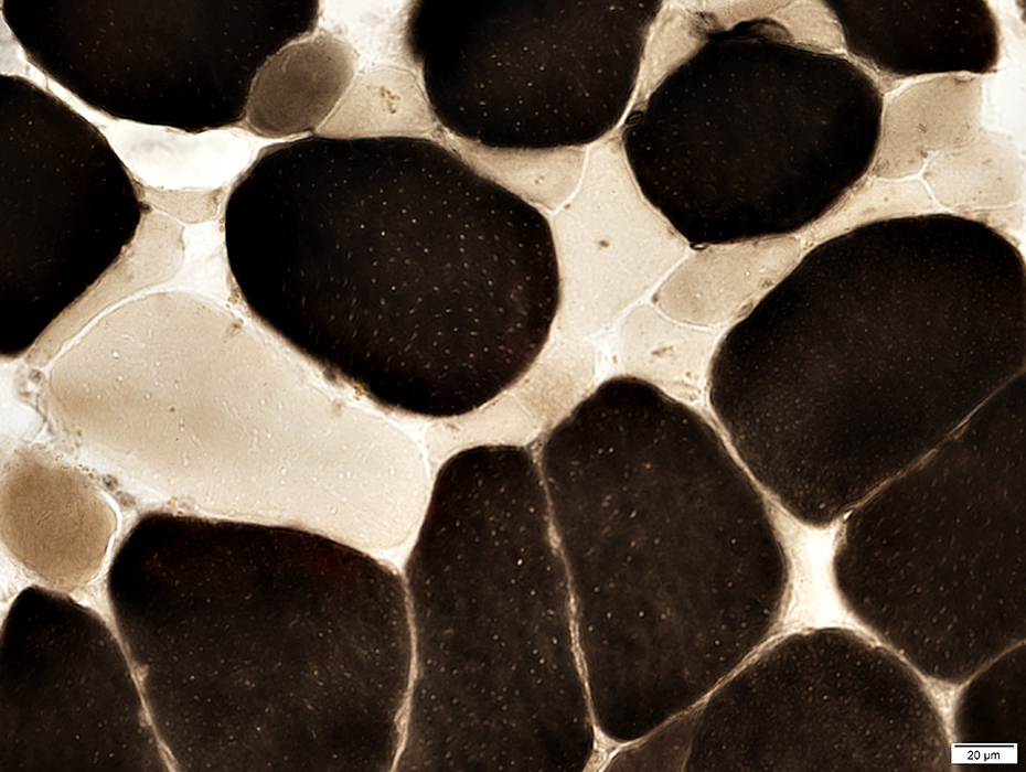
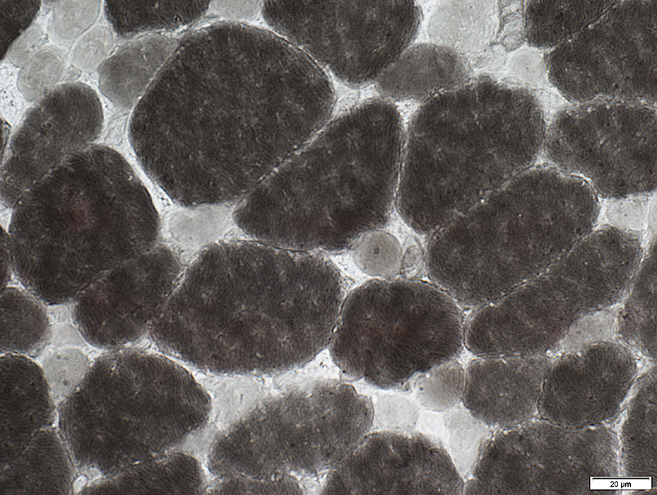
 ATPase pH 9.4 stain |
Shapes: Elongated; May wrap abound larger type 1 fibers
 ATPase pH 9.4 stain |
 ATPase pH 9.4 stain Type 2 Atrophy
|
 ATPase pH 4.6 stain Type IIB atrophy |
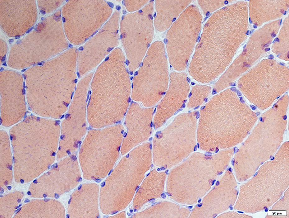
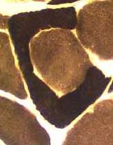
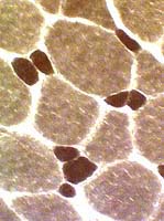
Childhood type II atrophy
Type 2 muscle fibersSmall
Polygonal or Round
 H&E stain |
Small muscle fibers: Polygonal or Round
 H&E stain |
 Gomori trichrome stain |
Small muscle fibers: Polygonal or Round
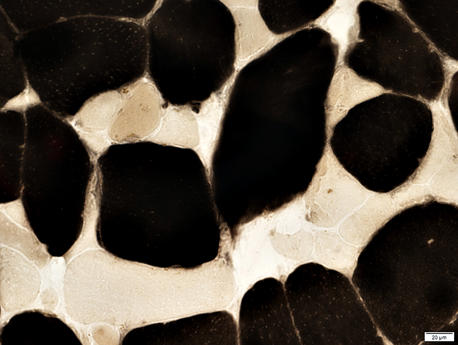
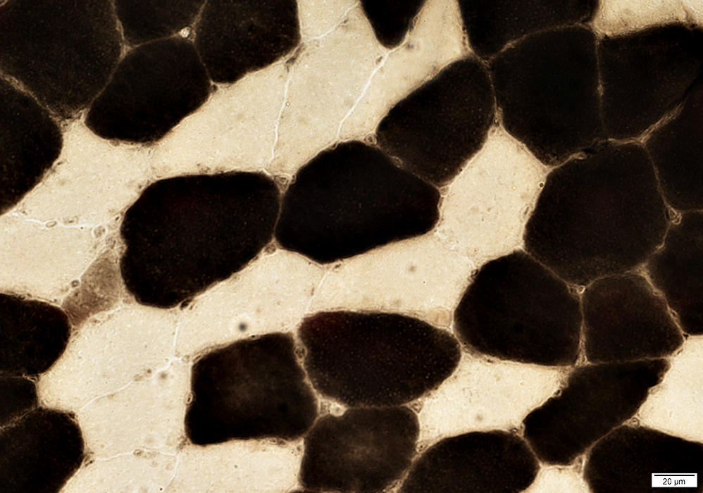
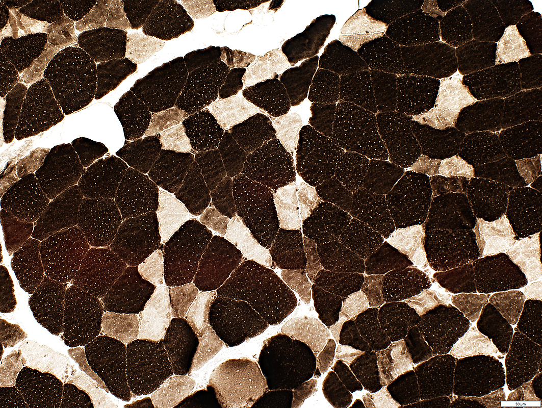
 ATPase pH 9.4 stain |
Small muscle fibers: Polygonal or Round
 ATPase pH 9.4 stain |
 ATPase pH 9.4 |
 ATPase pH 4.3 stain |
Pale stained
Type 2A & 2B
 ATPase pH 4.6 stain |
 NADH stain |
Pale on NADH, SDH & COX stain
 Cytochrome oxidase (COX) stain |
 SDH stain |
Type 2 muscle fibers
Darker on PAS stain
 PAS stain |
See
Fiber type disorders
Fiber type properties
Return to Neuromuscular Home Page
Return to Myopathies with wasting
References
1. Am J Physiol Cell Physiol 2012; Online August
2. Am J Physiol Cell Physiol 2015;308:C932-943
3. Muscle Nerve 2017;56:122-128
5/21/2021