MUSCLE FIBER ATROPHY & SMALLNESS
|
Early Type 1 fibers Type 2 fibers Severe Nuclear clumps Denervation Ultrastructure Dermatomyositis |
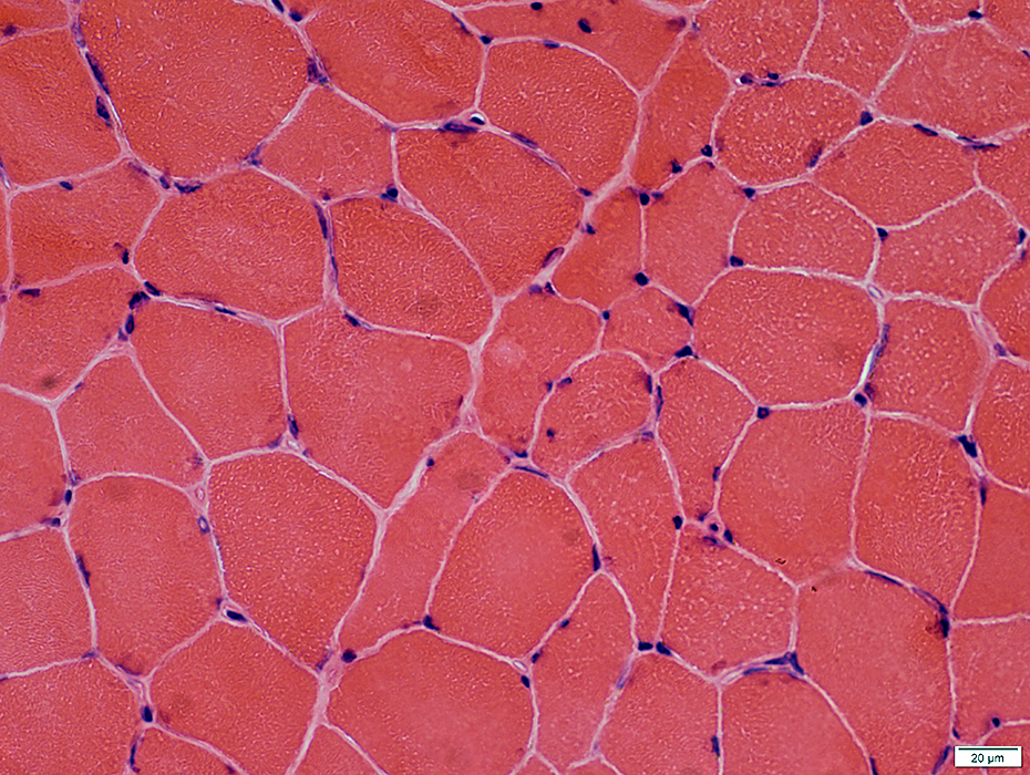
Atrophy, Severe & Diffuse
Muscle fiber atrophy
All muscle fibers are small
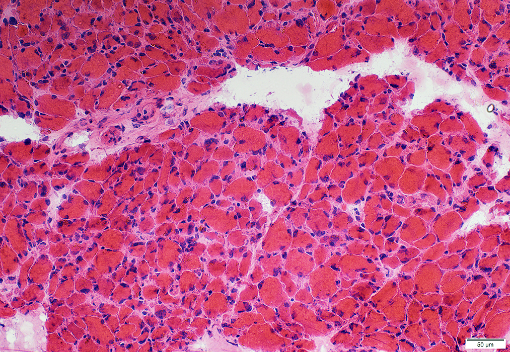 H&E stain |
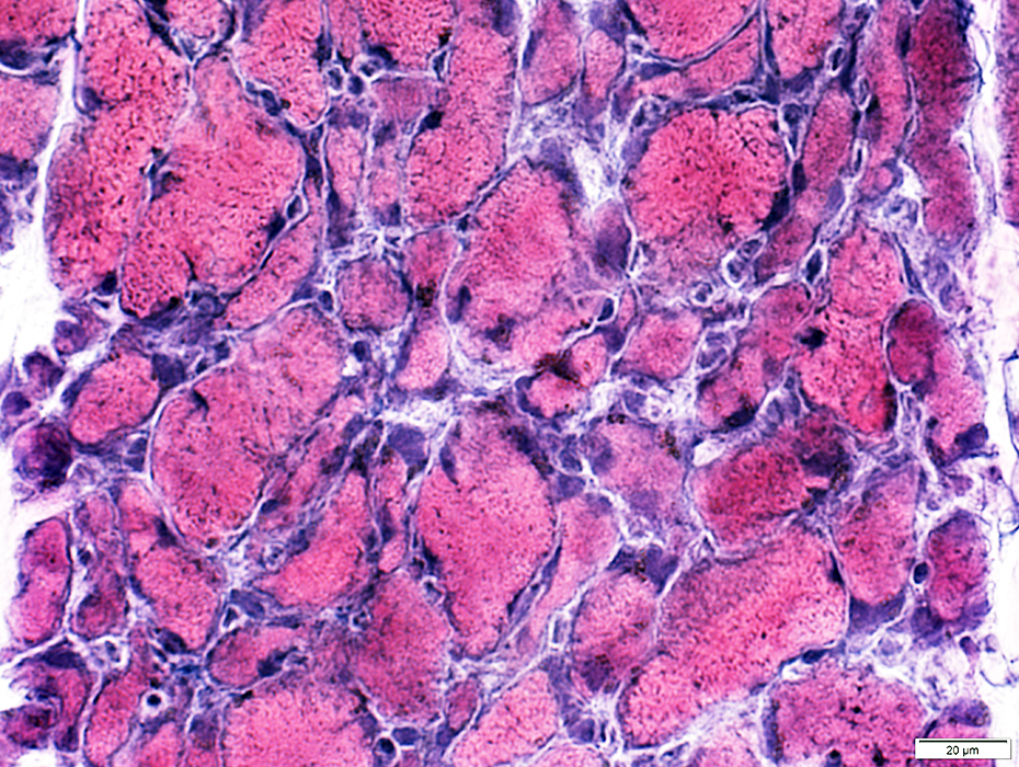
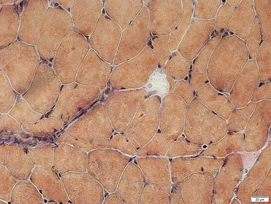
Small Muscle Fibers
Shapes: Varied
Surface: Irregular
Nuclei: Large; Pale
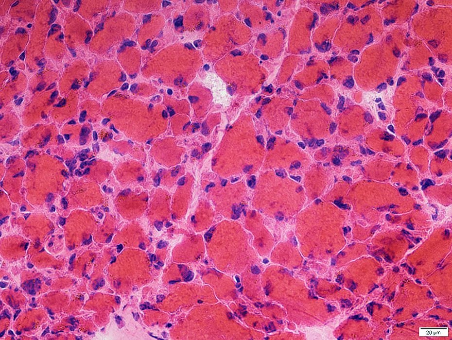 H&E stain |
 H&E stain |
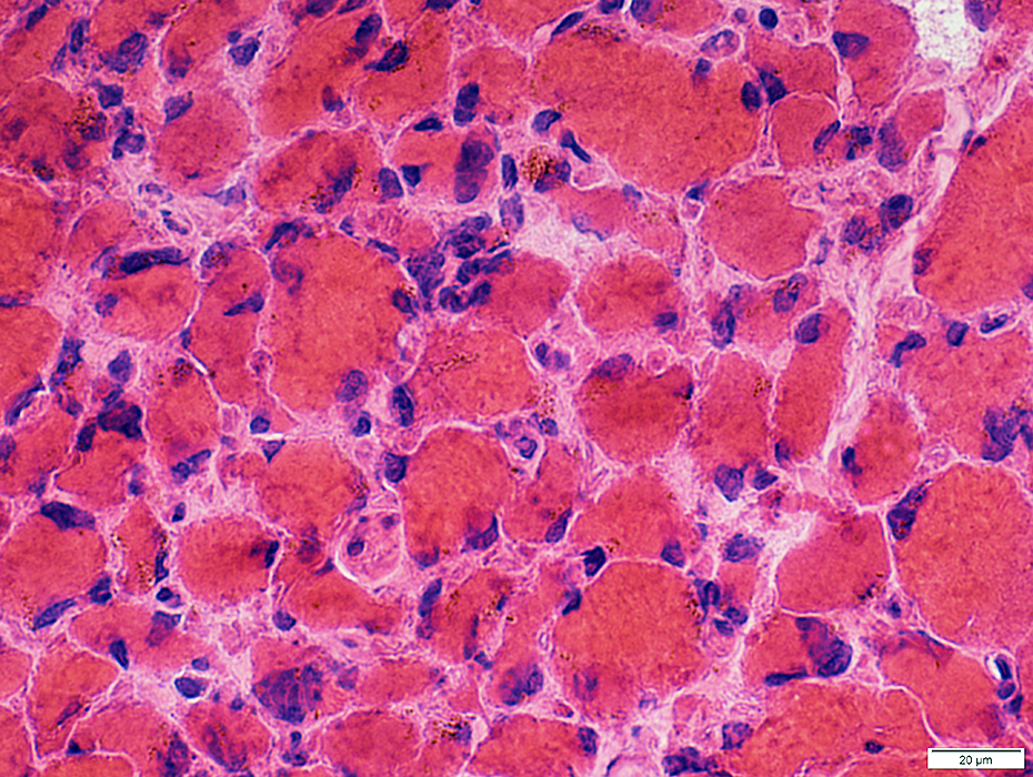
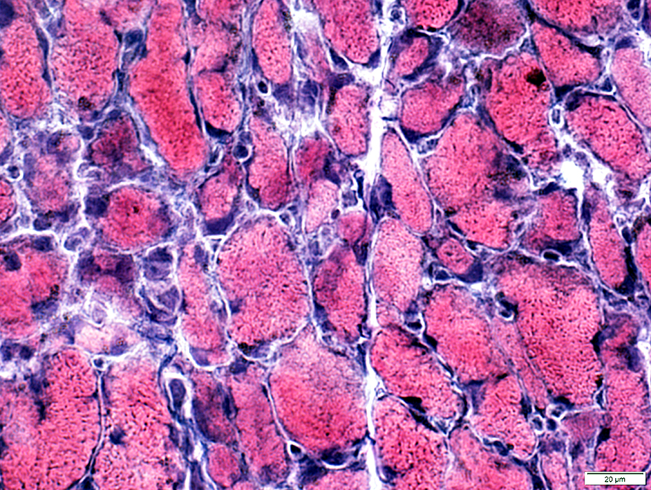
Shapes: Varied
Surface: Irregular
Nuclei: Large; Pale
Sizes: Varied
 H&E stain |
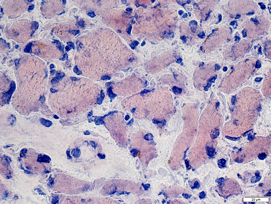 Congo red stain |
Shapes: Varied
Surface: Irregular
Nuclei: Large; Pale
Sizes: Varied
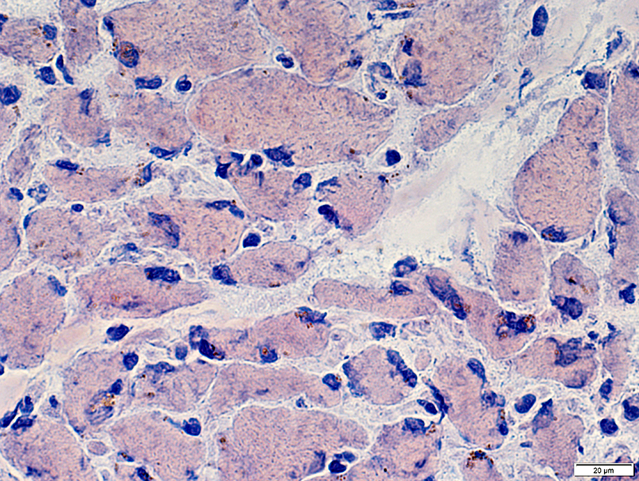 Congo red stain |
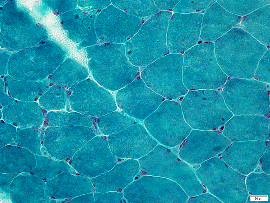
 VvG stain |
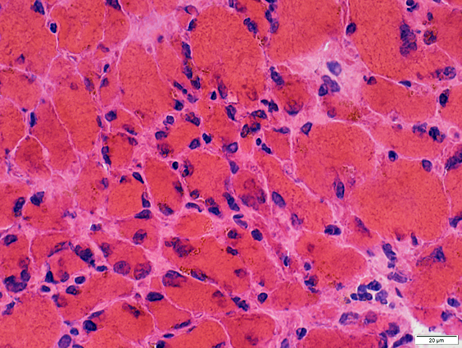
Shapes: Varied
Surface: Irregular
Nuclei: Large; Pale
Sizes: Varied
Internal architecture: Variable; Coarse
 VvG stain |
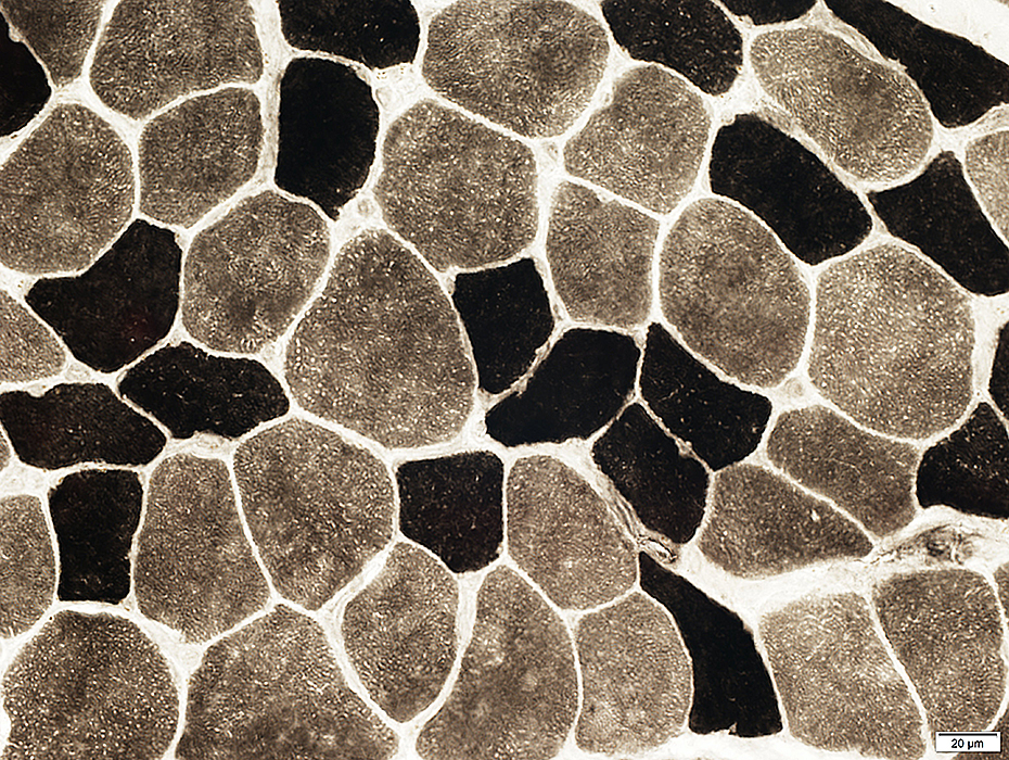
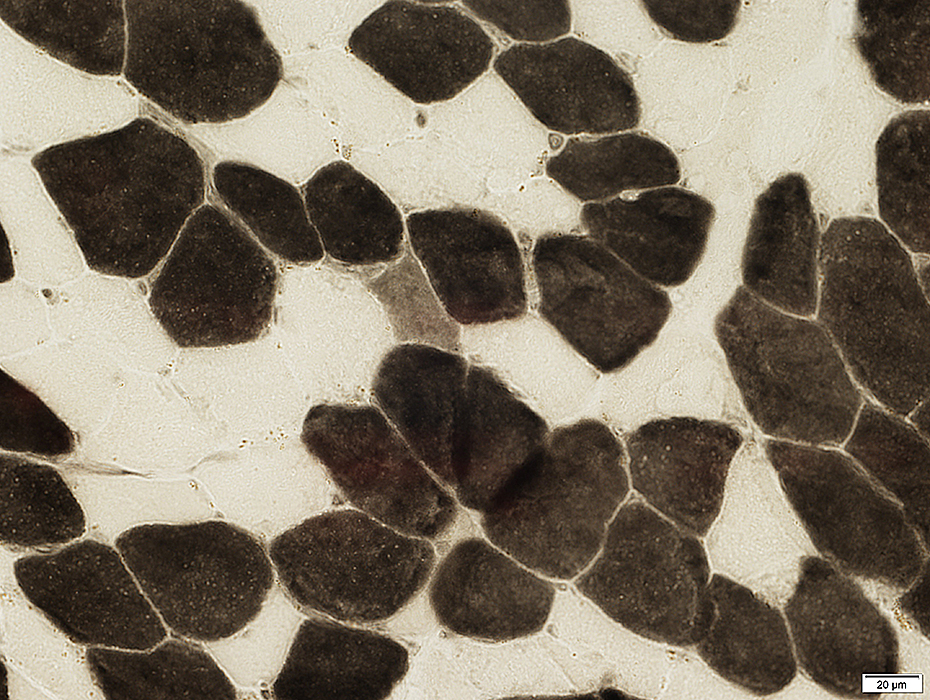
Muscle Fiber Atrophy
All fibers very small
Smallest muscle fibers: Type 2 (Dark)
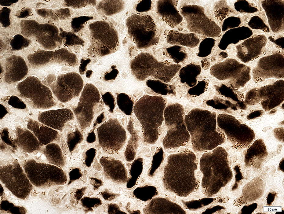 ATPase pH 9.4 stain |
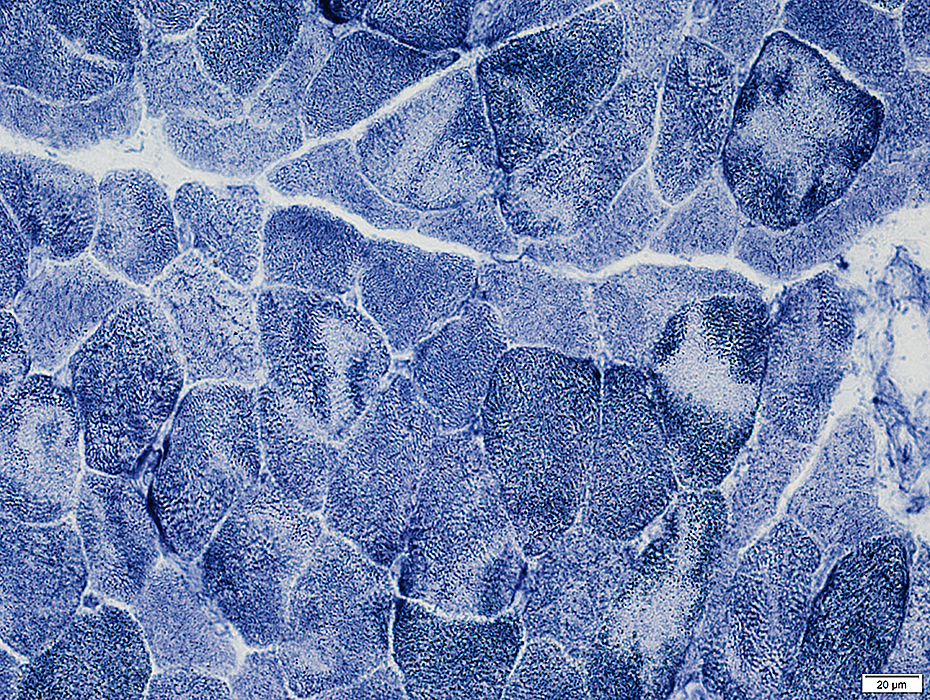
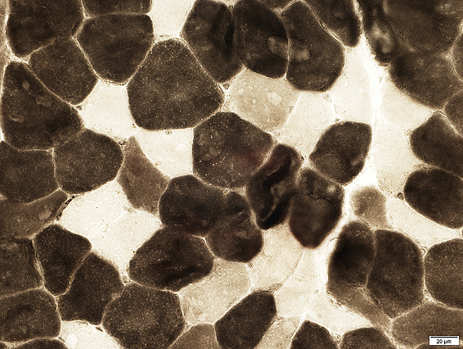
Muscle Fiber Atrophy
Internal architecture: Moderately coarse
Smallest muscle fibers: Intermediate stained
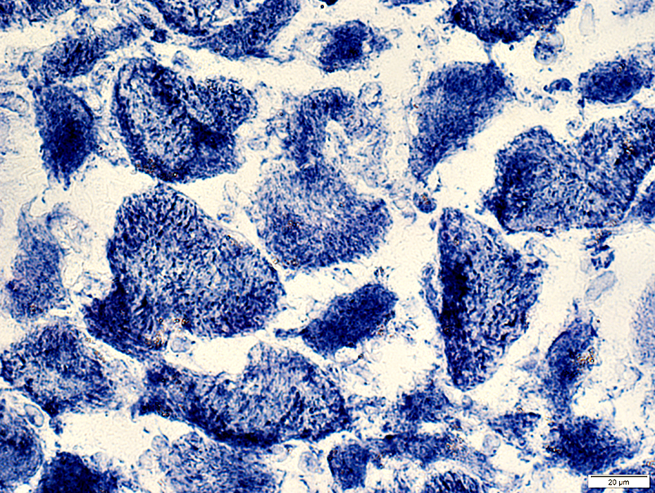 NADH stain |
Muscle Fiber Atrophy
Clustered lipofuscin granules
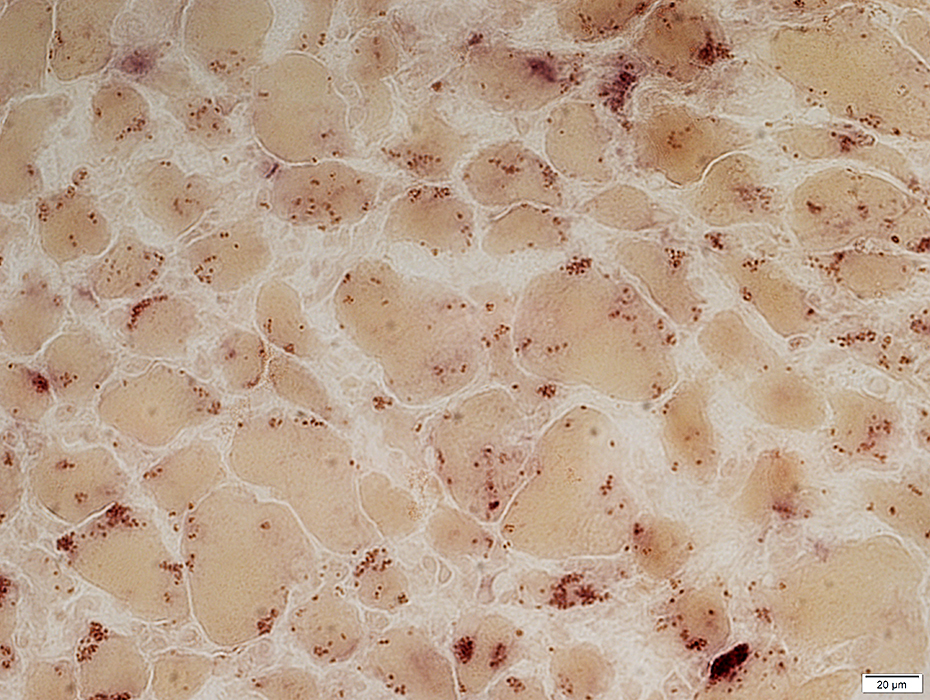 Acid phosphatase stain |
See
Fiber type disorders
Fiber type properties
Muscle Fiber Atrophy: Diffuse, Early
 H&E stain |
Mildly small
Bimodal distribution
Fiber Shapes
Polygonal
Surface membrane: Irregular
 Gomori trichrome stain |
 VvG stain |
Polygonal
Surface membrane: Irregular
Internal Architecture
Irregular
 NADH stain |
 ATPase pH 9.4 stain |
Type 2 fiber atrophy > Type 1
 ATPase pH 4.3 stain |
 ATPase pH 4.6 stain |
Neuromuscular Junctions
Remain dark stained
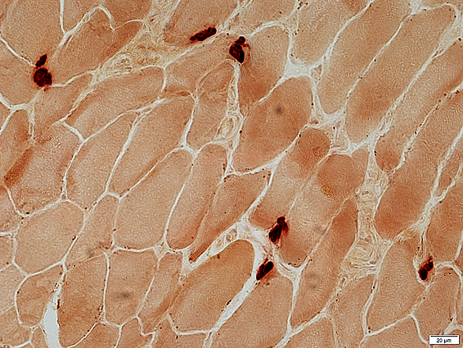 Esterase stain |
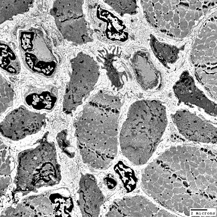
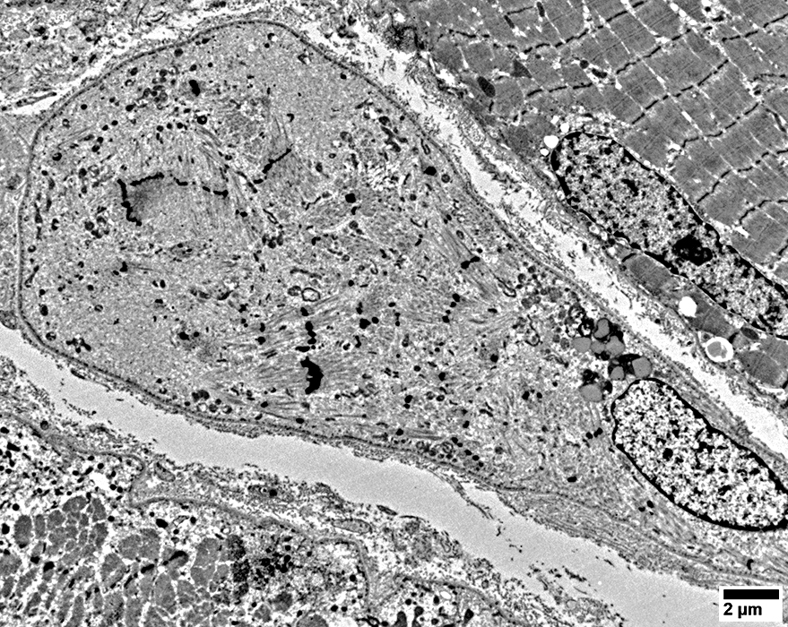
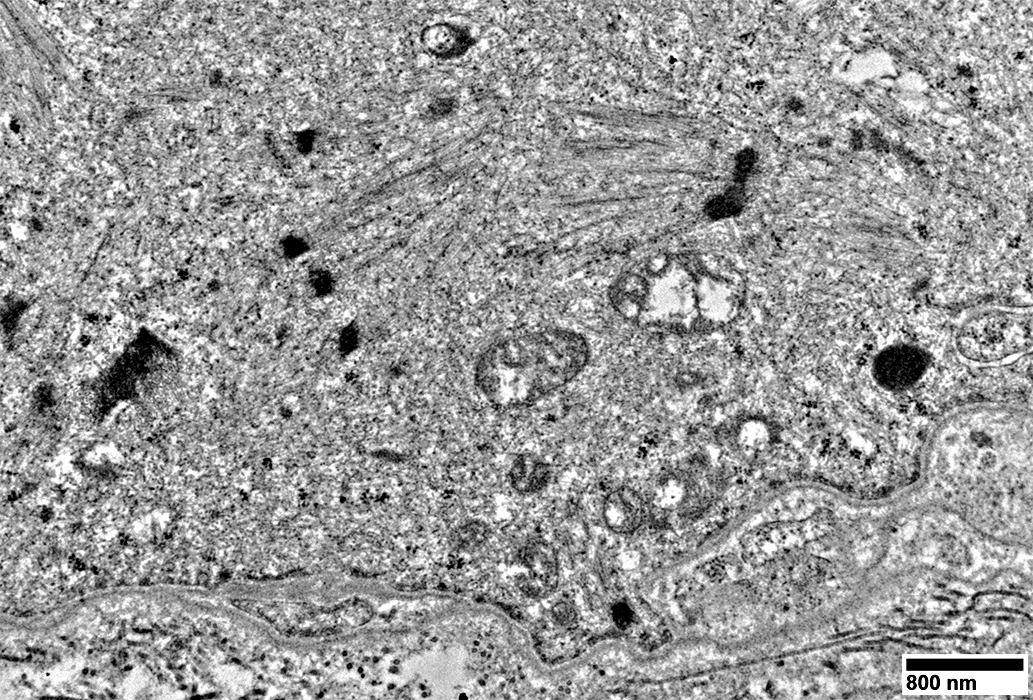
Atrophic Muscle fibers: Ultrastructure
|
Nuclear clumps Small muscle fibers |
Atrophic Muscle Fibers: General Features
- Size: Small
- Internal architecture
- Myofibril & Sarcomeres
- Disorganization
- Z-bands: Fragmentation; Streaming
- Loss
- Myofibril & Sarcomeres
- Muscle fiber surface
- Basal lamina: May project empty sleeves or redundant loops from fiber surface
- May be smooth or undulating
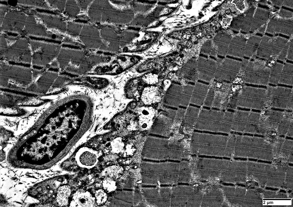
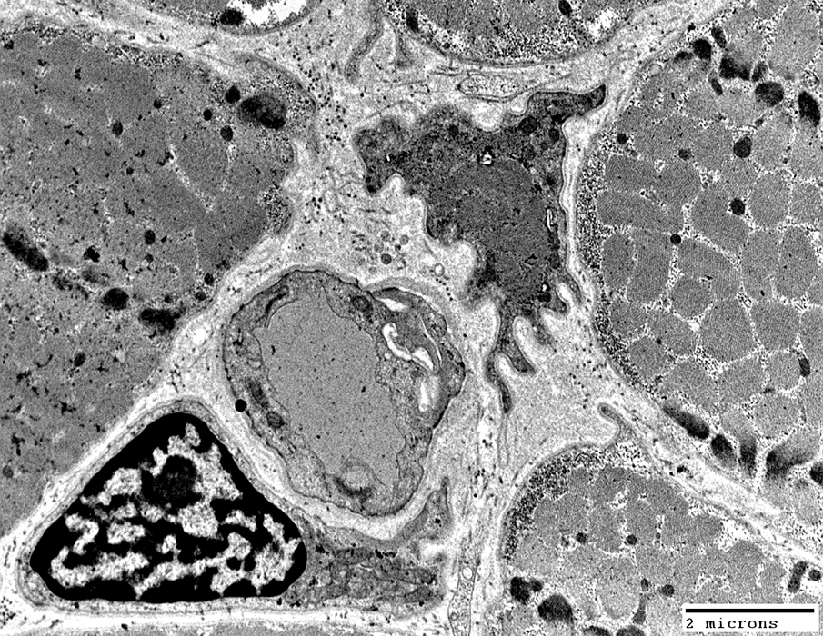
Muscle Fiber Atrophy: Sarcolemma
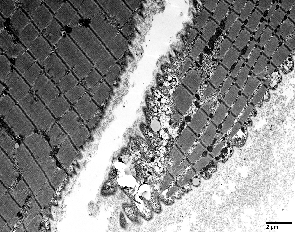 |
Undulating Basal lamina & Sarcolemma
 |
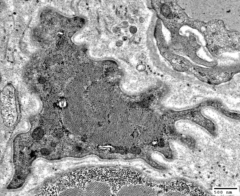
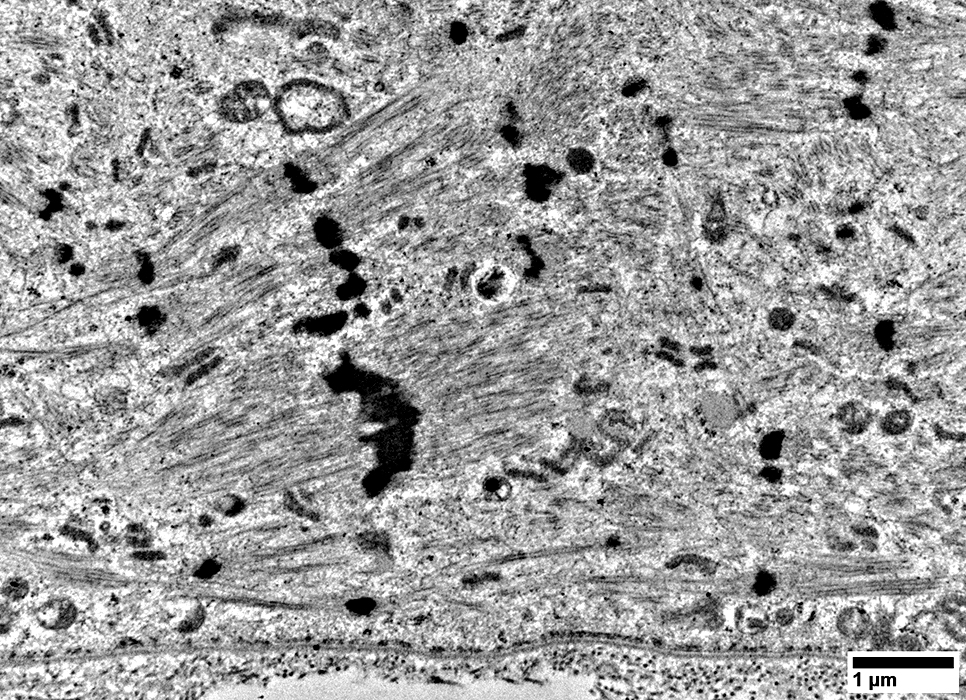
Muscle Fiber Atrophy: Sarcomere Structure
Sarcomere structure (Right): Disorganized
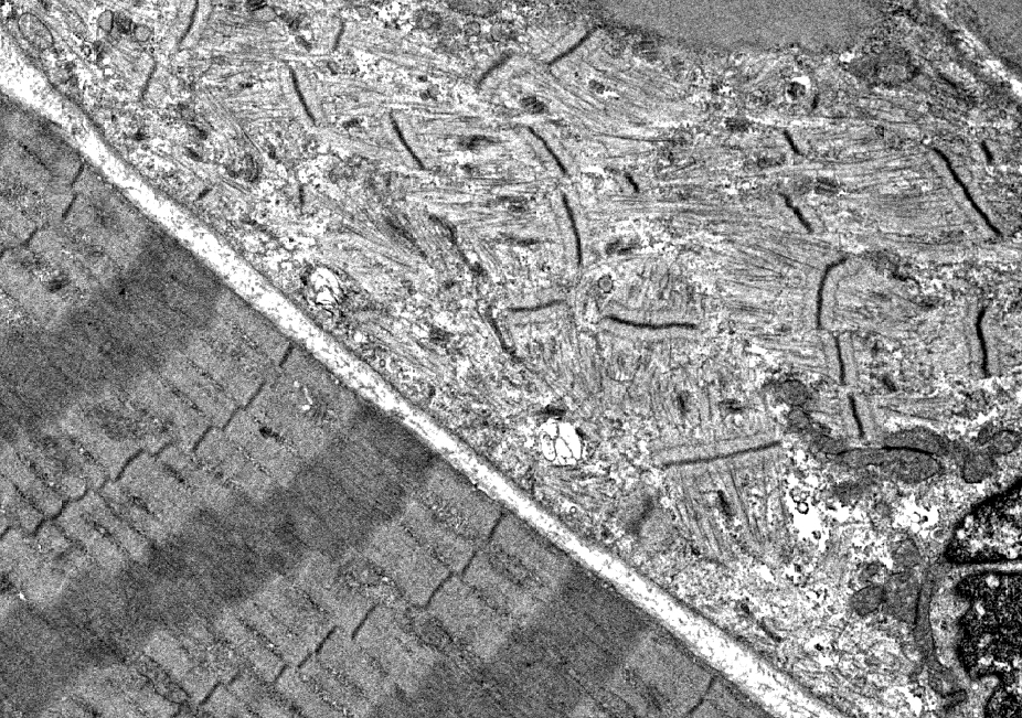 From: Sean Ferris |
Sarcomere structure (Left): Disorganized
Lipid Droplet (Right; Arrow)
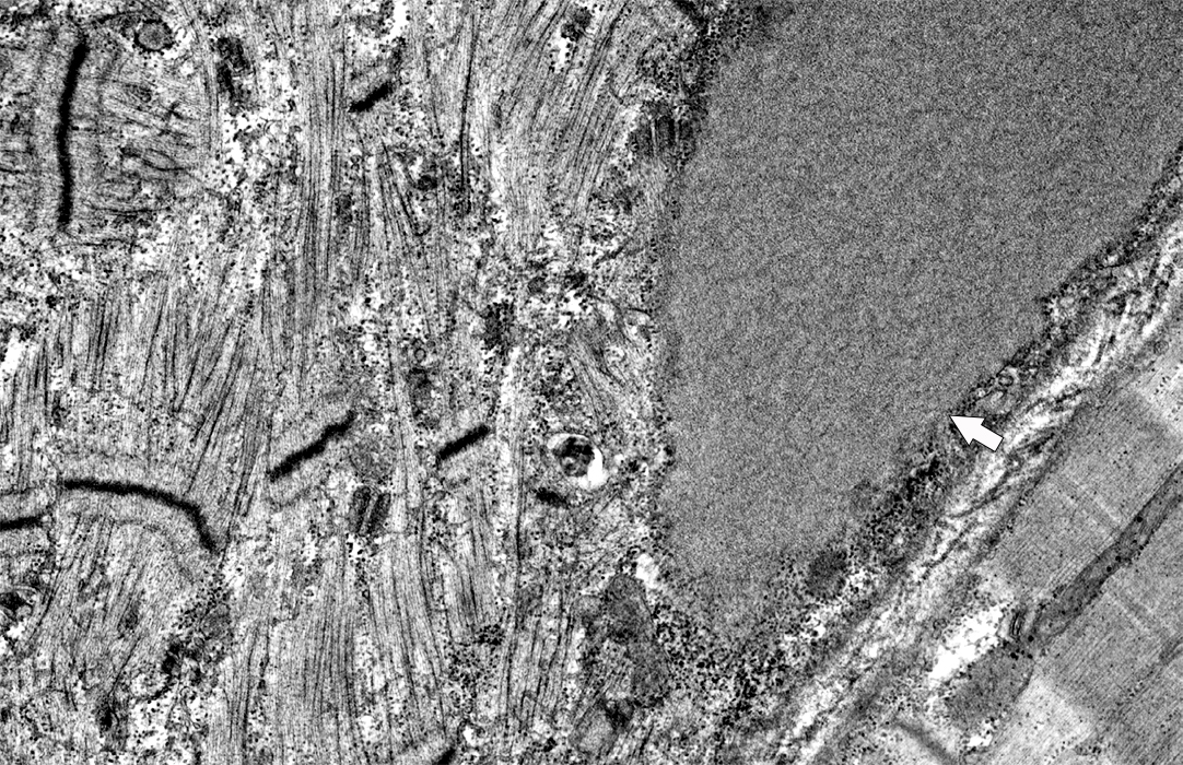 From: Sean Ferris |
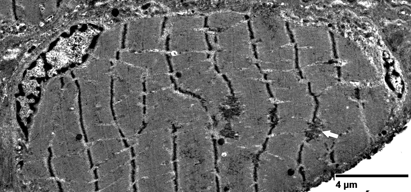 Atrophic Muscle fiber Z-Band streaming (Arrow) |
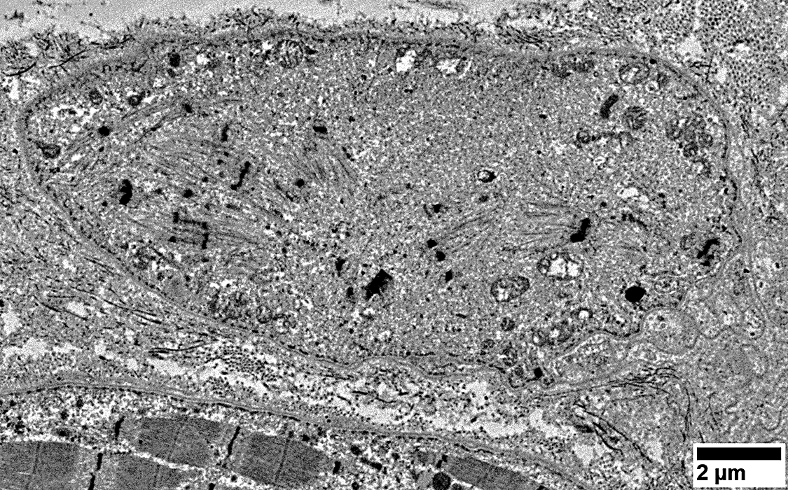
Muscle Fiber Atrophy: Severe
 From: R Schmidt |
 From: R Schmidt |
 From: R Schmidt |
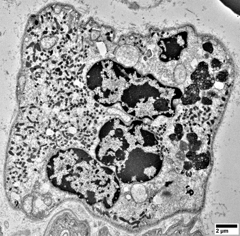
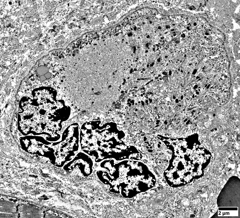
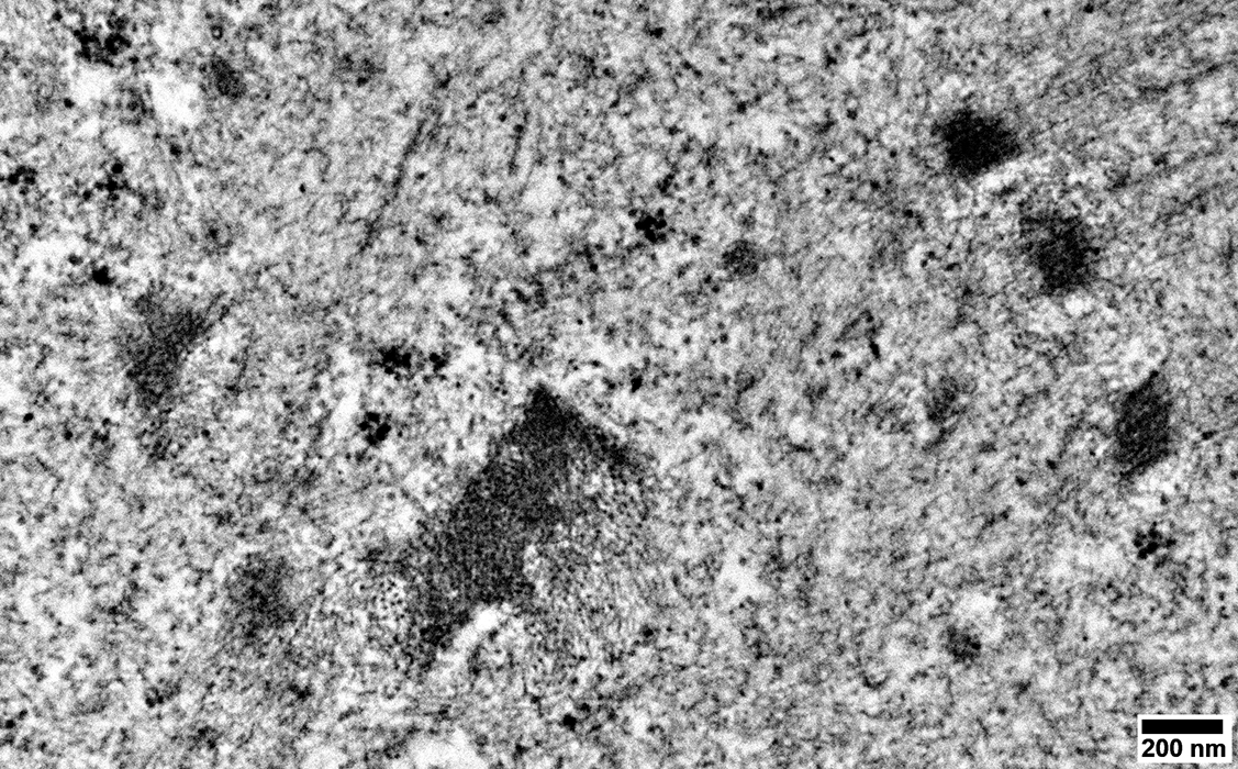
Pyknotic Nuclear Clumps: Ultrastructure
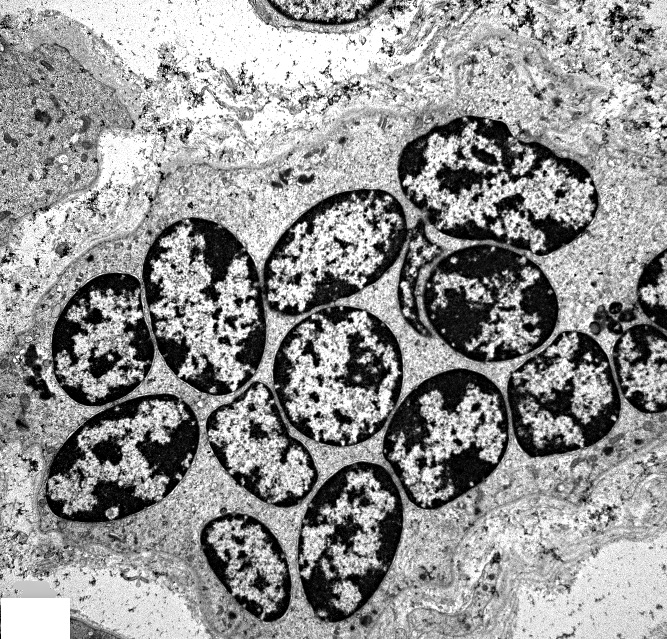 From: Cory Toth MD |
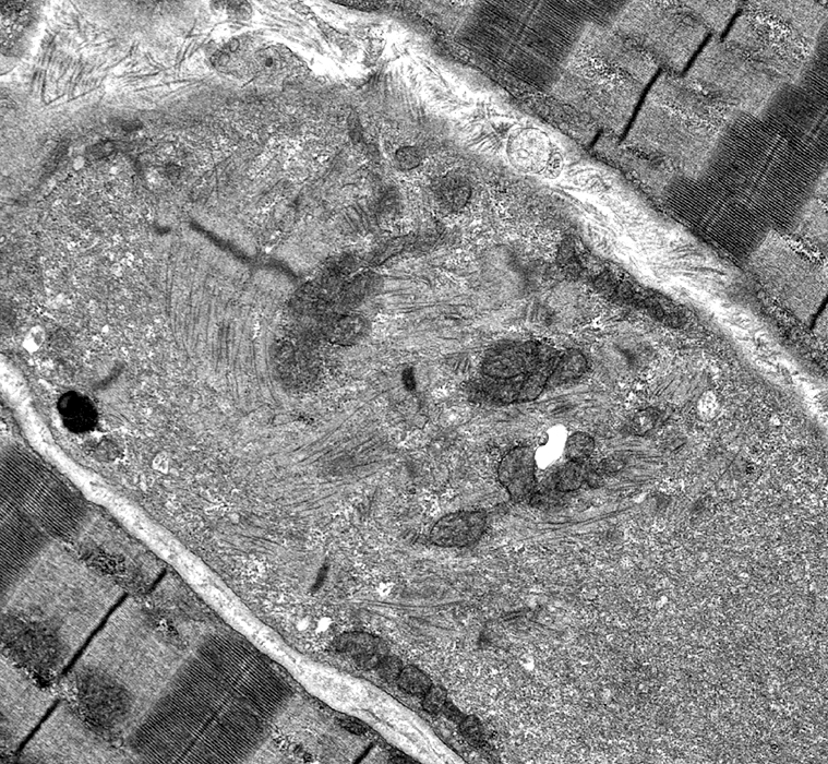
Muscle Fiber Cytoplasm
Sarcomeres: Lost or Disorganized
Mitochondria & Lipopigment: May be Increased (Below)
 From: R Schmidt |
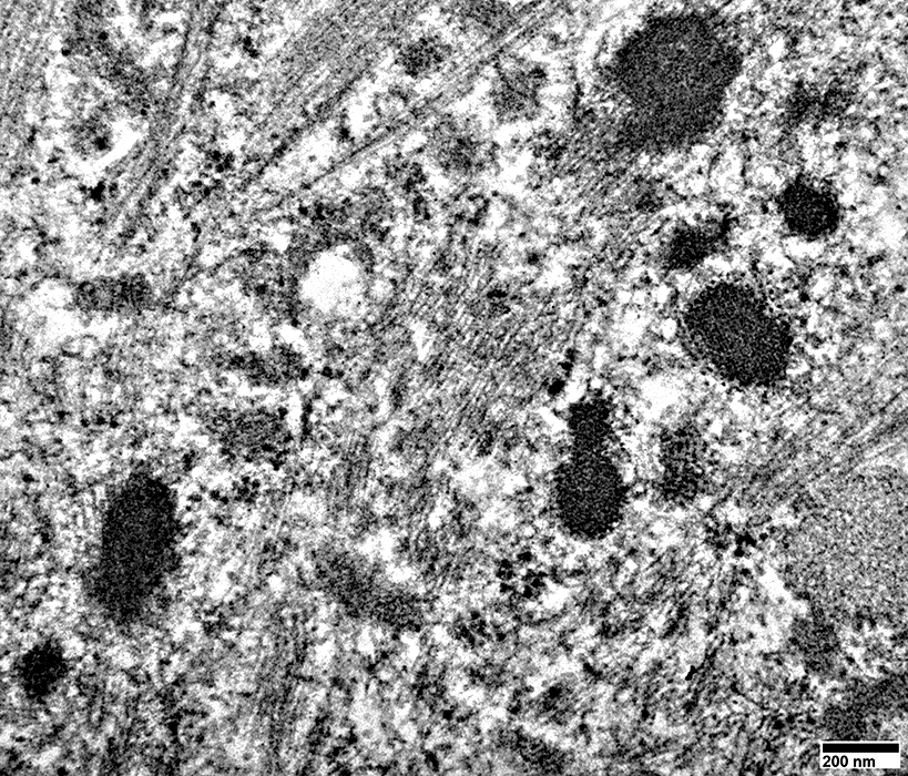
Mitochondria: Clustered in Atrophic Muscle Fiber
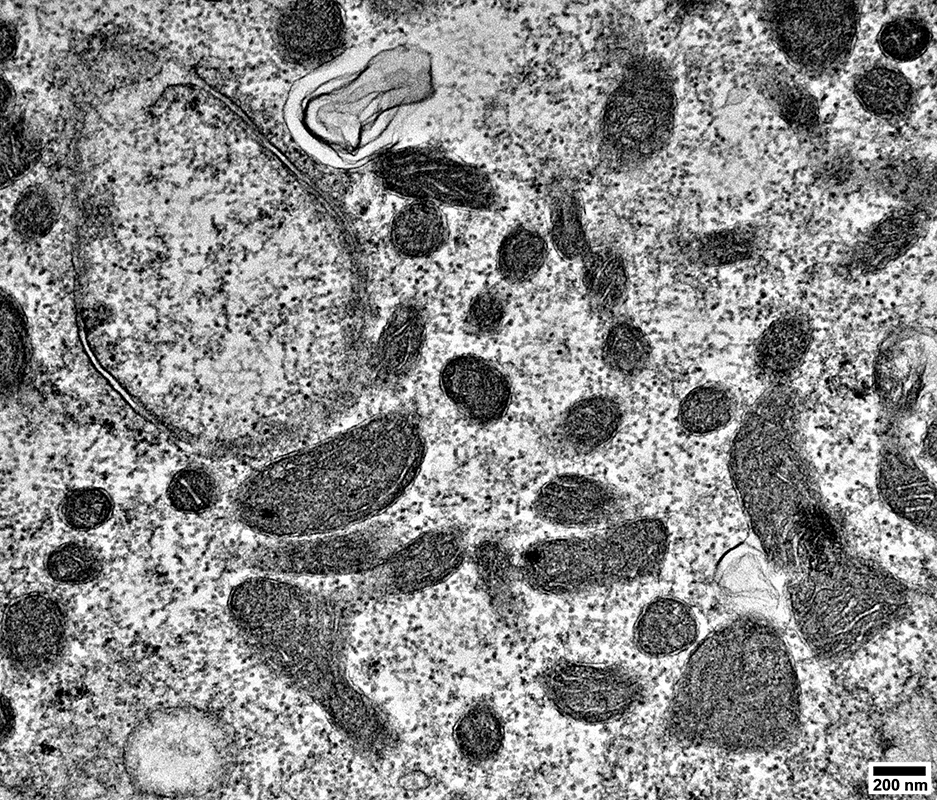 From: R Schmidt |
Lipopigment: Clustered in Atrophic Muscle Fiber
 From: R Schmidt |
Muscle Fiber Atrophy: Severe
 From: Sean Ferris |
Sarcomere structure: Absent or Disorganized
 From: R Schmidt |
 From: R Schmidt |
Z-bands
Fragmented
Fragments similar to Cytoplasmic bodies or Rods
 From: R Schmidt |
 From: R Schmidt |
Fragmented
Fragments similar to Cytoplasmic bodies or Rods
 From: R Schmidt |
Muscle Fiber Atrophy: Disorganized Sarcomeres
 From: R Schmidt |
 From: R Schmidt |
 From: R Schmidt |
Basal Lamina: Redundant
External link: Radboudou
Return to Neuromuscular Home Page
Return to Myopathies with wasting
References
1. Am J Physiol Cell Physiol 2012; Online August
5/24/2026