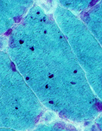
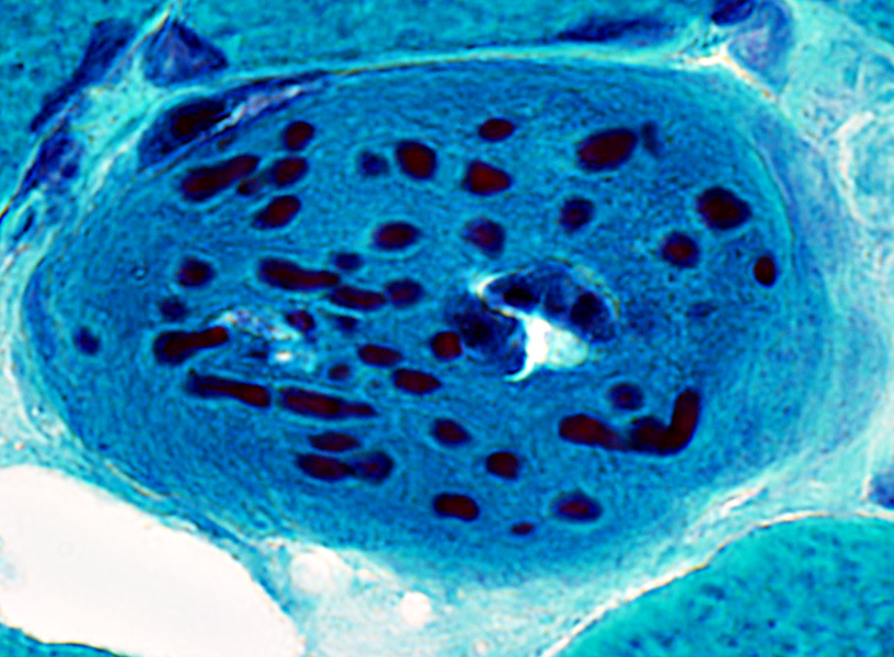
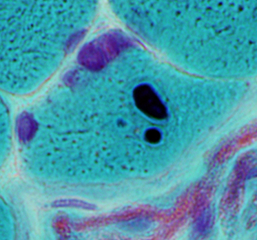
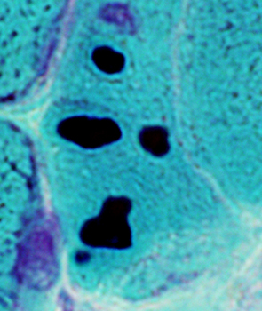
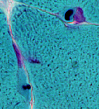
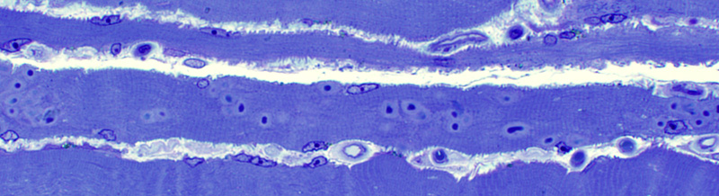
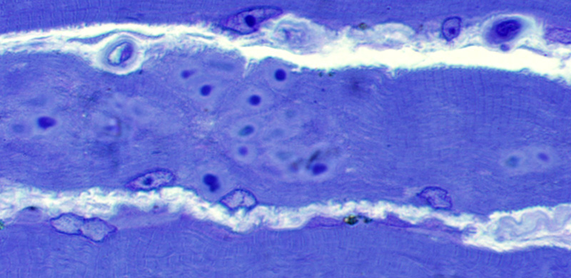
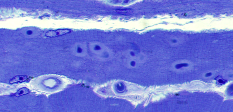
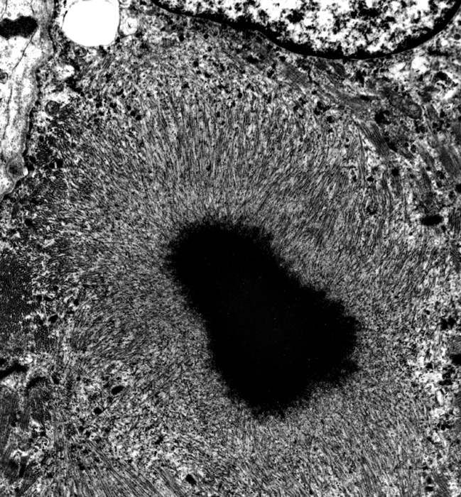
Cytoplasmic bodies
- Structure
- Central region: Homogenous; Dark stain on Gomori trichrome similar to rods
- Outer zone: Radially oriented filaments; May contain actin
- Location
- Within cytoplasm: Central or subsarcolemmal
- Early stage: May be associated with Z-Band
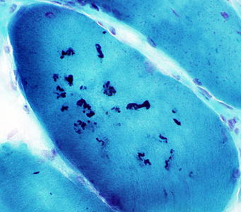
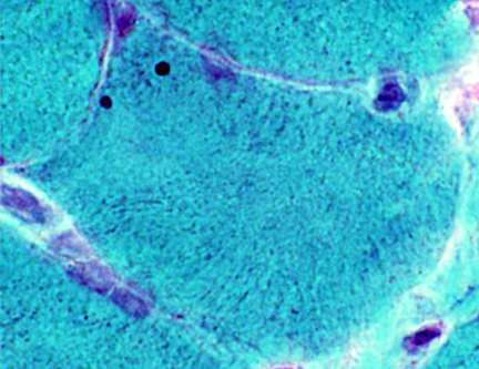
- Pathology
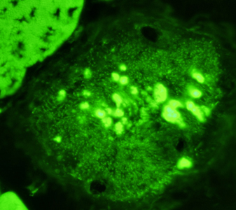
- Histochemical properties
- Stain strongly
- Gomori trichrome
- Phalloidin
- No Stain
- Myofibrillary ATPase
- NADH
- Fiber types
- May occur selectively in type I or II fibers
- Type I fibers in distal type
- Type I or II fibers in limb-girdle syndromes
- Molecular contents
- General: Actin; Stain for Phalloidin
- Other variable: Desmin, Dystrophin, Sarcoglycans, β-Amyloid precursor, N-CAM, LC3
- May be PAS+
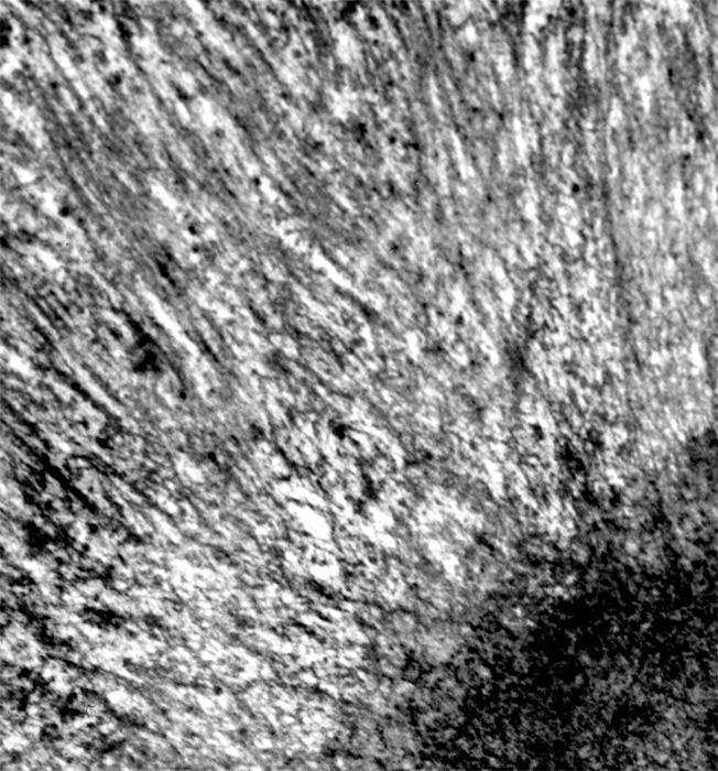
- Ultrastructure: Variable
- Foci of myofibrillary destruction: Z-disk derived bodies & streaming
- Cytoplasmic or spheroid bodies: Thick & thin filament remnants
- Aggregates of Z-disk filaments
- Differential diagnosis: Nemaline rods
- Color: Similar
- Also associated with abnormal Z-band structure
- Usually smaller
- No halo
- Many per muscle fiber
- Often clustered
- Disease associations: "Cytoplasmic bodies" described in
|