HIV: Immune Reconstitution Inflammatory Syndrome (IRIS)
|
General Perimysial pathology Endomysial pathology Muscle fibers & Immune cells |
 H & E stain |
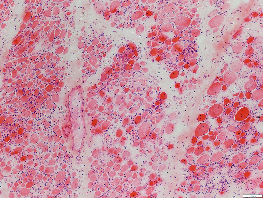
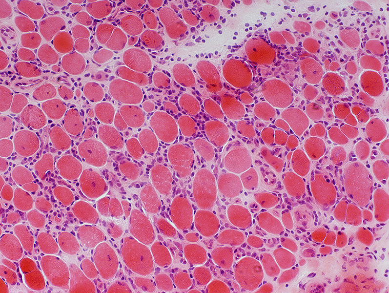
Endomysium: Increased; Histiocytic cells
Muscle fibers: Varied size; Small
 H & E stain |
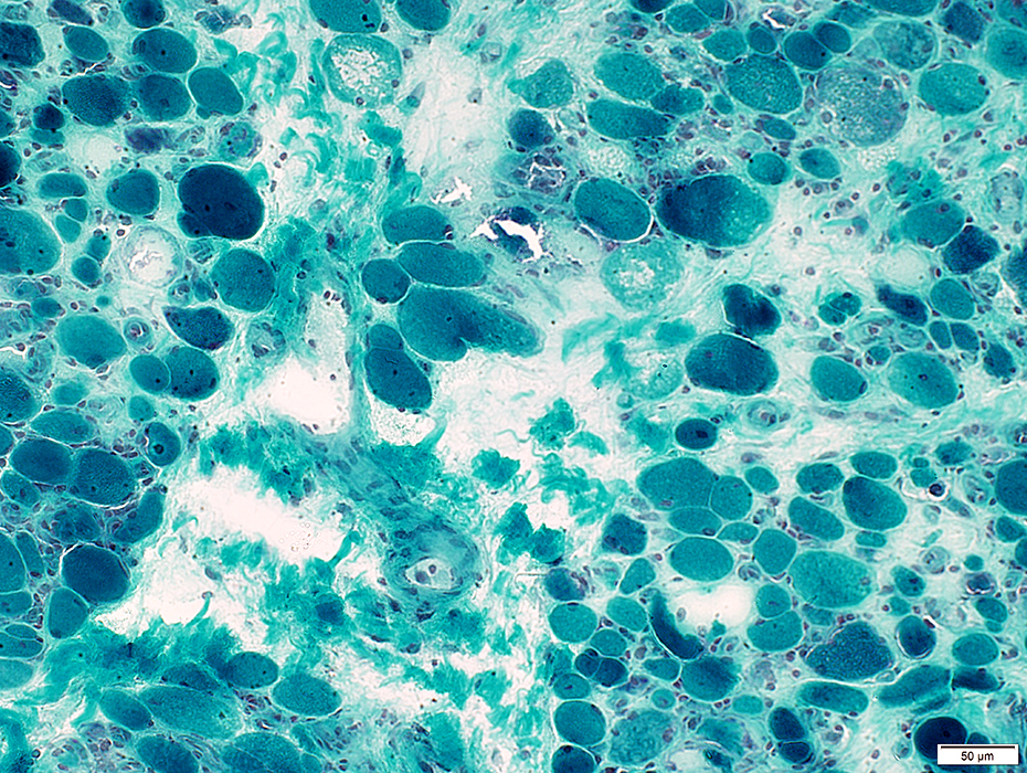
 Gomori trichrome stain |
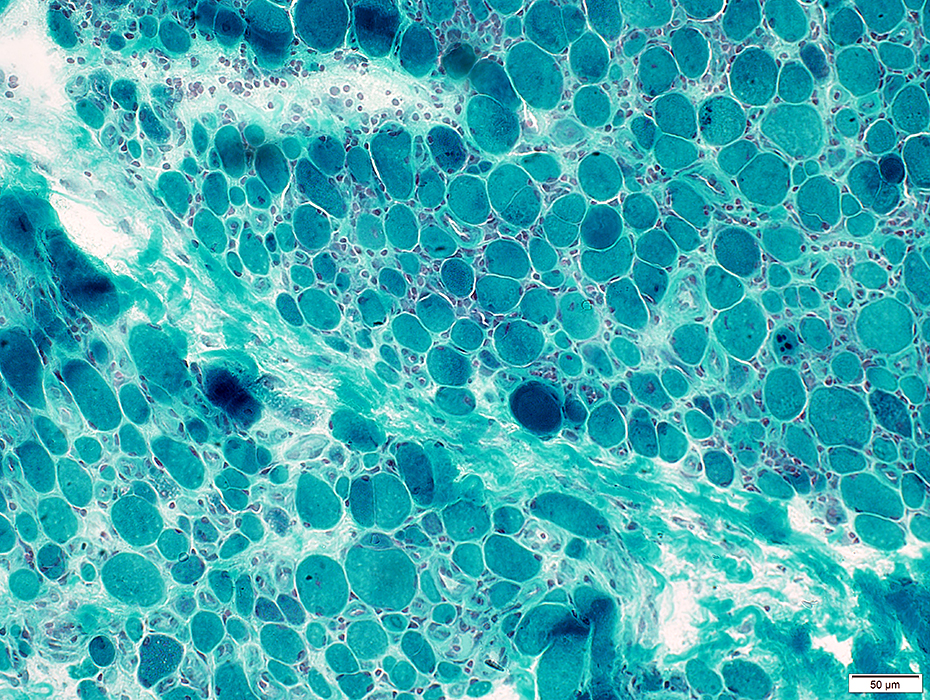
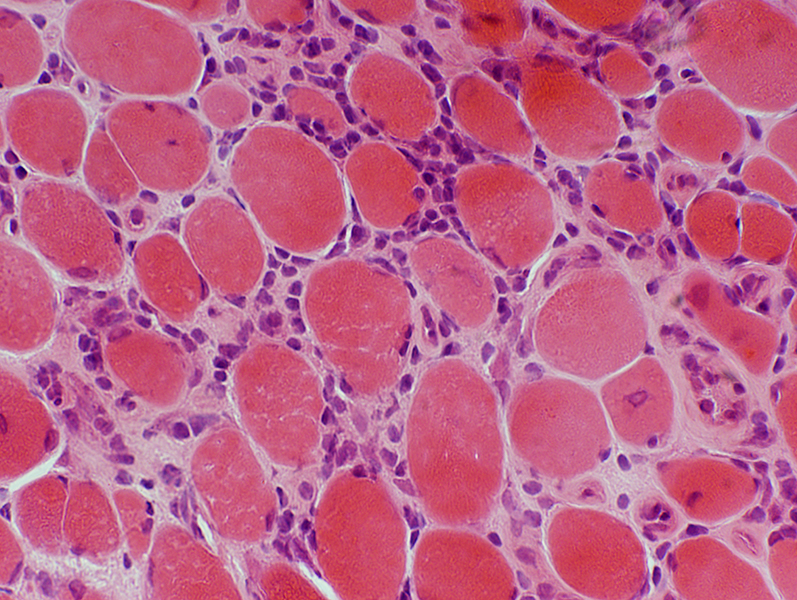
Endomysium: Increased; Histiocytic cells
Muscle fibers: Varied sizes; Small fibers round
 VvG stain |
IRIS: Perimysial Pathology
 Gomori trichrome stain |
 Esterase stain |
 Acid phosphatase stain |
 Esterase stain |
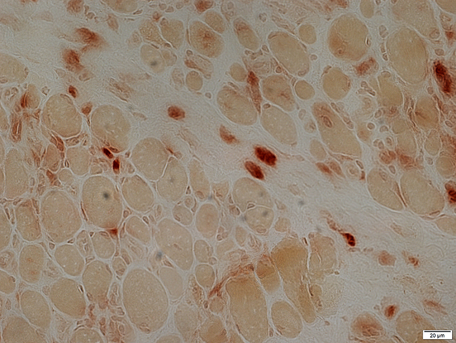 Acid phosphatase stain |
IRIS: Endomysial Histiocytic Cells
 Esterase stain |
|
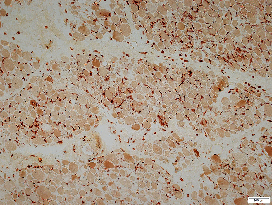
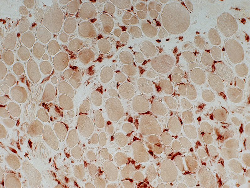
Histiocytes: Large, irregular shaped cells are scattered in the endomysium Endomysial connective tissue: Increased between muscle fibers 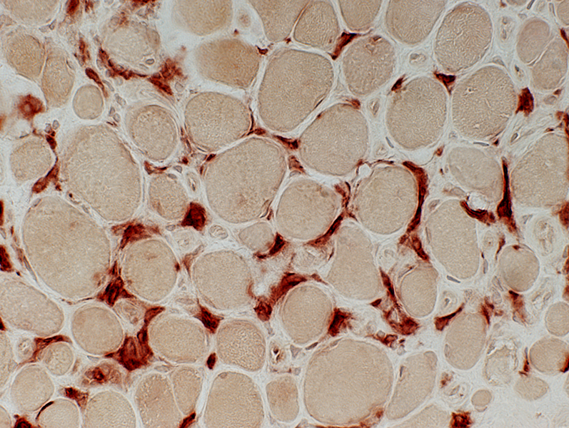 Esterase stain |
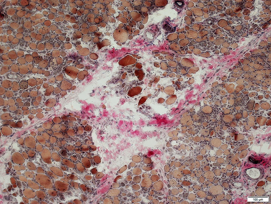
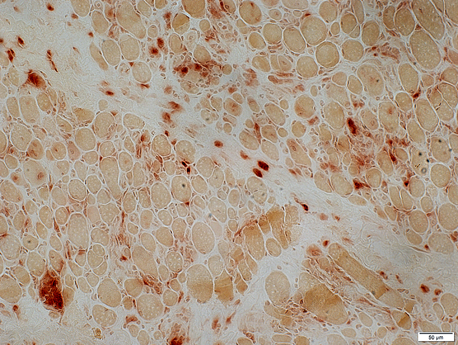
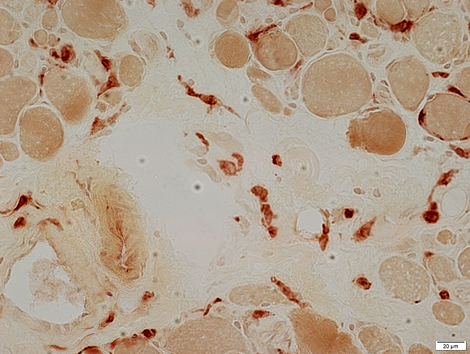
Histiocytes: Stain with acid phosphatase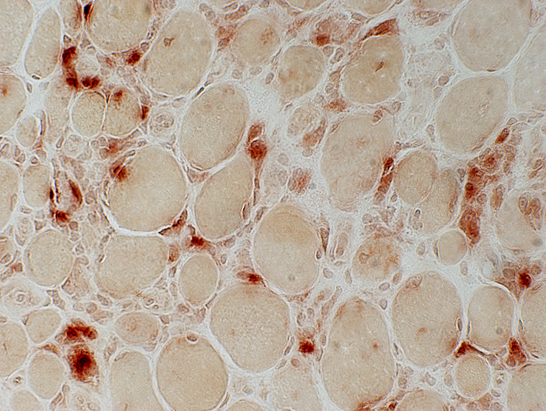 Acid phosphatase stain |
IRIS: Muscle fiber pathology & Histiocytic cellularity
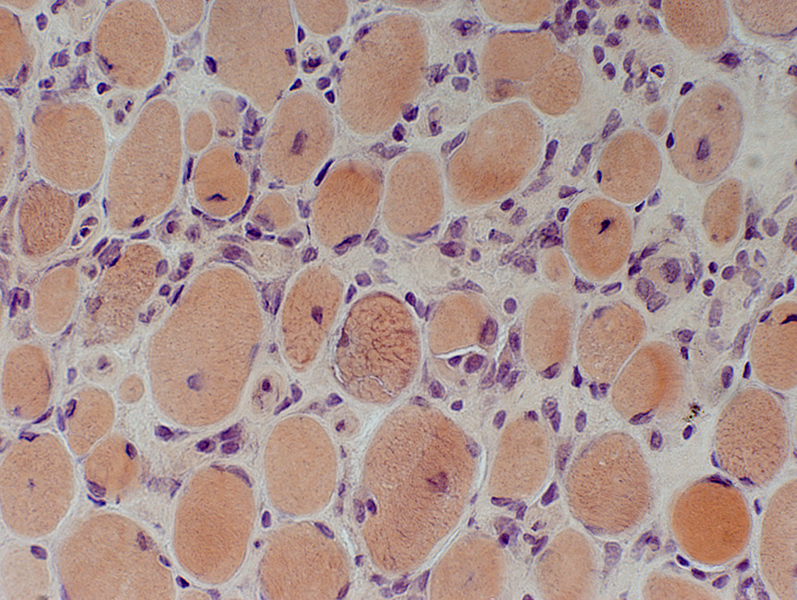
 H & E stain Muscle fibers: Atrophy; Internal nuclei Inflammation: Large cells in endomysium |
 H & E stain |
 H & E stain |
|
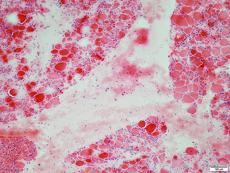
Endomysial vessels: Large size; Thick walls; Large endothelial cells (Arrow) 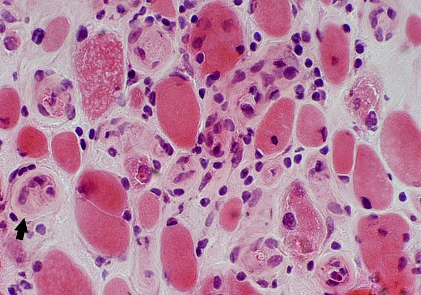 H & E stain |
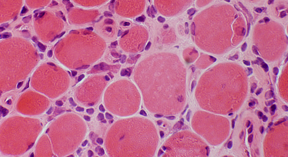
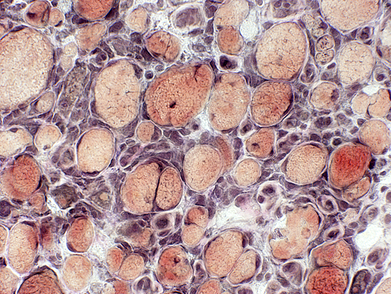
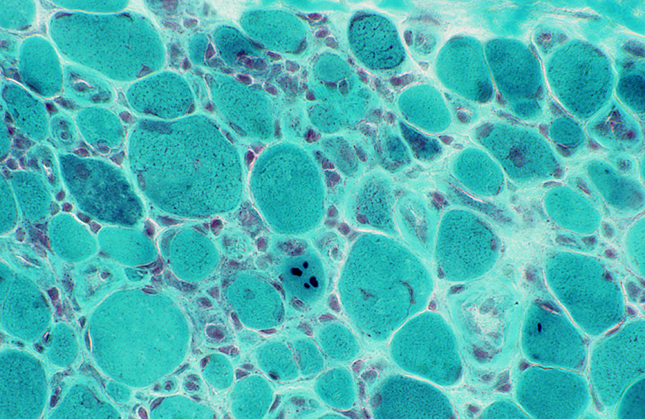 Gomori trichrome stain Endomysial connective tissue: Mildly increased Muscle fibers: Varied size; Cytoplasmic bodies in some fibers |
 Gomori trichrome stain |
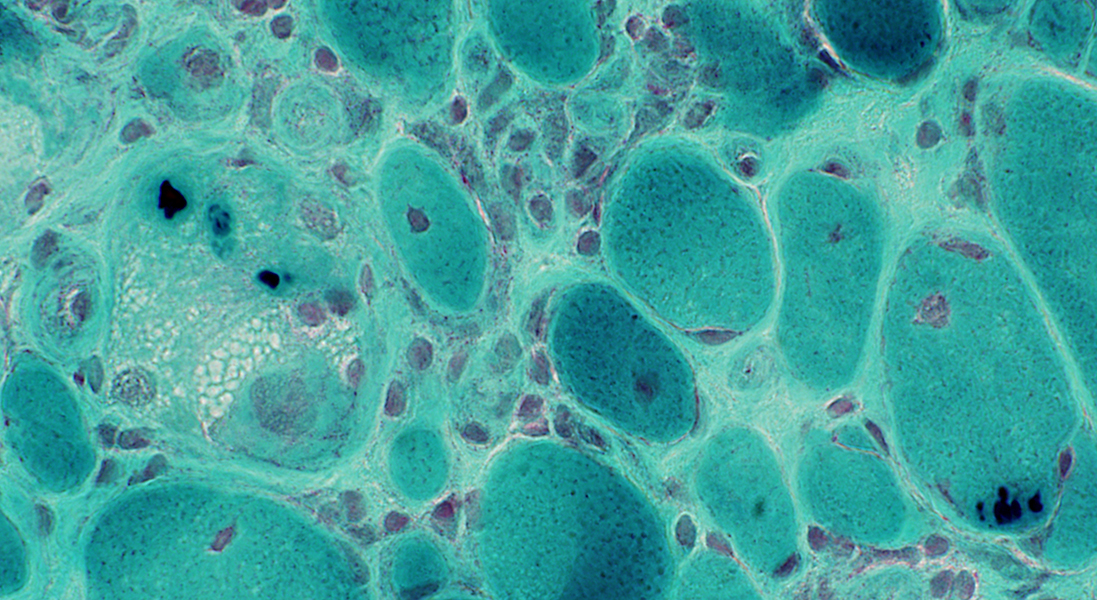
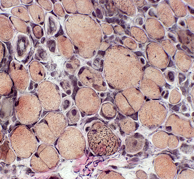
Cytoplasmic bodies: Many, clustered, in one muscle fiber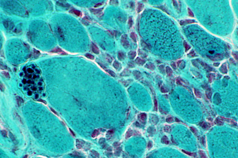 Gomori trichrome stain |
Endomysial capillaries: Large size; Thick wall; Enlarged endothelial cells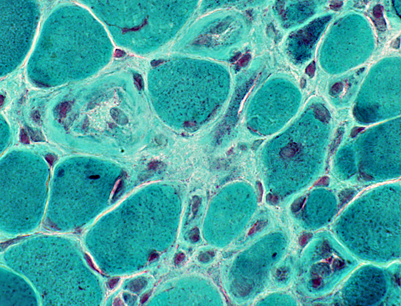 Gomori trichrome stain |
 VvG stain Muscle fibers: Varied sizes Inflammatory cells: Scattered in endomysium between muscle fibers |
 VvG stain |
Inflammatory cells: Scattered in endomysium between muscle fibers
 Congo red stain |
Immature muscle fibers: Intermediate stained (2C)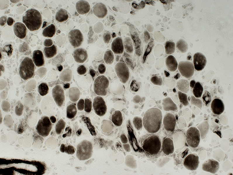 ATPase pH 4.3 stain |
Immature muscle fibers: Coarse internal architecture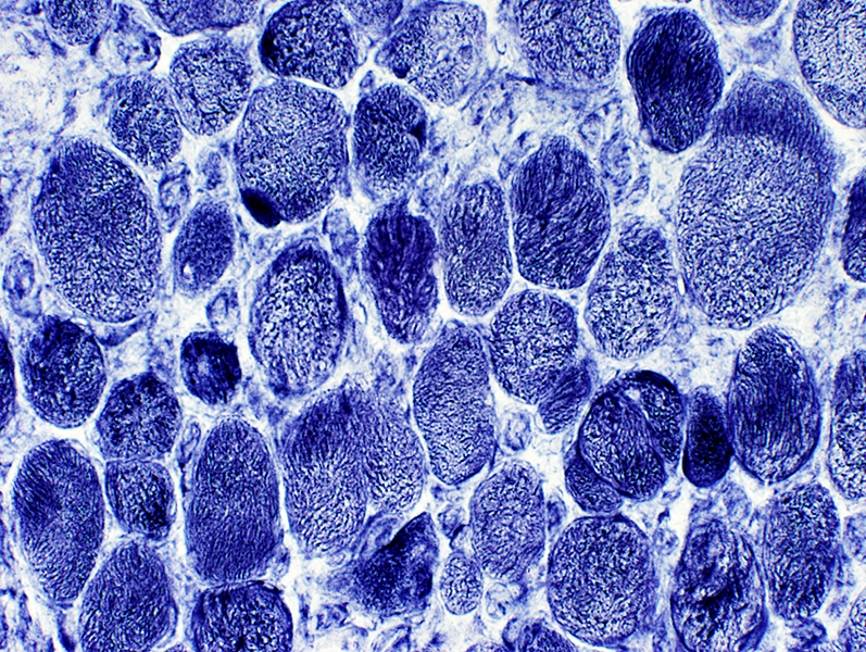 NADH stain |
Immature Muscle Fibers: Some small fibers stain for alkaline phosphatase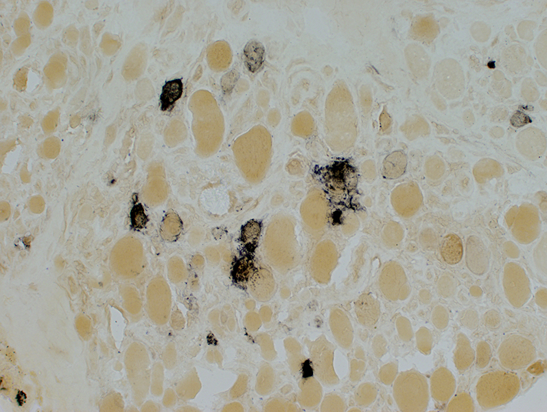 Alkaline phosphatase stain |
Mitochondrial proliferation: Increased SDH staining in scattered muscle fibers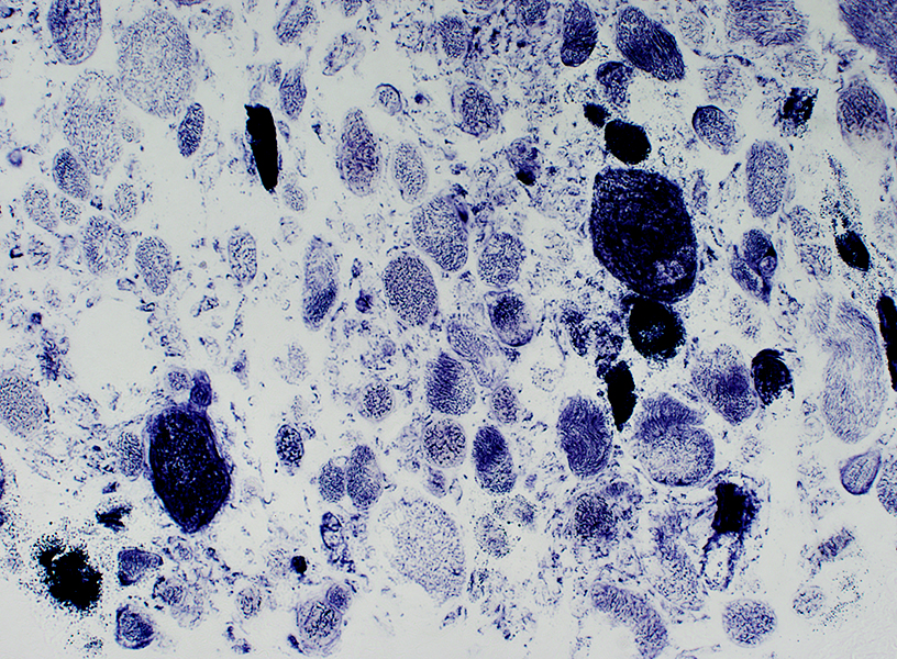 Succinate Dehydrogenase (SDH) stain |
COX negative muscle fibers: Some fibers have reduced, or absent, staining for cytochrome oxidase (COX)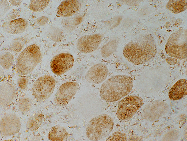 Cytochrome oxidase (COX) stain |
Return to HIV
1/22/2025