Plectin Mutations: Myopathy + Epidermolysis Bullosa
From: Chunyu (Hunter) Cai
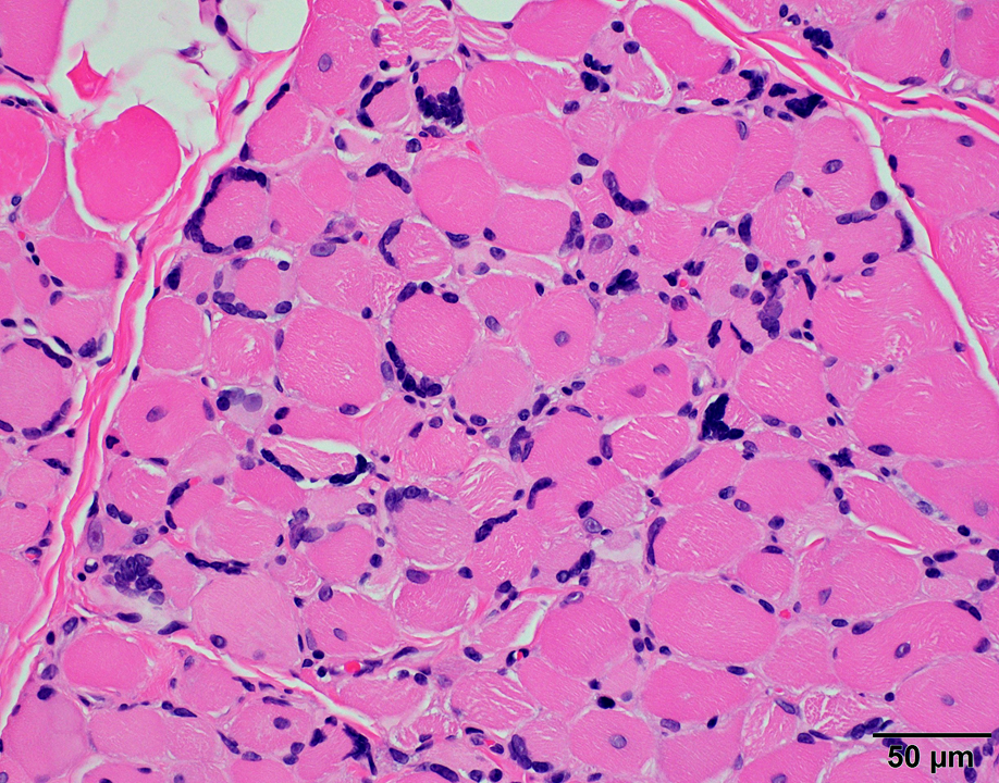 H&E stain |
Muscle Fibers
Necrosis: Scattered
Size: Varied
Nuclei: Internal; Often clustered
Cytoplasm: Irregular clear regions
Endomysial connective tissue: Increased
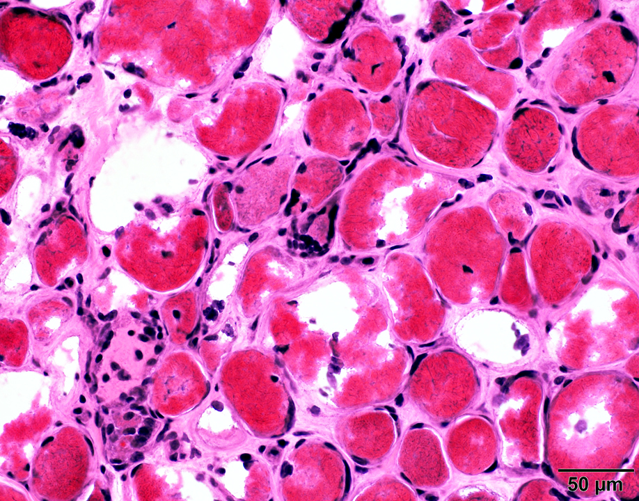 H&E stain |
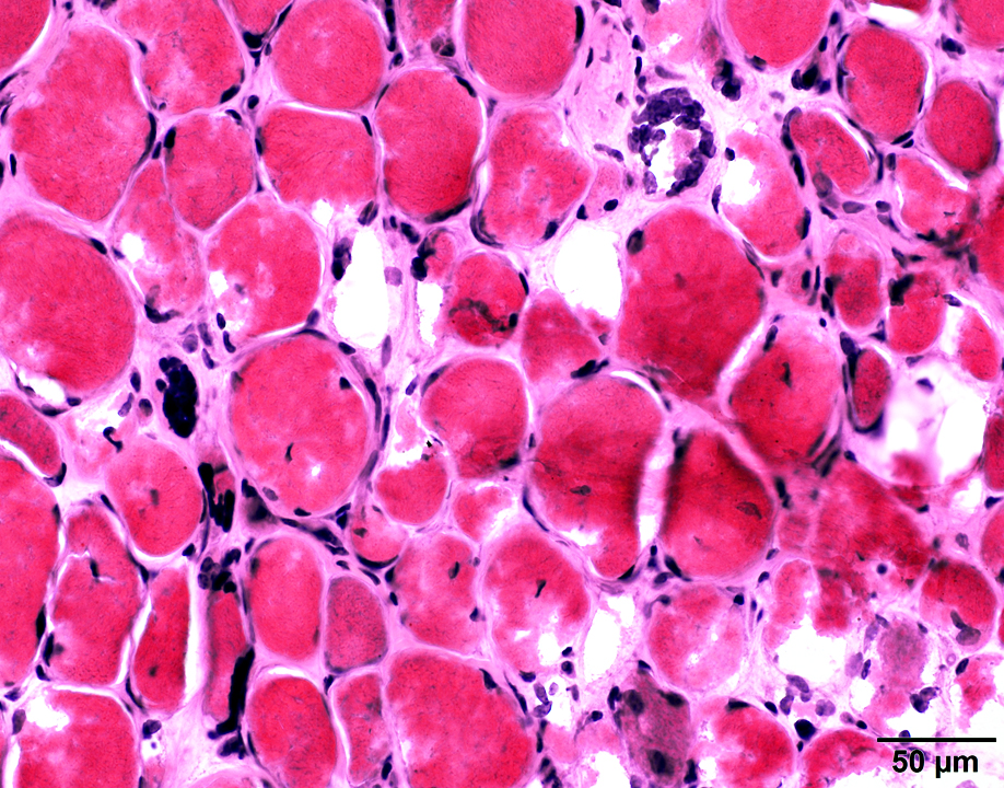 H&E stain |
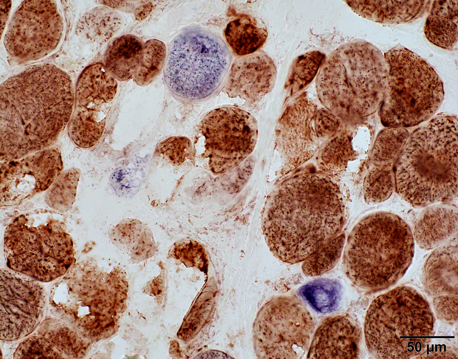
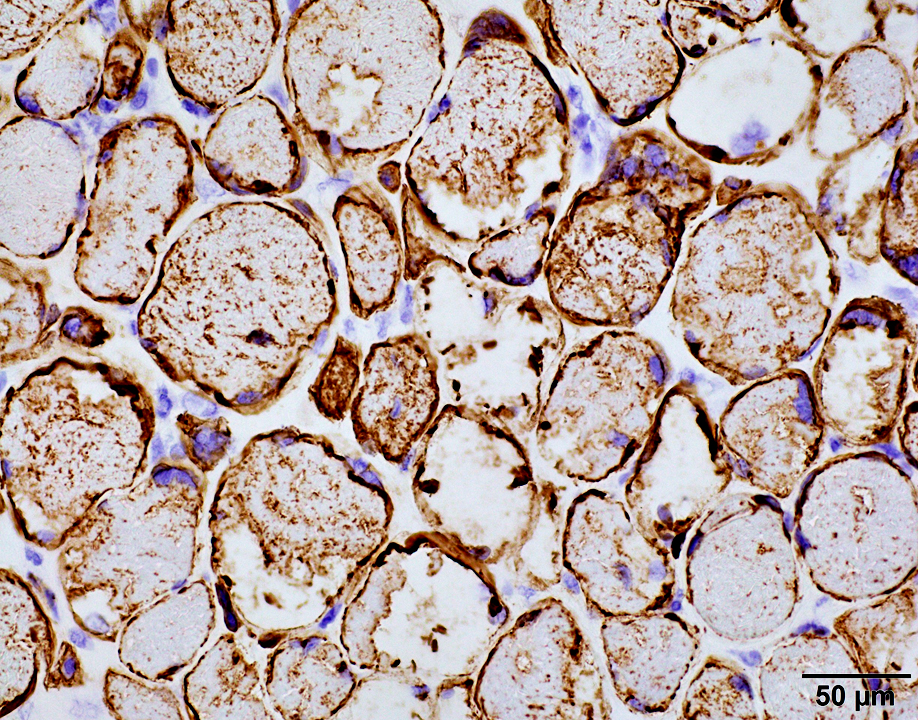 Desmin stain |
Punctate cytoplasmic staining for Desmin & Dysferlin
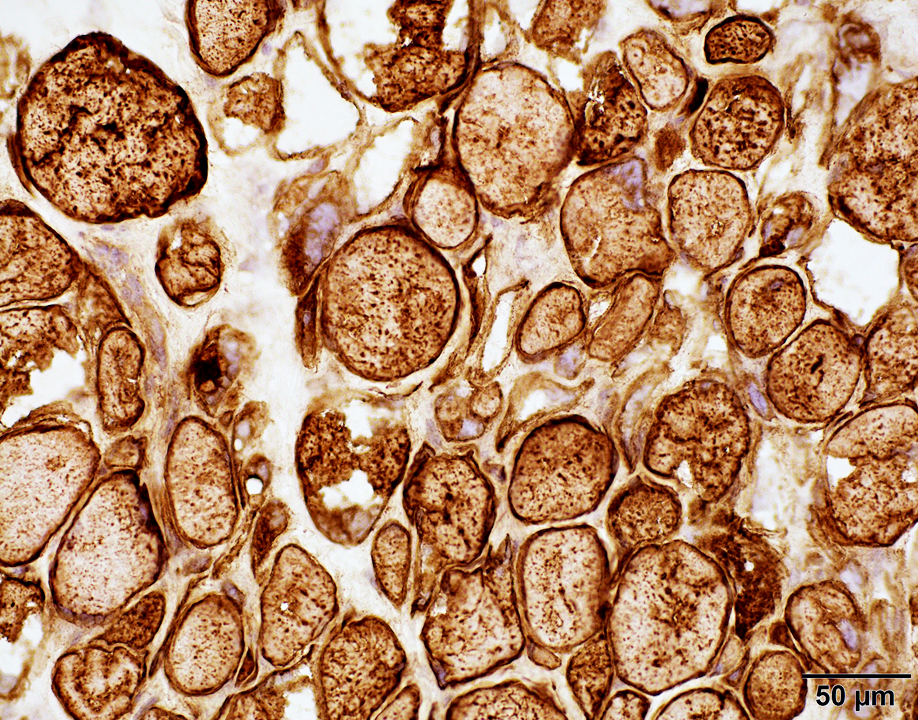 Dysferlin stain |
 |
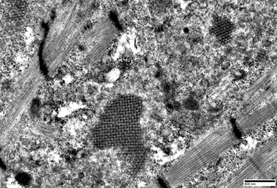 |
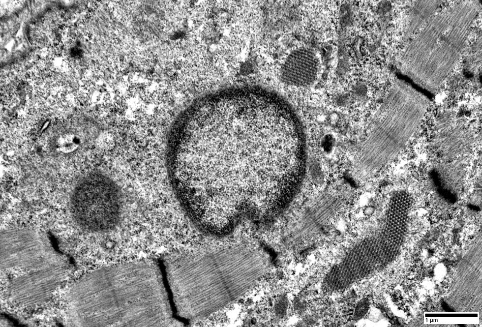
 Muscle fiber Aggregates Cytoplasmic body |
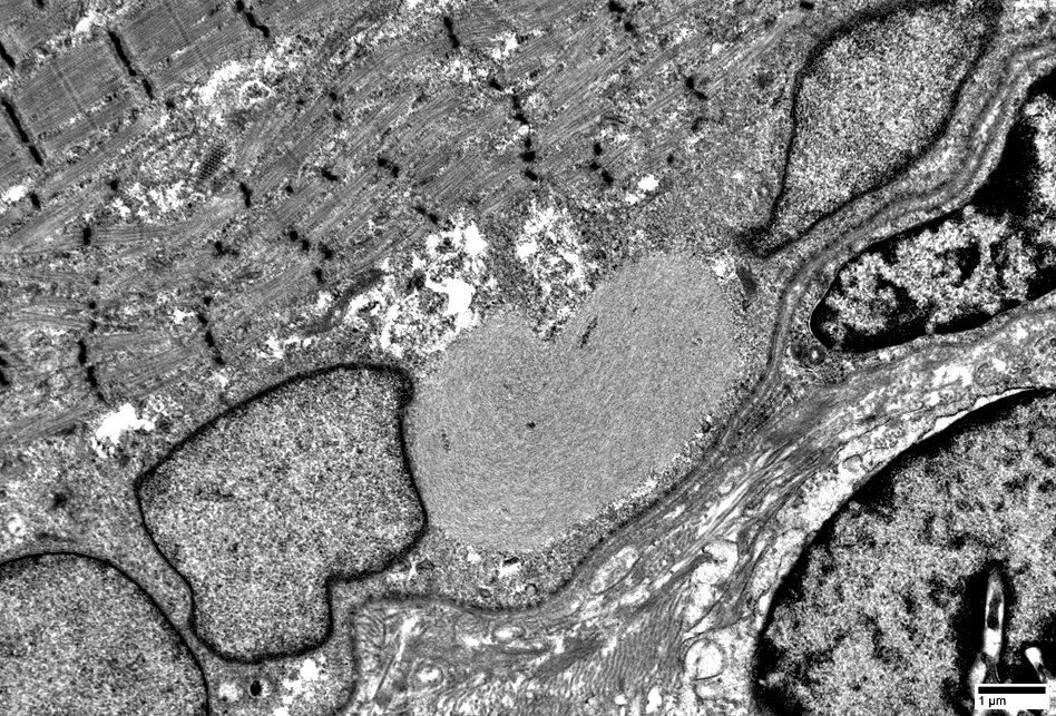 |
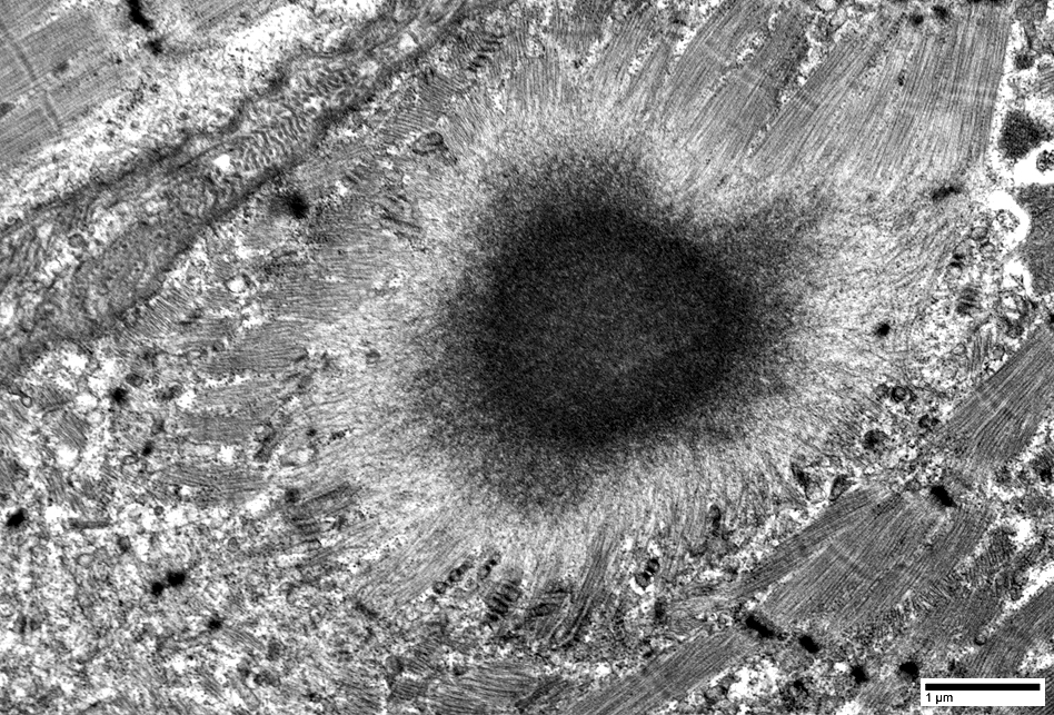
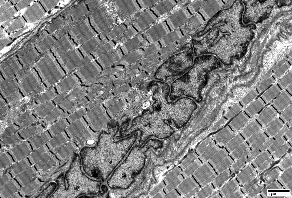
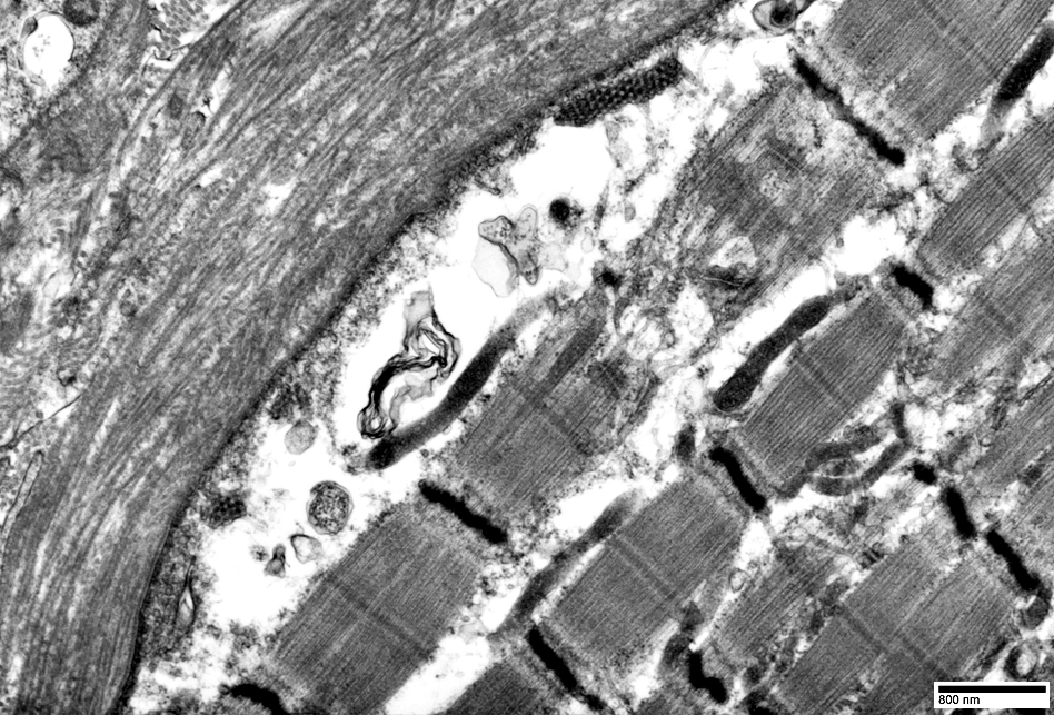
Subsarcolemmal filamentous collections
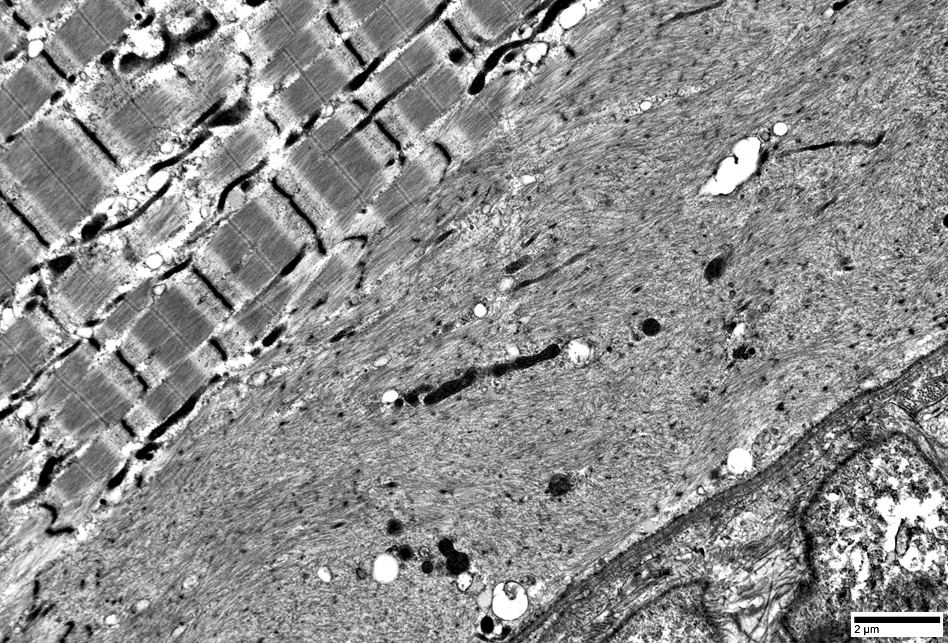 |
 |
Subsarcolemmal Z-band disruption & separation from surface membrane
|
Chains
|

|
Clusters
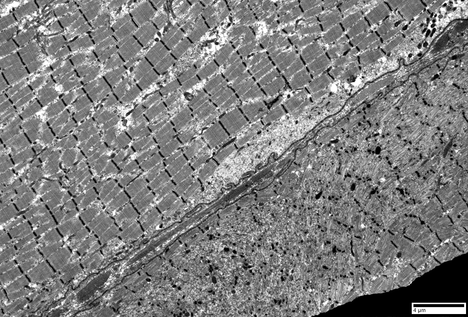
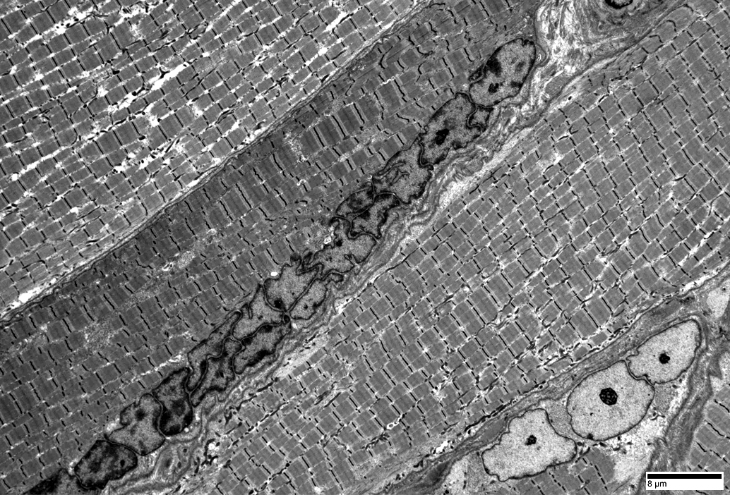
Mitochondrial Pathology
 SDH + COX stain |
COX stain: Reduced
SDH stain: Increased
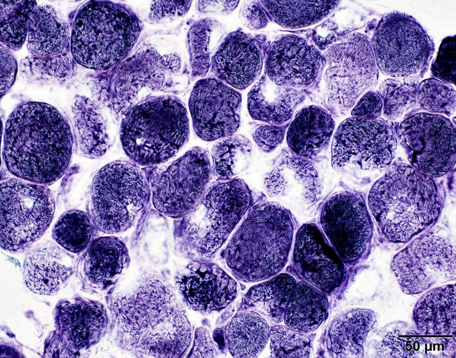 SDH stain |
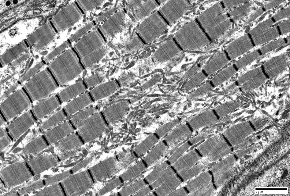 |
Elongated mitochondria
 |
Return to Neuromuscular Home Page
Return to Muscle autoantibodies
4/21/2018