OCULOPHARYNGEAL MUSCULAR DYSTROPHY
|
Aggregates Cytoplasmic bodies Nuclear inclusions Vacuoles |
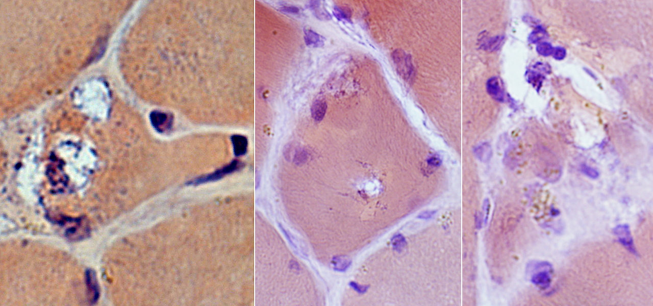
 H&E stain |
 H&E stain |
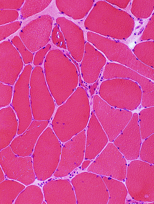
Size: Varied
Small fibers: Rounded or Angular; Some are basophilic
Vacuoles: Occur in small fibers
Internal nuclei: Occasional muscle fibers
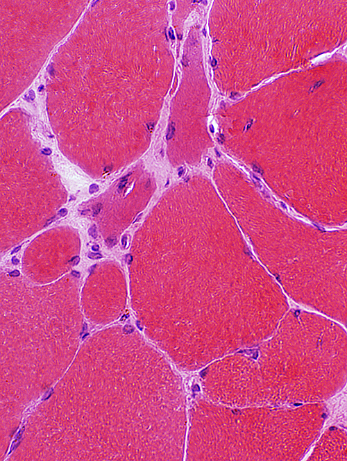
Vacuoles
 H&E stain |
 H&E stain |
|
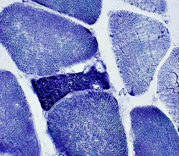
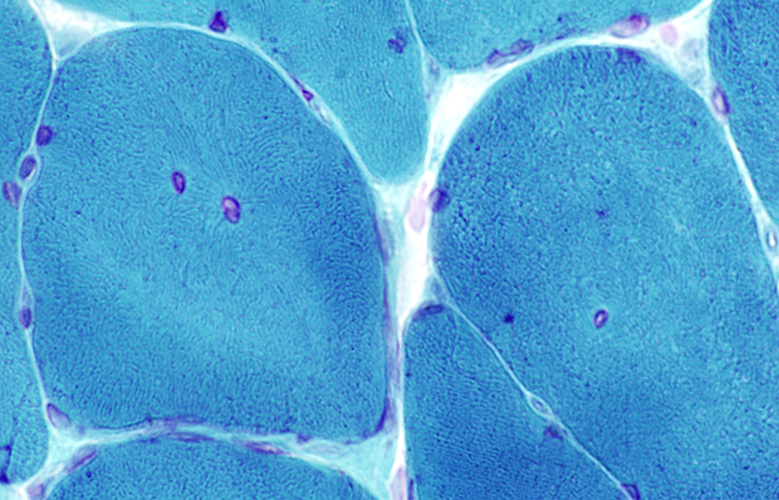
 Gomori trichrome stain |
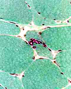
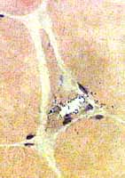
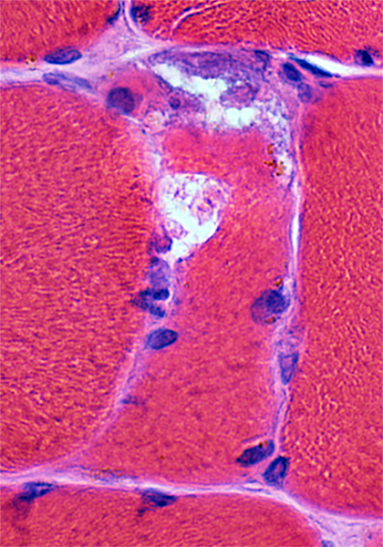
Location: In small polygonal or angular muscle fibers
Shape: Irregular
Subcellular location: Peripheral (Sub-Sarcolemmal) or Central
May be clear or contain red stained (GT) or granular material (Congo red)
 Gomori trichrome stain |
 Congo red stain |
 From: R Schmidt |
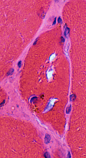
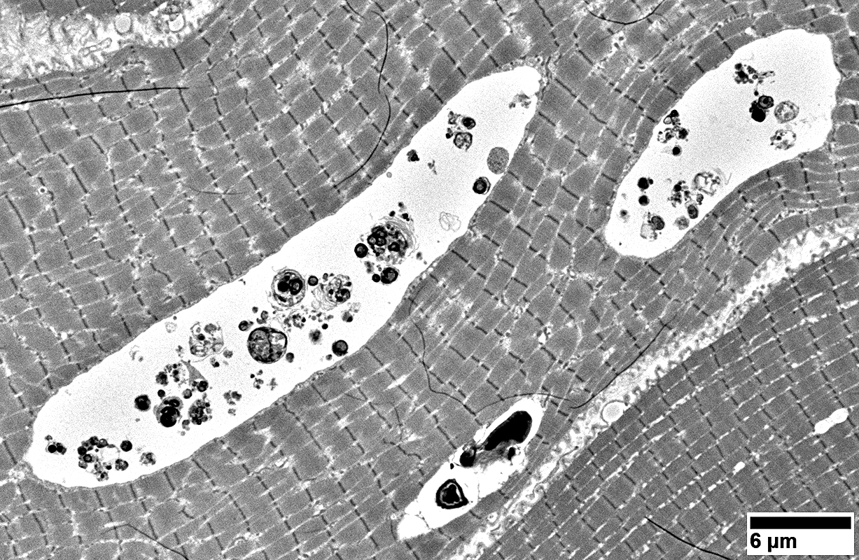
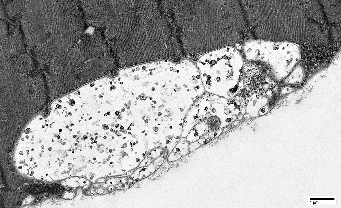
Location: Cytoplasm
Contents: Autophagic debris of varied types scattered within pale membrane-lined vacuole
|
 NADH stain Small fibers stain darkly on NADH |
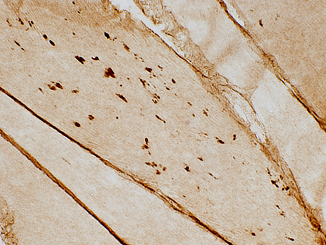 Caveolin-3 stain |
|
Cytoplasmic aggregates |
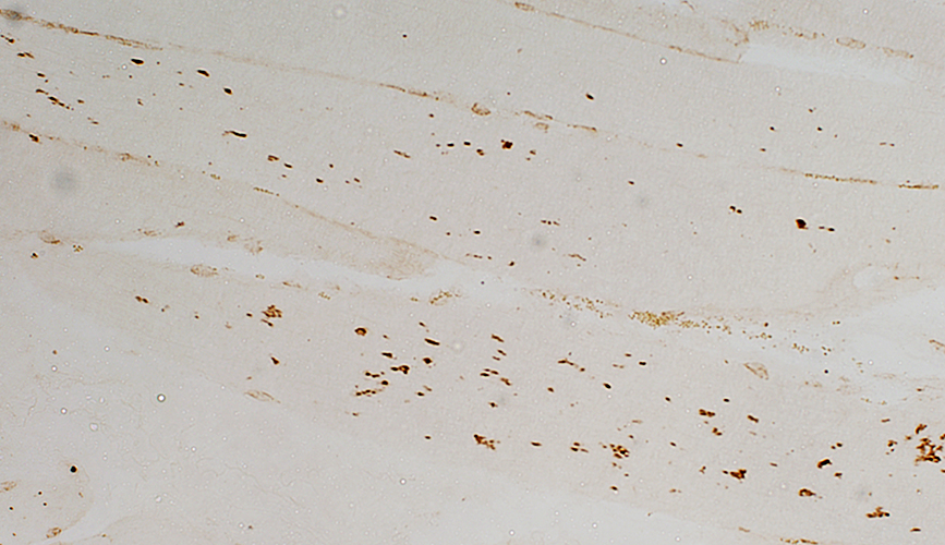 LC-3 stain |
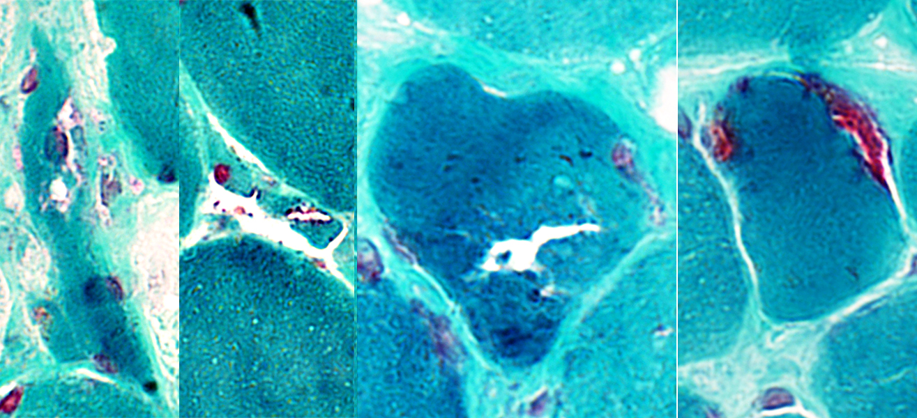
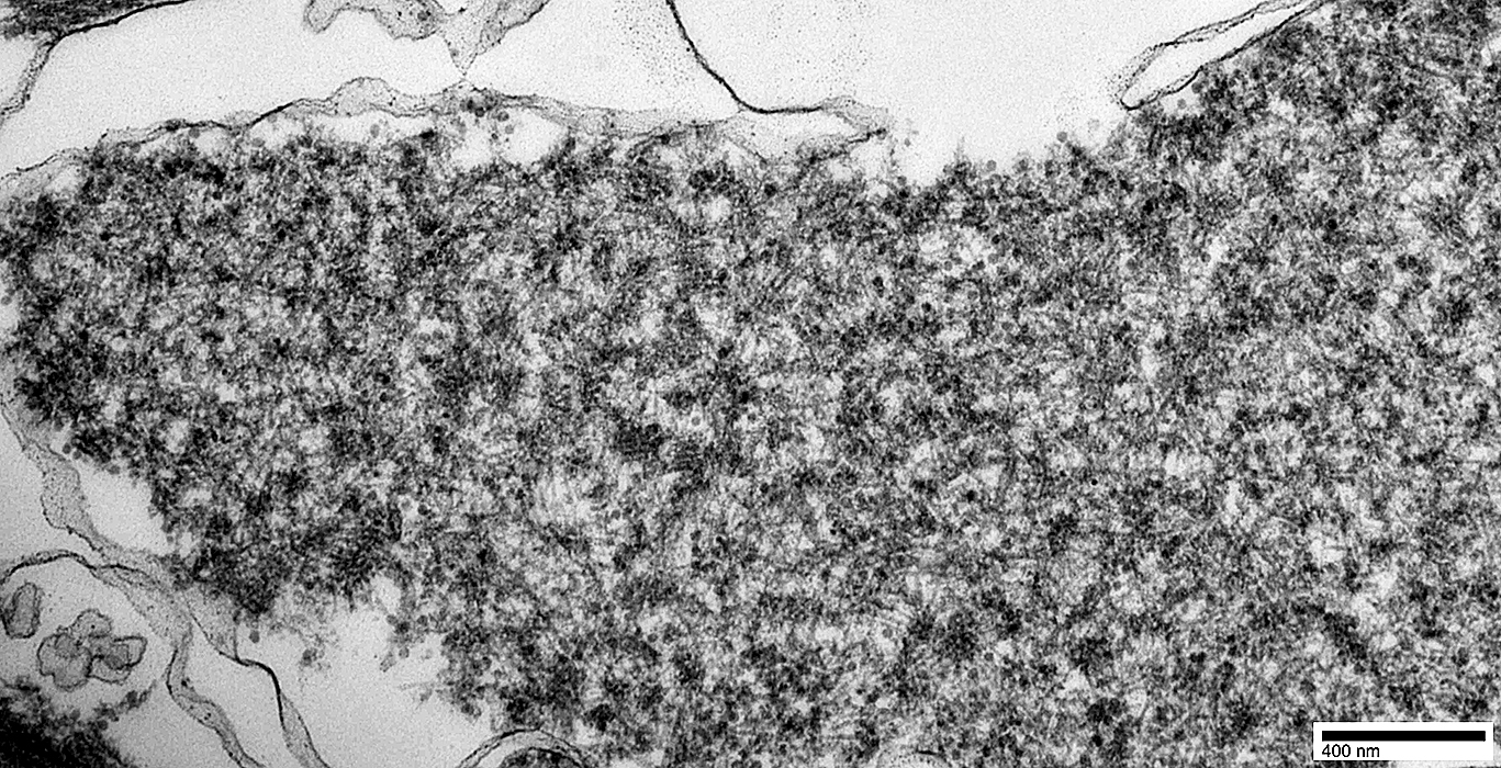
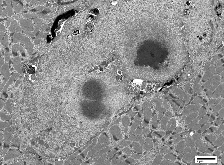
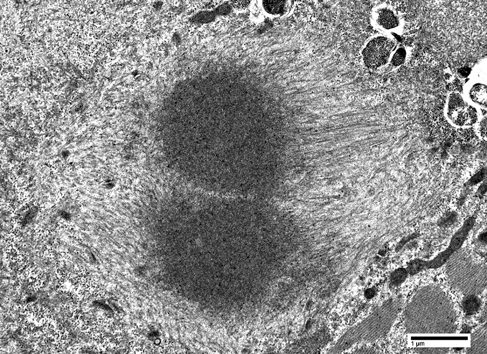
Nuclei: Tubulofilamentous Inclusions
 Gomori trichrome stain Internal nuclei: May have clear centers |
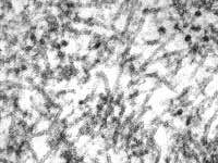 Nuclear inclusions, Tubulofilamentous · Form tangles & palisades · Size: 8.5 nm diameter; Up to 0.25 μm length · ? Specific marker for disease |
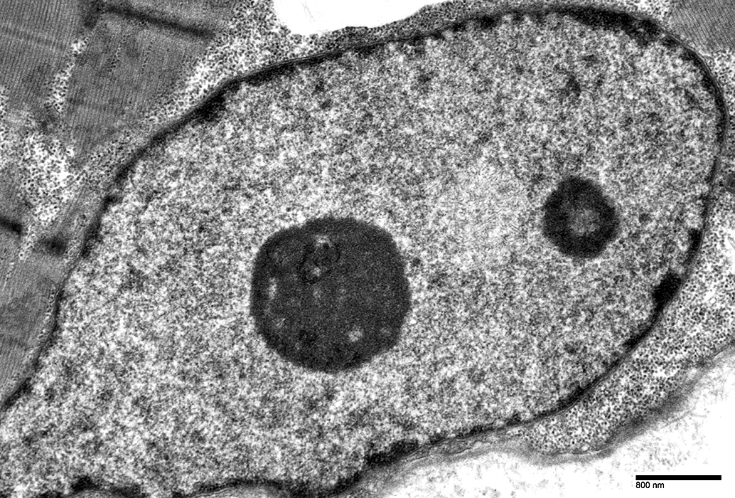 From Chunyu (Hunter) Cai |
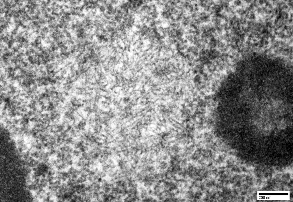 From Chunyu (Hunter) Cai |
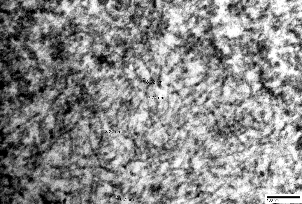 From Chunyu (Hunter) Cai |
 From Chunyu (Hunter) Cai |
 From Chunyu (Hunter) Cai |
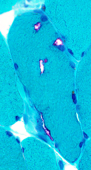
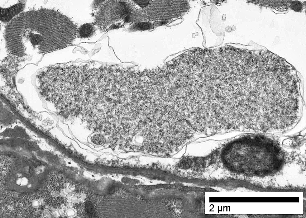
OPMD: Cytoplasmic Bodies
 Gomori trichrome stain Cytoplasmic bodies: May be seen in some small muscle fibers |
 From Chunyu (Hunter) Cai |
 From Chunyu (Hunter) Cai |
Return to Neuromuscular Home Page
Return to Pathology index
Return to Oculopharyngeal Muscular dystrophy
4/9/2021