Myofibrillar (Desmin) Myopathy
|
Desmin Aggregates Caps Myopathy (I402N) 5th decade 3rd decade Myopathy (R454W) AVSF Ultrastructure ZASP |
|
Cytoplasmic aggregates Eosinophilic Homogeneous 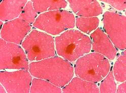 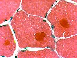 H & E stain |
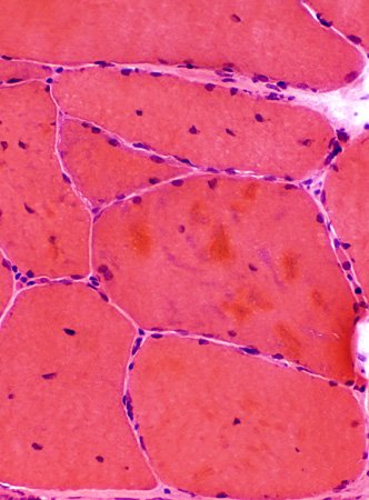 H & E stain |
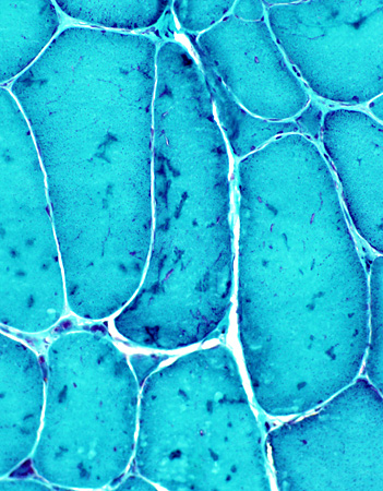
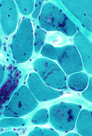 Gomori trichrome stain |
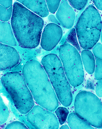
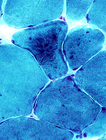 Gomori trichrome stain |
Sarcoplasmic membranes
Absent from regions with desmin-containing cytoplasmic bodies.
Irregular (moth-eaten) internal architecture is present in several muscle fibers
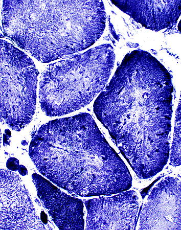
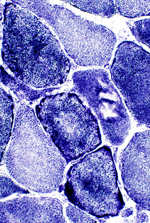 NADH stain |
Myofibrillar apparatus
Absent from regions with desmin-containing cytoplasmic bodies
Aggregates are in both fiber types
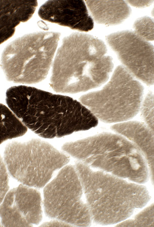
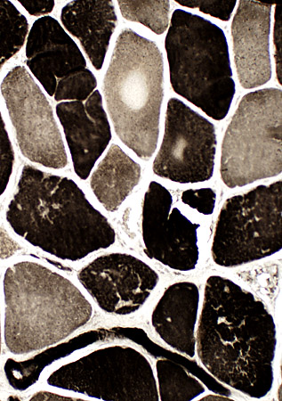 ATPase, pH 9.4 |
|
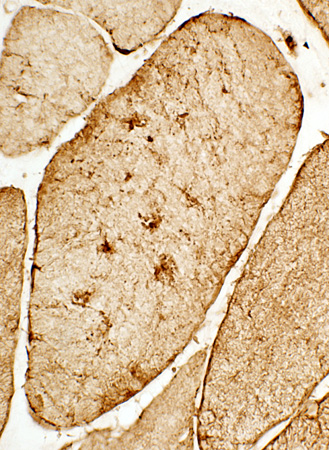
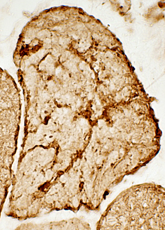
Desmin aggregates Cytoplasmic accumulations Various shapes & sizes Other desmin staining Diffuse in regenerating fibers 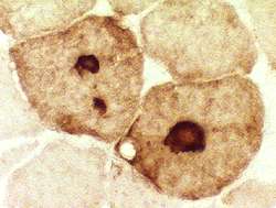 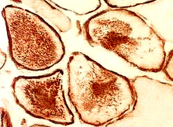 |
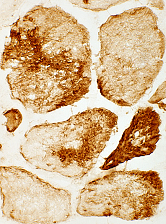 Anti-desmin antibody |
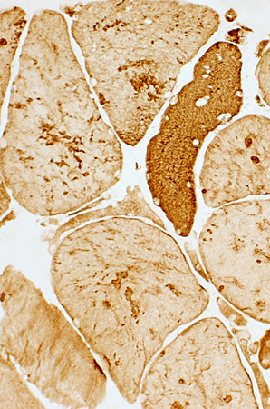
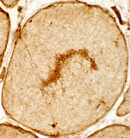
|
|
Myofibrillar Myopathy: Desmin mutations
I402N Desmin mutation: 46 year old patient
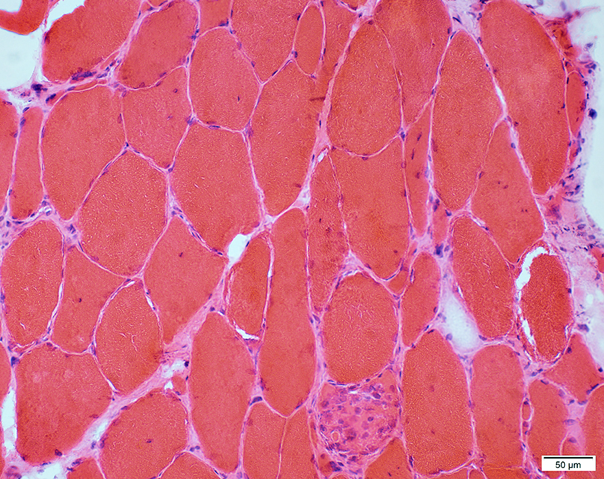 H&E stain |
Necrosis: Few fibers
Internal nuclei
Endomysial connective tissue: Mildly increased
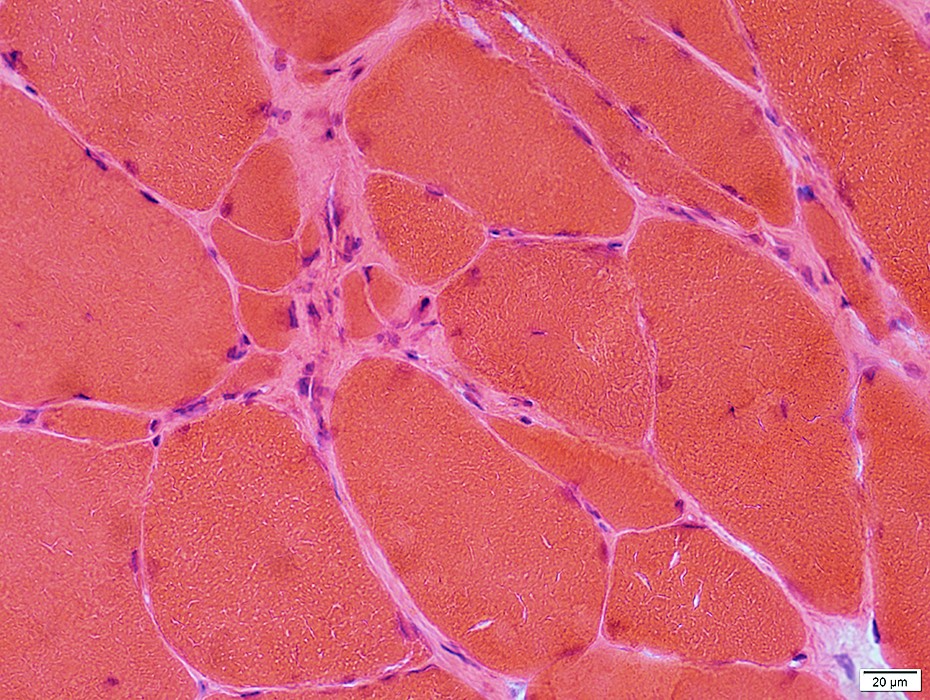 H&E stain |
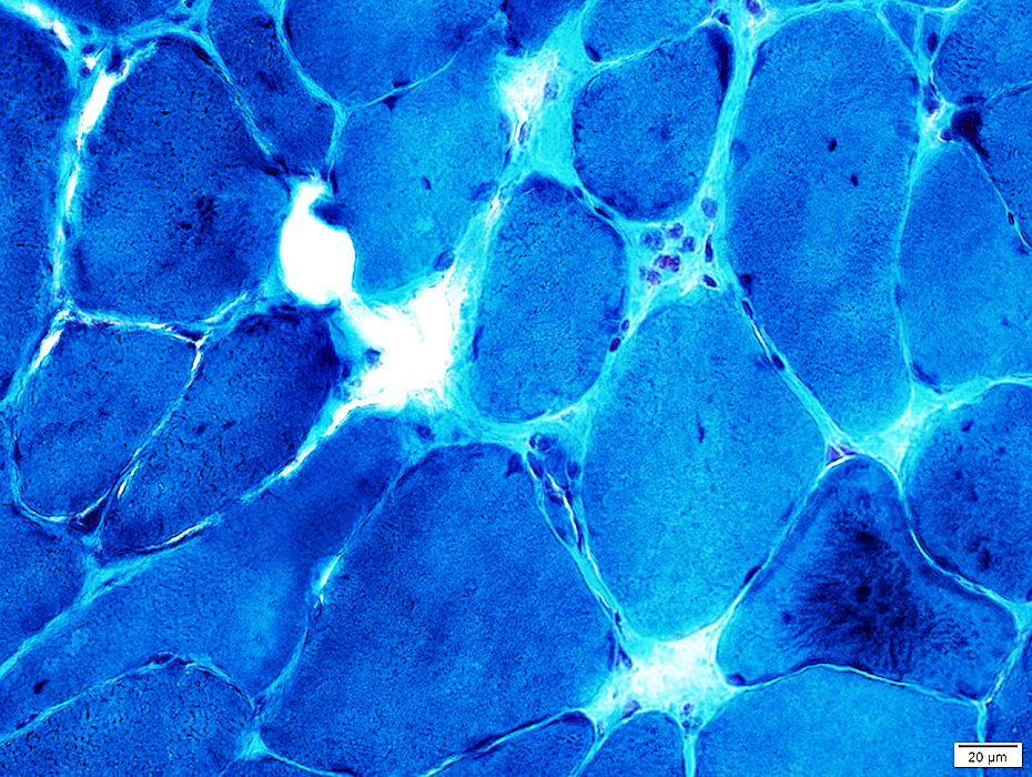 Gomori trichrome stain |
Internal architecture: Aggregates; Irregular
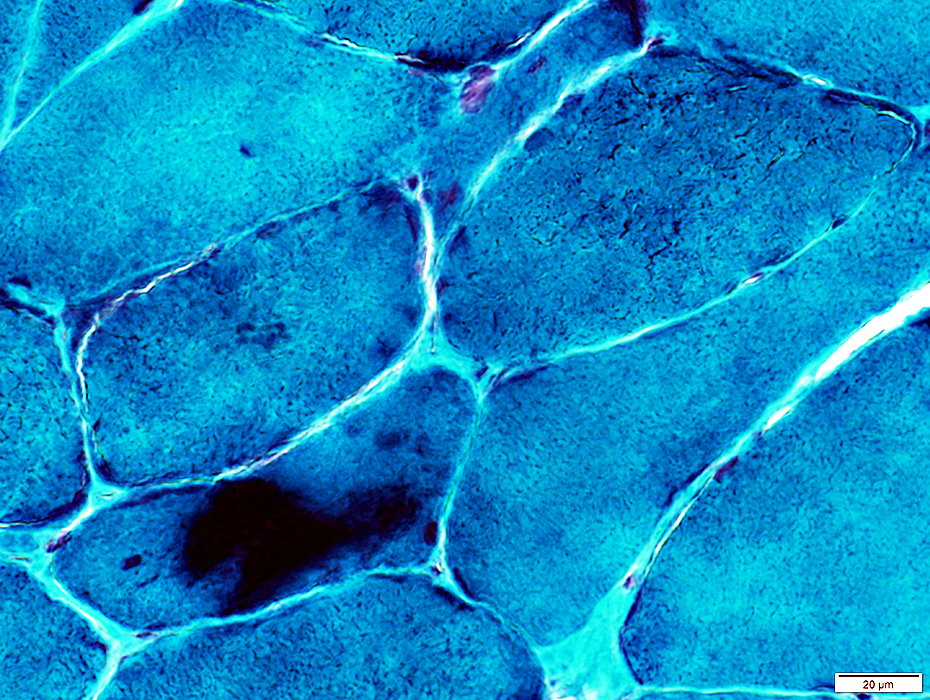 Gomori trichrome stain |
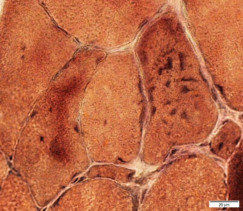 VvG stain |
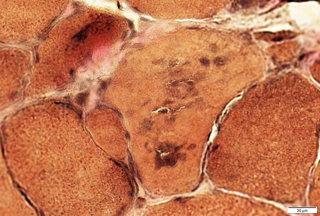 VvG stain |
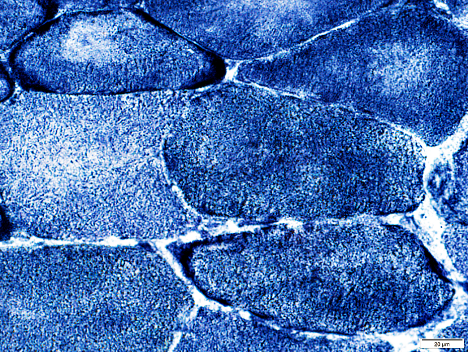 HADH stain |
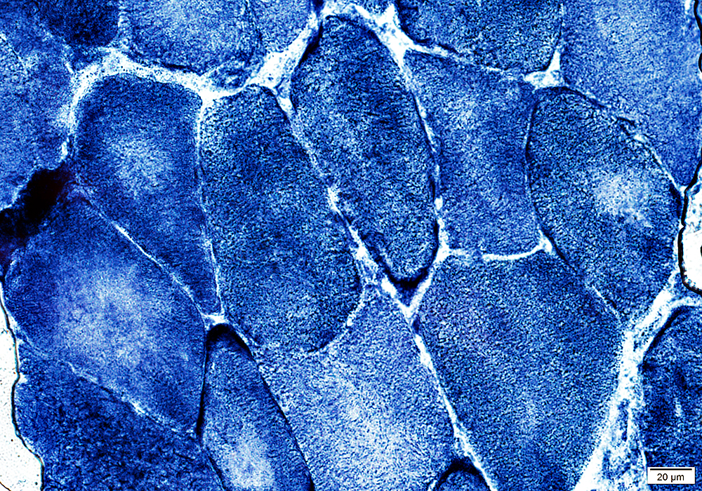 HADH stain |
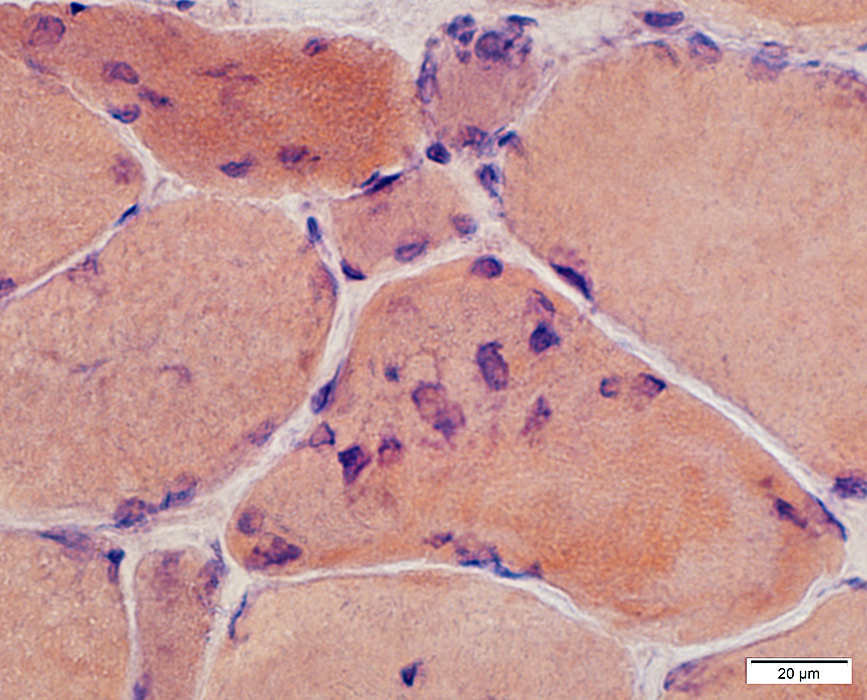 Congo red stain |
Cytoplasm: Congophilic aggregates
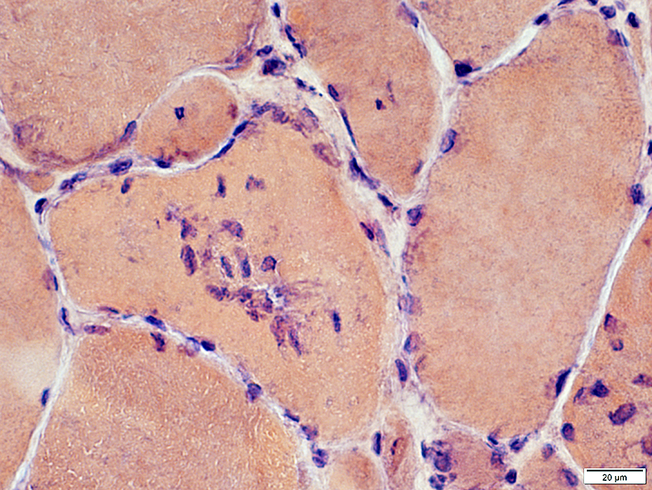 Congo red stain |
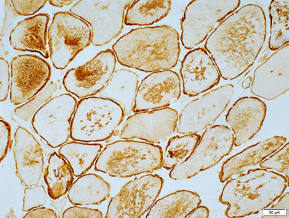 Desmin stain |
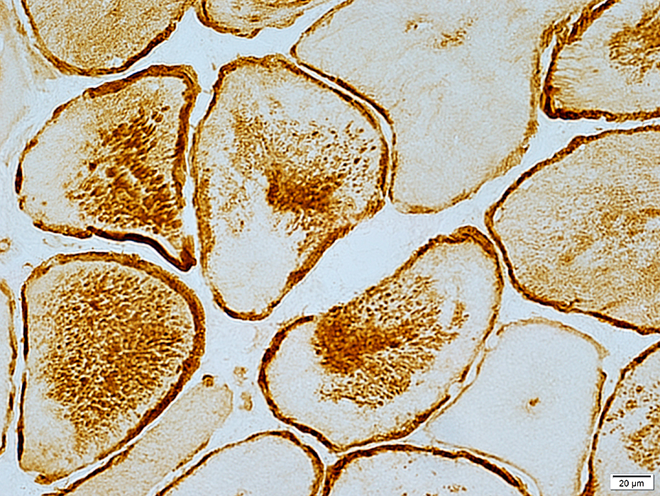 Desmin stain |
I402N Desmin mutation: 24 year old patient
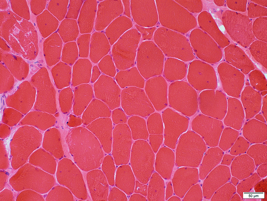 H&E stain |
Endomysial connective tissue: Mildly increased
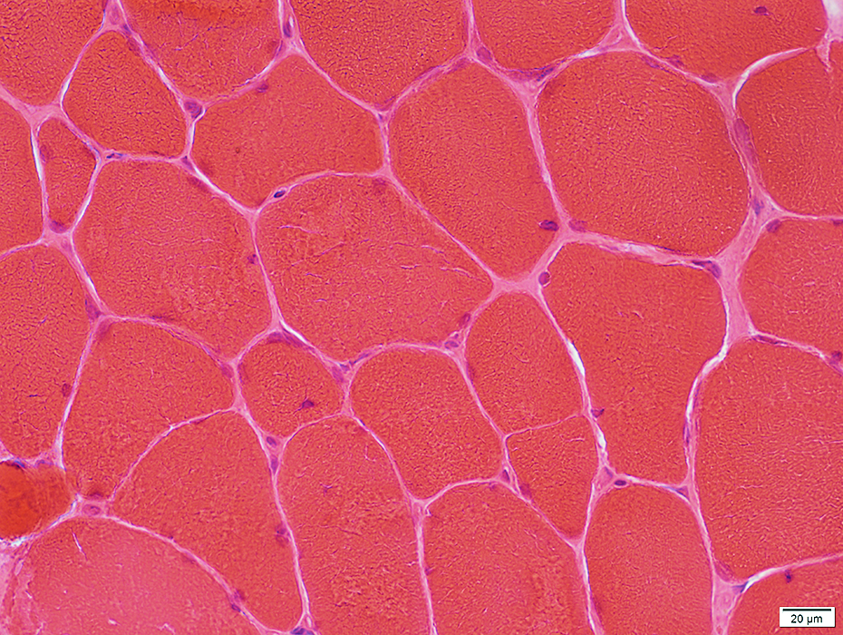 H&E stain |
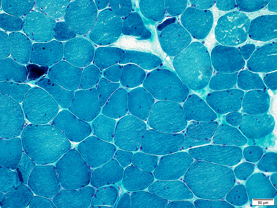 Gomori trichrome stain |
Endomysial connective tissue: Mildly increased
Internal nuclei
Aggregates: Irregular; Dark stained
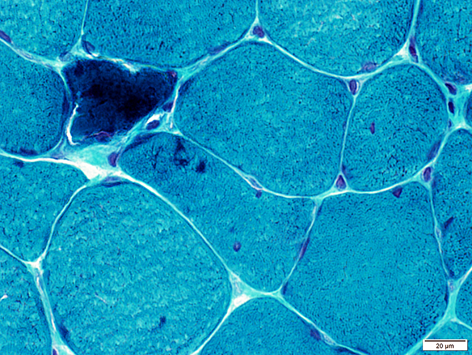 Gomori trichrome stain |
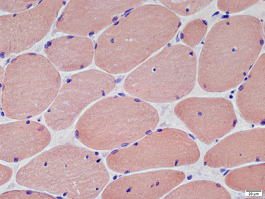 Congo red stain |
Internal nuclei
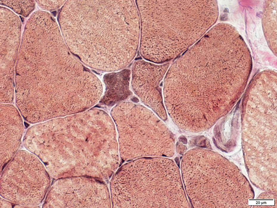 Congo red stain |
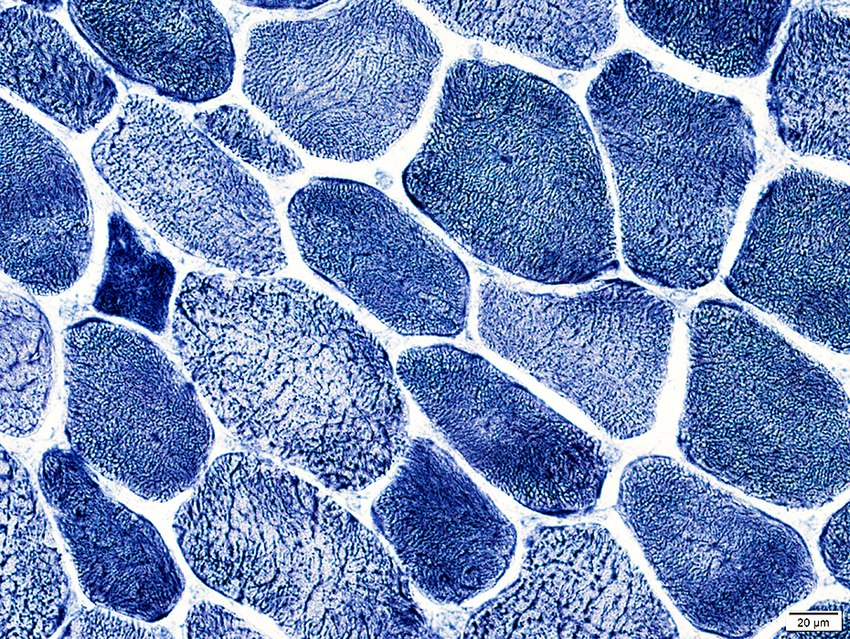 NADH stain |
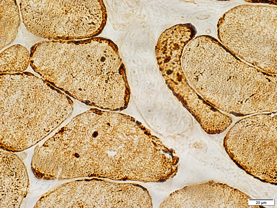 Desmin stain |
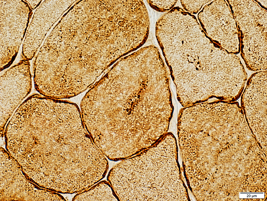 Desmin stain |
Desmin Myopathies with Sarcolemmal Vacuoles
R454W Desmin tail domain mutation|
Histochemistry Vacuoles with Sarcolemmal Features Ultrastructure |
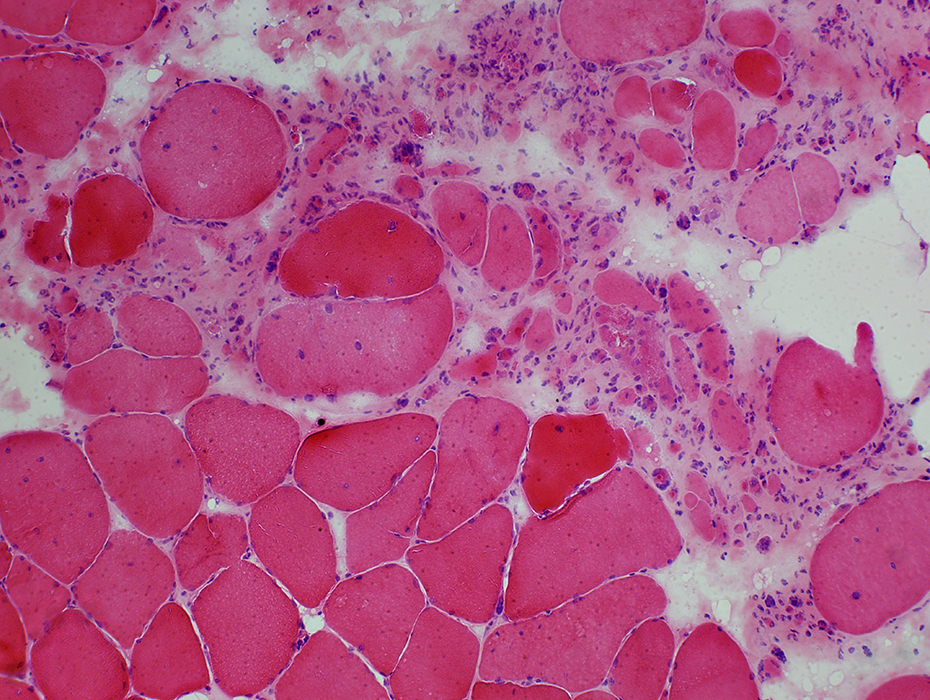 H&E stain Myopathy Multifocal pathology Muscle fibers Atrophy: Often in groups Hypertrophy Clear, irregular vacuoles Endomysial connective tissue: Increased |
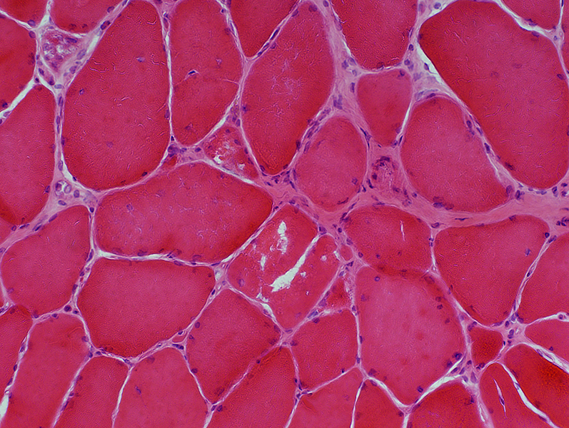 H&E stain |
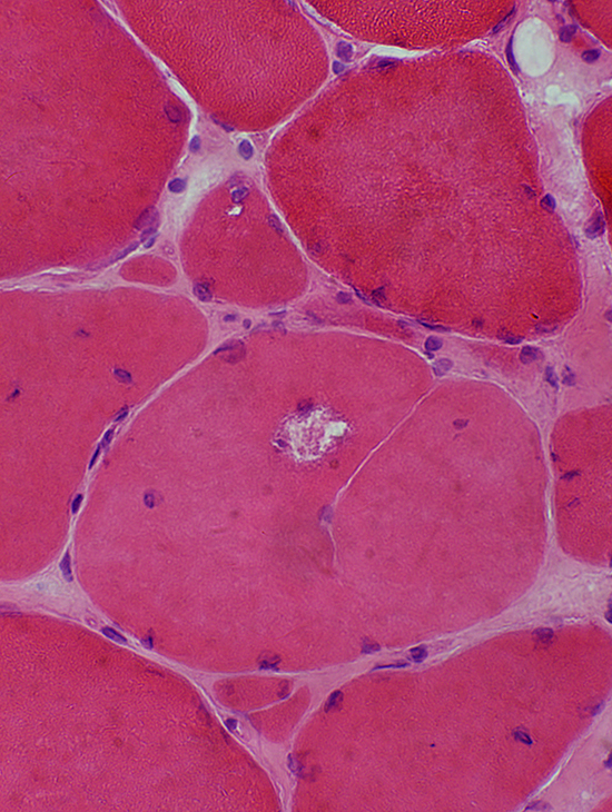 H&E stain |
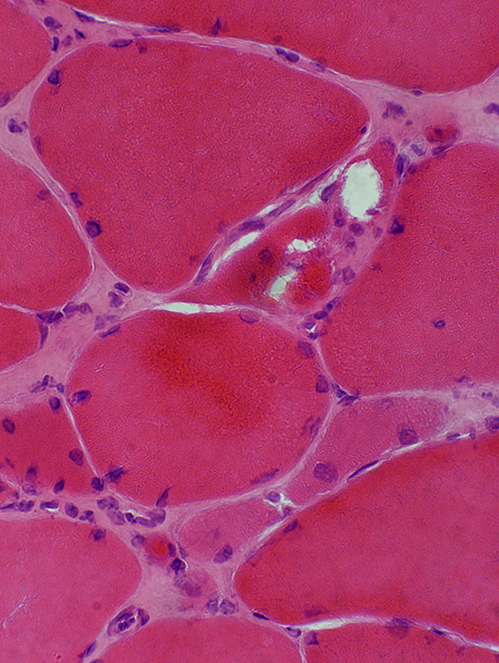 H&E stain |
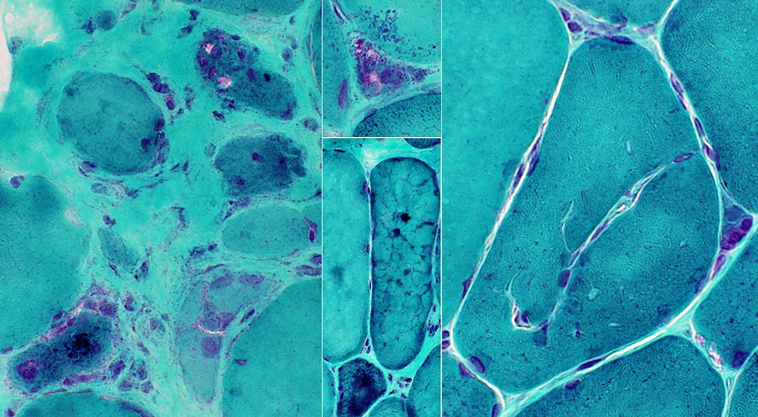 Gomori trichrome stain |
|
Vacuoles Irregular shapes Contain red-staining material Subsarcolemmal or Central |
Invaginations Branched Extend deeply into muscle fibers May contain capillaries |
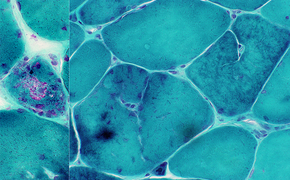 Gomori trichrome stain |
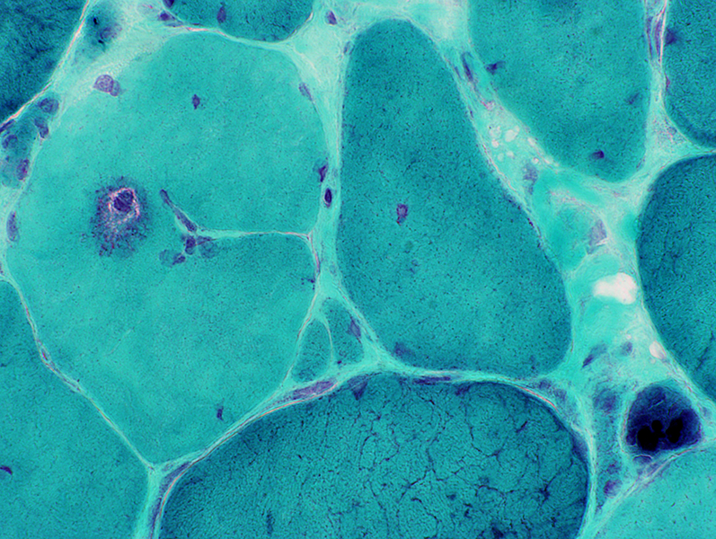 Gomori trichrome stain |
Cytoplasmic bodies
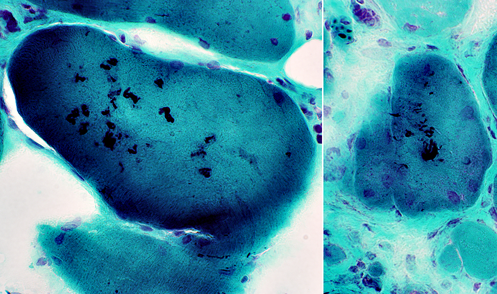 Gomori trichrome stain |
|
Myopathy Muscle fibers Varied size Internal architecture Irregular Aggregates Vacuoles Endomysium Connective tissue Increased |
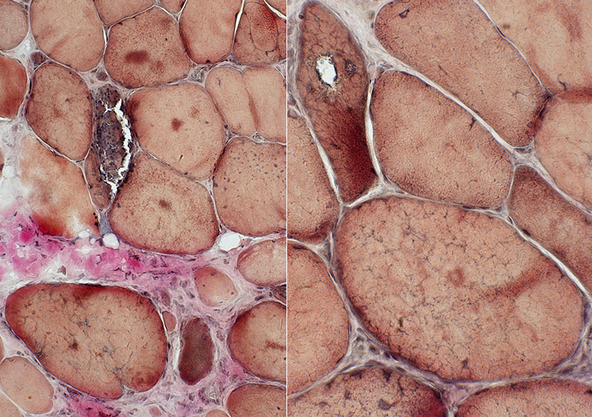 VvG stain |
Internal architecture: Irregular
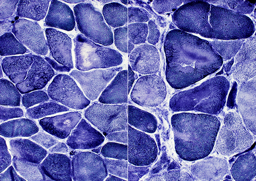 NADH stain |
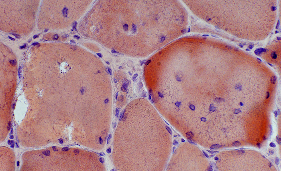 Congo red stain |
Splits & Invaginations: Extensive; Branched
Nuclei: Large; Irregular shapes; Some have clear centers
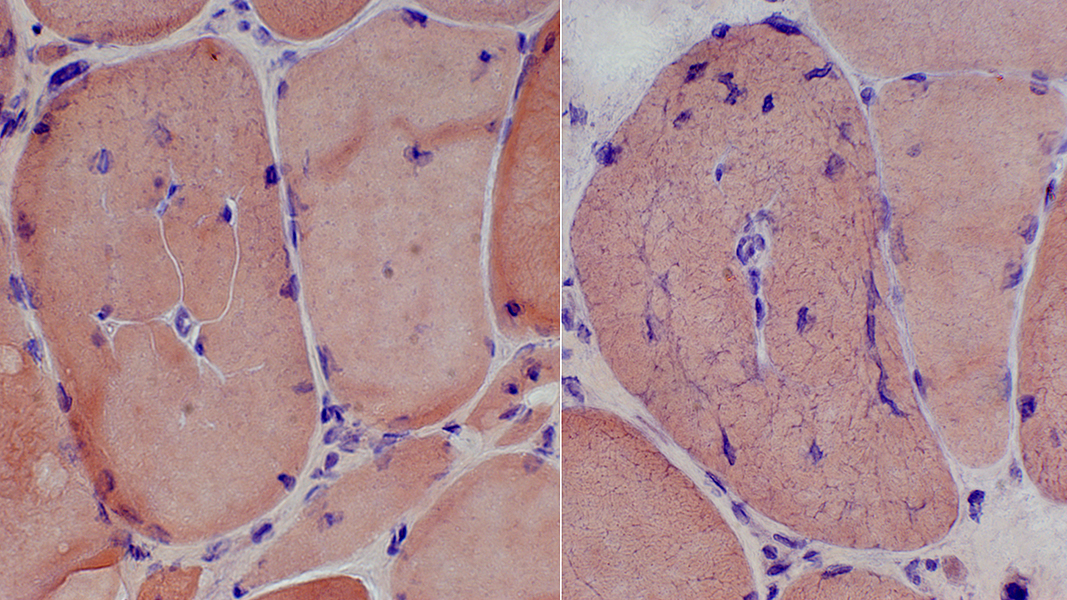 Congo red stain |
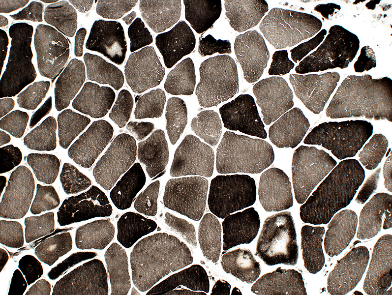 ATPase pH 9.4 stain Aggregates (Pale areas): In Type I & II fibers |
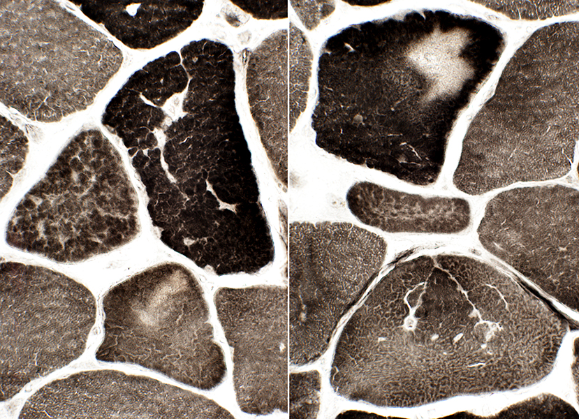 ATPase pH 9.4 stain |
Type IIC fibers: Common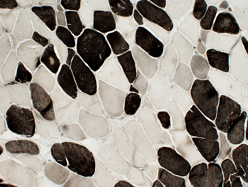 ATPase pH 4.3 stain |
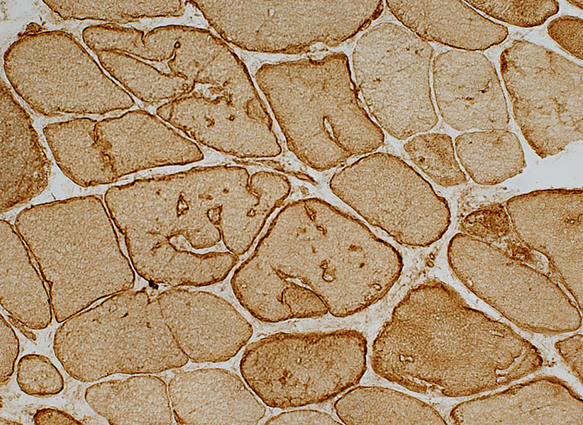 Caveolin-3 stain Invaginations & Vacuoles: Lined with Caveolin-3 on membrane |
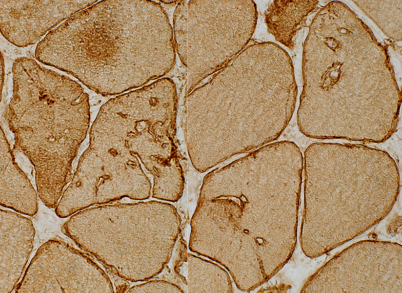 Caveolin-3 stain |
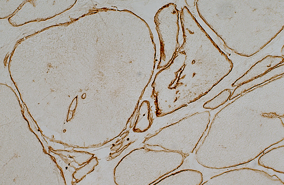 Dystrophin stain Invaginations & Vacuoles: Lined with Dystrophin on membrane |
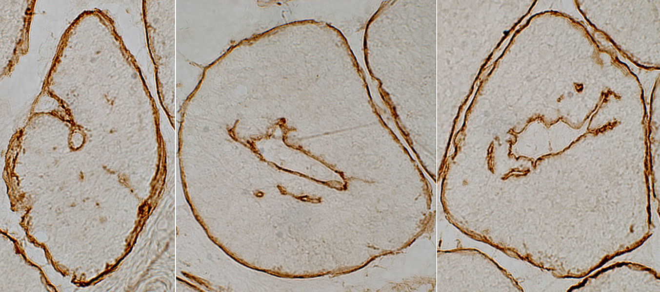 Dystrophin stain |
Aggregates: Stain for desmin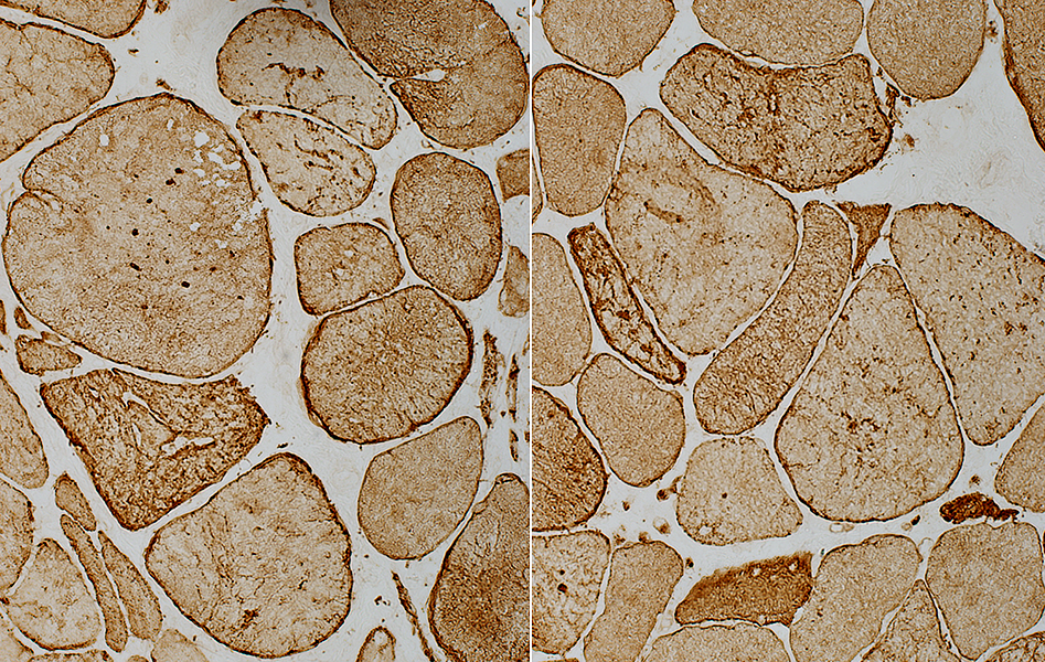 Desmin stain |
Desmin Myopathy: Ultrastructure (R454W)
|
Aggregates Filaments Organelles Tubules Vacuoles Z-Disk |
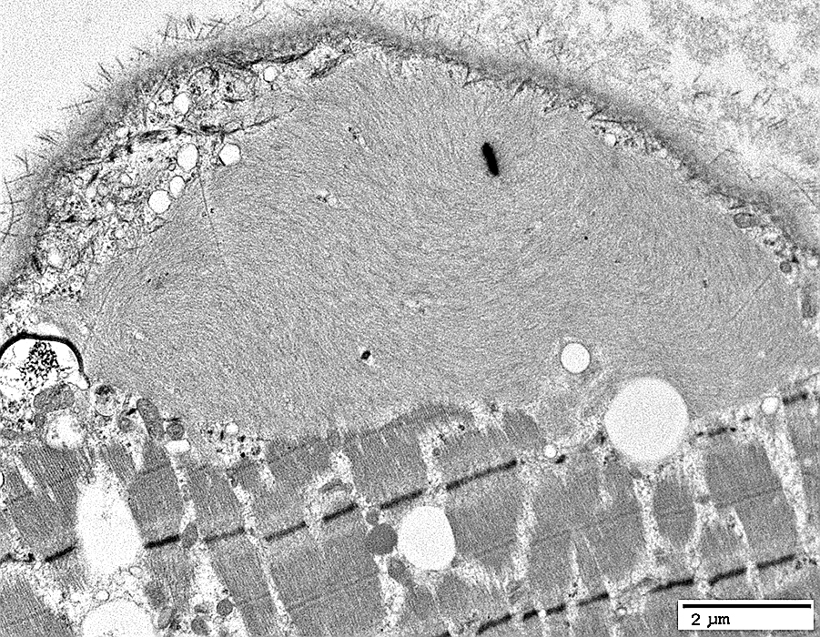 From: R Schmidt |
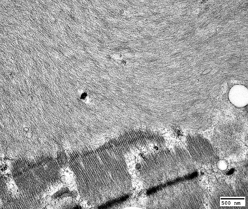 From: R Schmidt |
Desmin Myopathy: Filament Aggregates
Loose filament cluster with additional organelles & Z-Band-like material
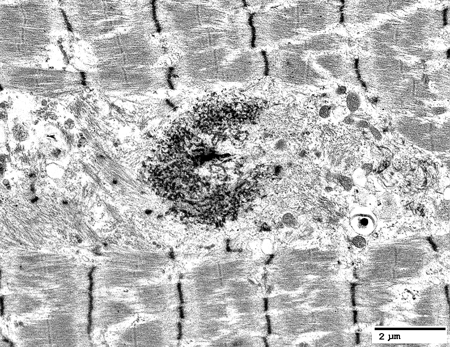 From: R Schmidt |
Desmin Myopathy: Organelle Aggregates
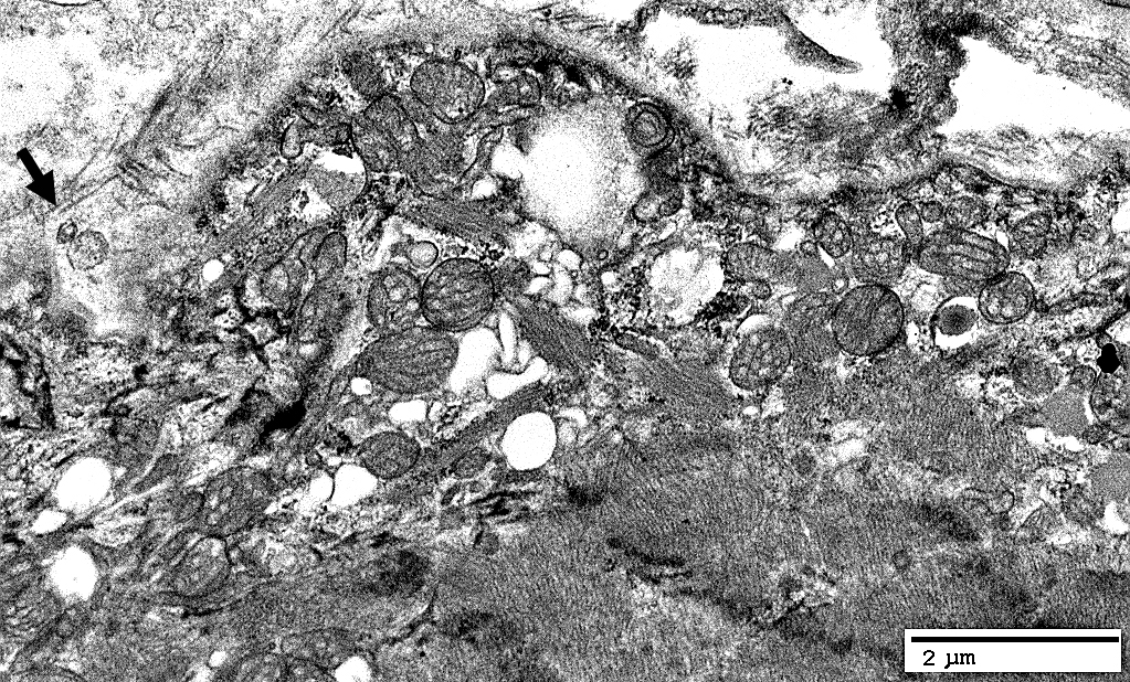 From: R Schmidt |
Membrane & Basal lamina invagination with Extracellular debris (Arrow)
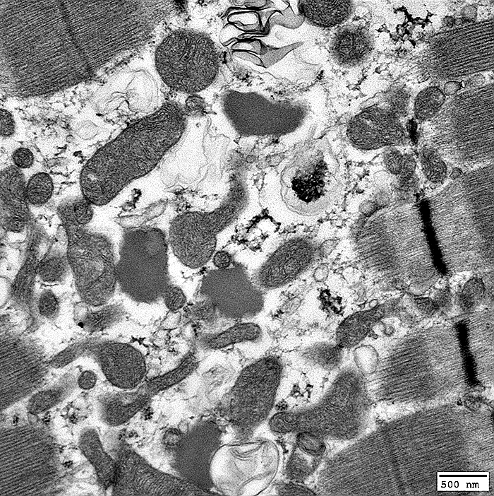 From: R Schmidt |
Contents include Mitochondria, Vacuoles with irregular membranes & Droplets of abnormal lipid
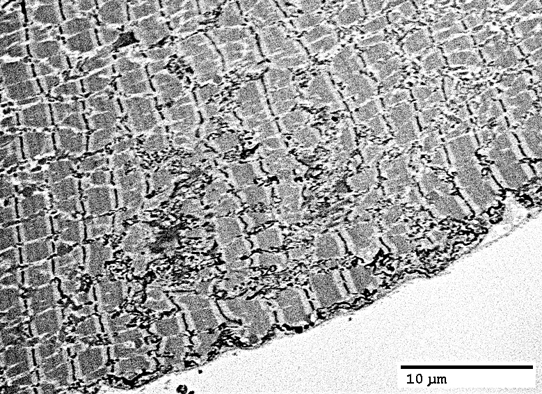
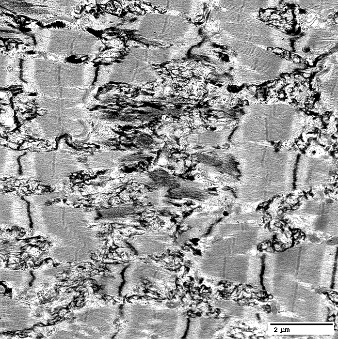
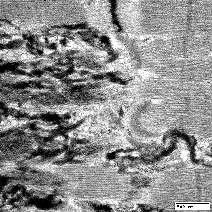
Desmin Myopathy: Z-Disk pathology
 From: R Schmidt |
 From: R Schmidt |
General: Extension of Z-Disk material along long axis of muscle fiber
Also see: Rod myopathy
 From: R Schmidt |
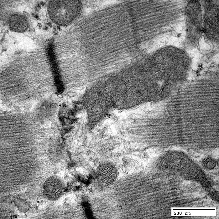
Z-band Disruption
Mitochondria: Large; Abnormal structure
 From: R Schmidt |
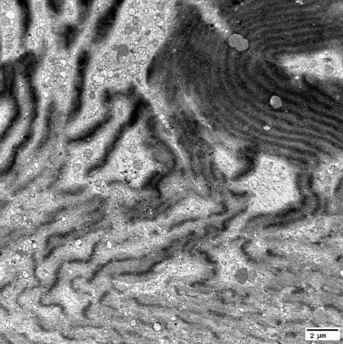 From: R Schmidt |
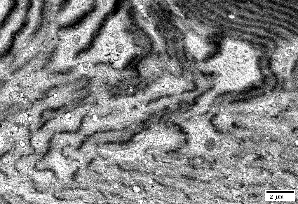 From: R Schmidt |
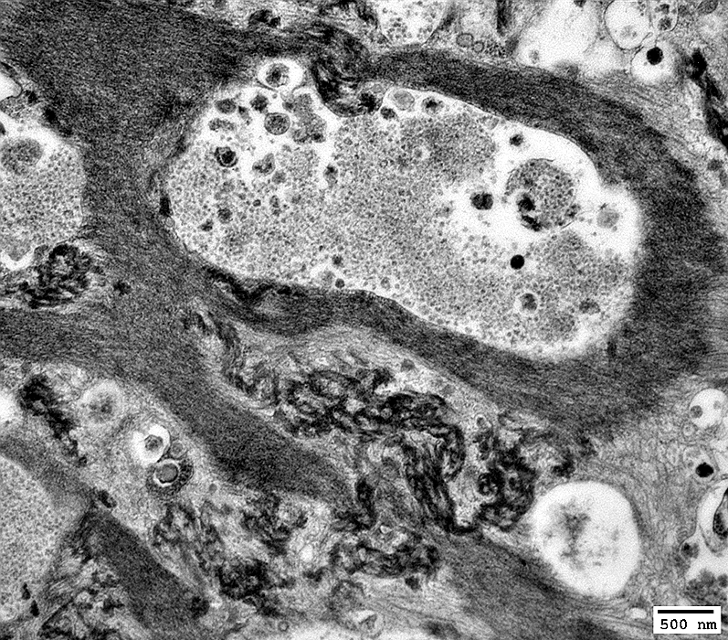
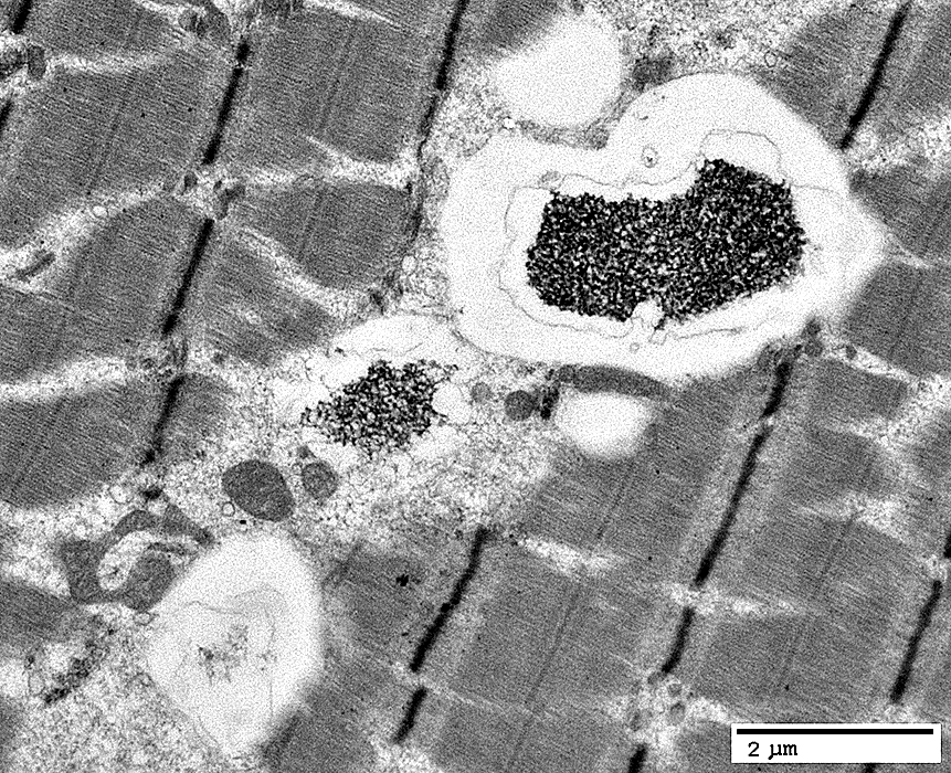
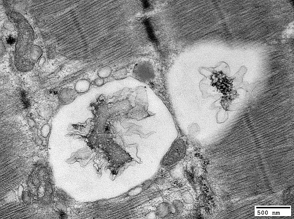
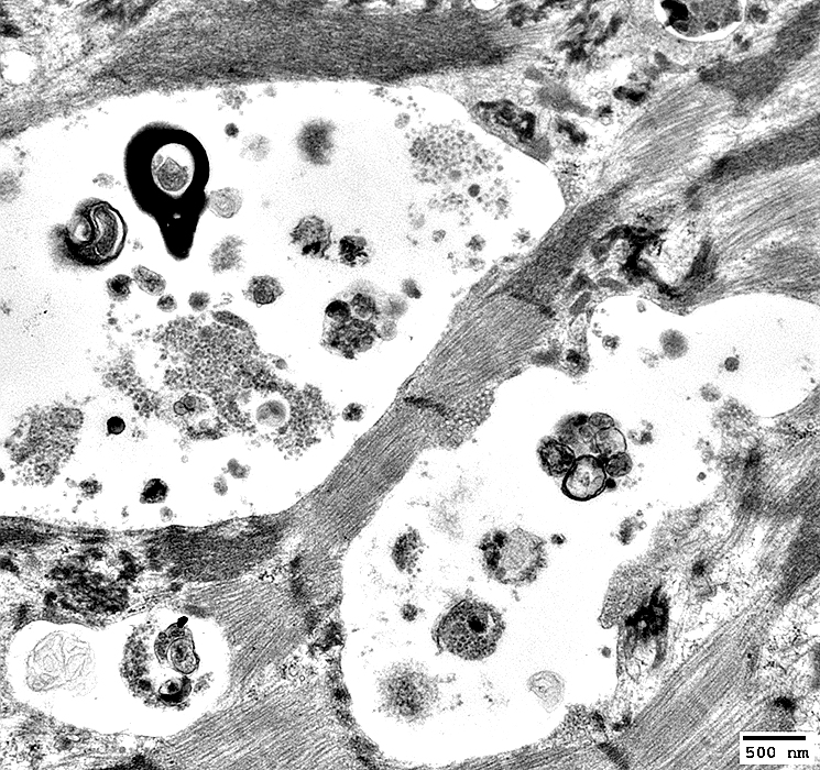
Autophagic Vacuoles
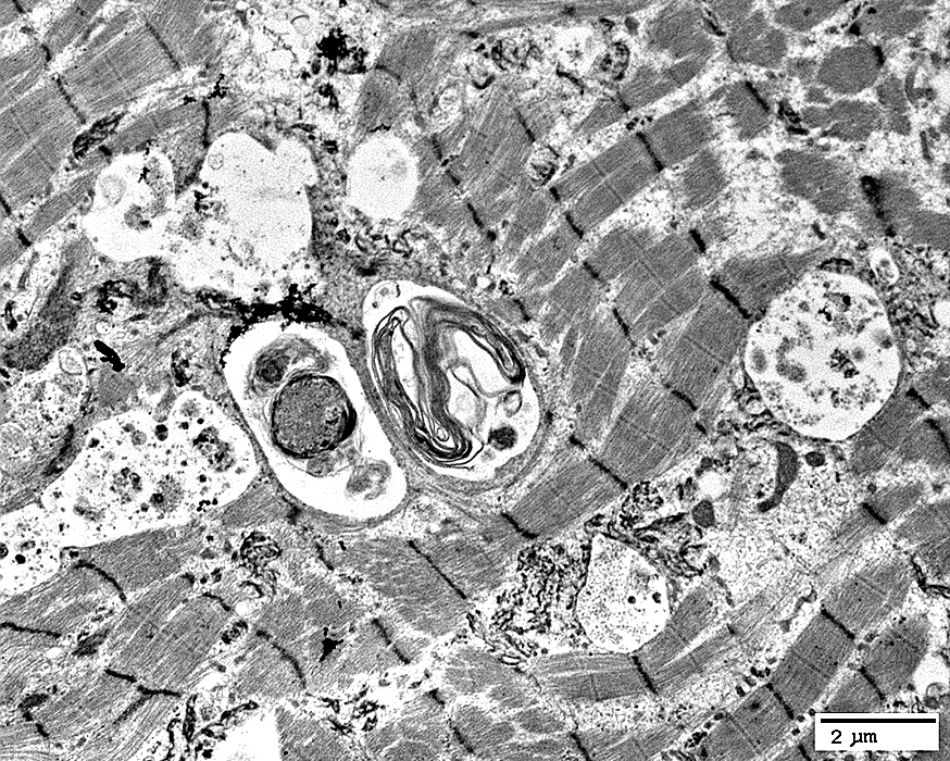 From: R Schmidt |
Vacuoles: Have varied contents
Aggregated material: May surround vacuoles
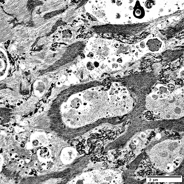 From: R Schmidt |
Contents: Varied
May be surrounded by dark aggregated material
 From: R Schmidt |
 From: R Schmidt |
 From: R Schmidt |
 From: R Schmidt |
 From: R Schmidt |
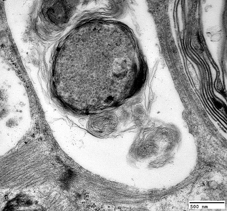 From: R Schmidt |
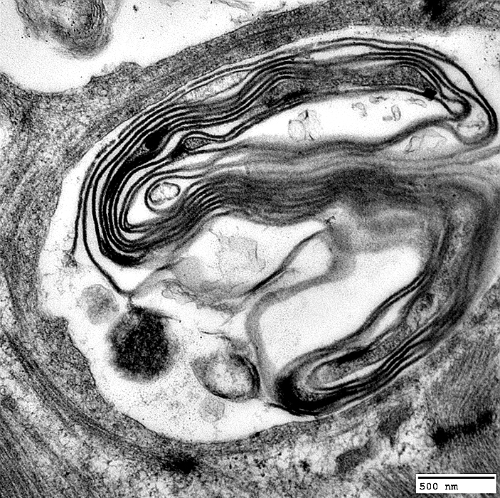 From: R Schmidt |
Sural nerve: Mildly reduced number of myelinated axons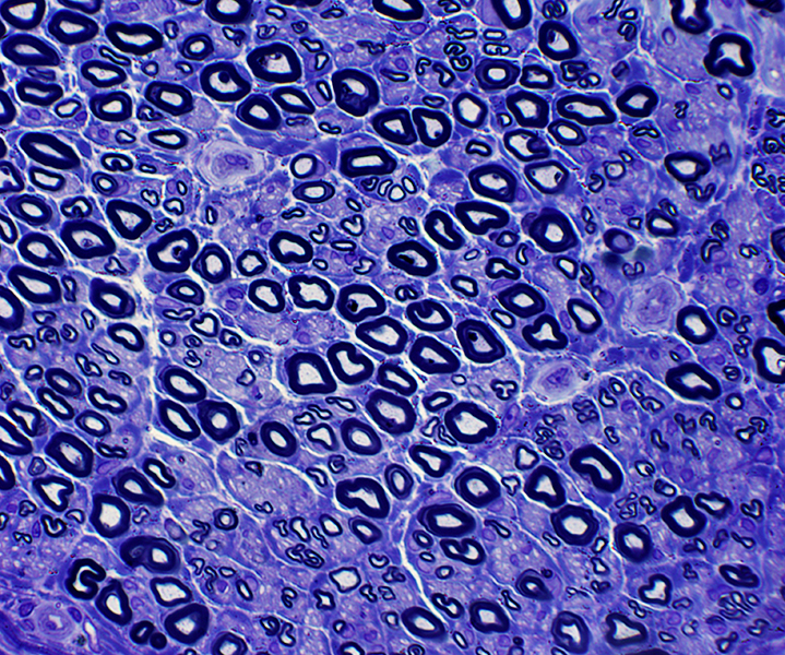 Toluidine blue stain |
Return to Myofibrillar/Desmin myopathies
Return to Muscle biopsies
4/20/2021