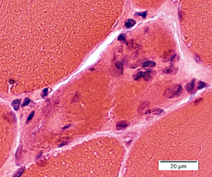
Myofibrillar Myopathy 4 (MFM4): ZASP
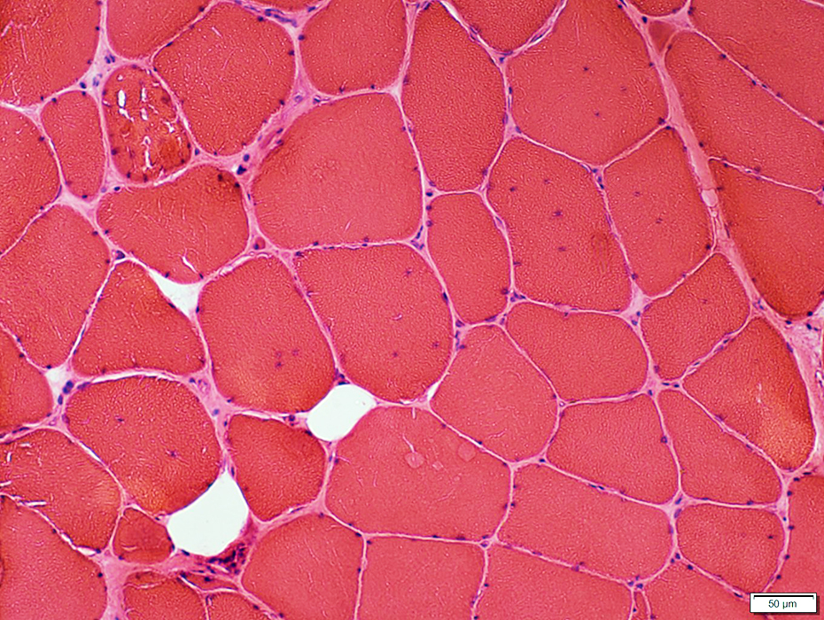 H&E stain |
Fiber sizes
Varied
Hypertrophy & Moderate atrophy
Small fibers may occur in clusters (Below)
Internal nuclei
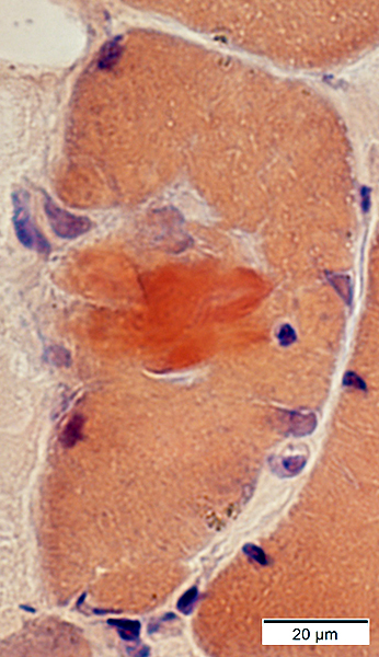
Vacuoles & Aggregates (Arrow): In scattered muscle fibers
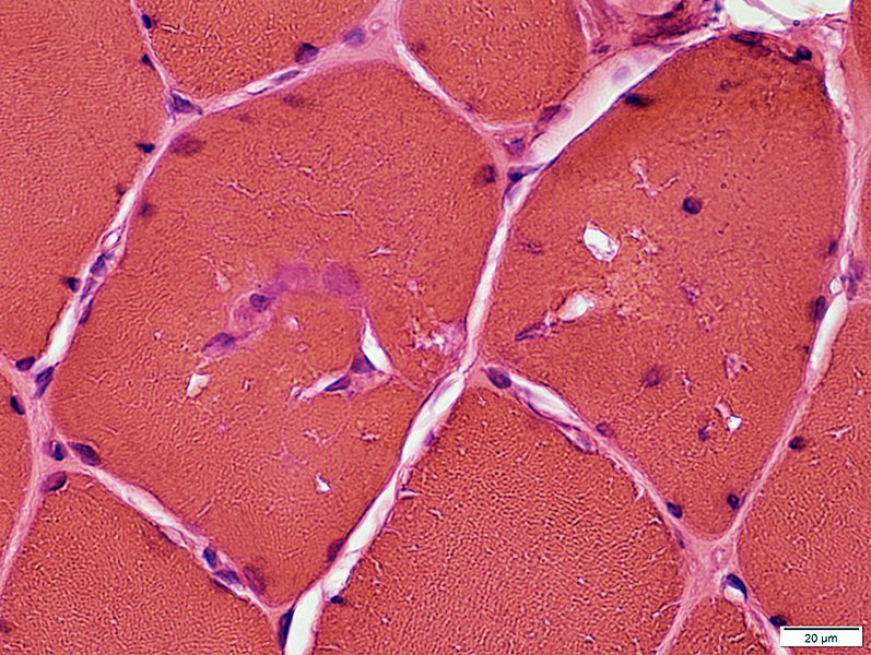
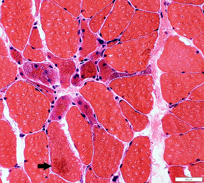 H&E stain |
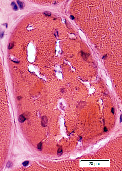 H&E stain |
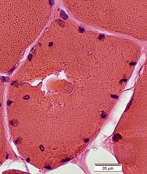 H&E stain |
Vacuoles
Aggregates
Myonuclei: Irregular shapes; Increased numbers at edge of fibers
 H&E stain |
 H&E stain |
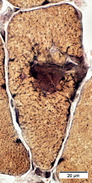
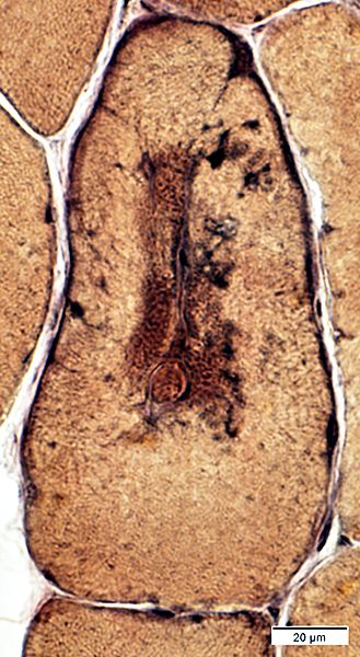
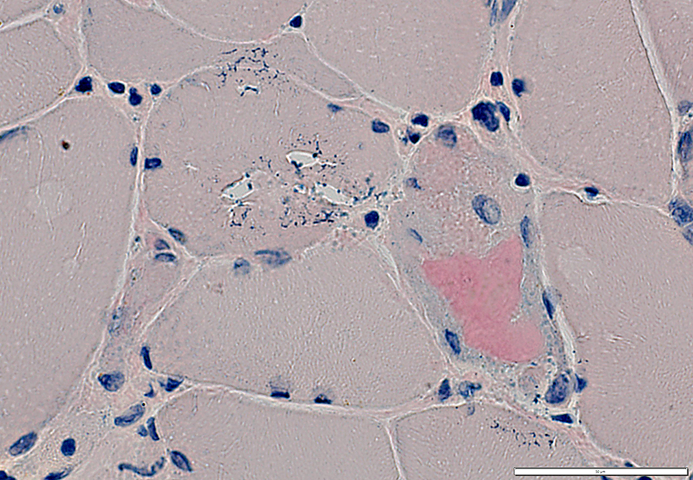
 Gomori trichrome stain |
 Gomori trichrome stain |
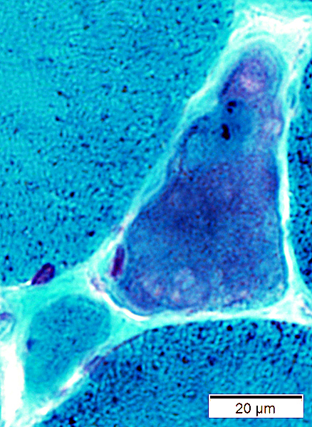 Gomori trichrome stain |
 Gomori trichrome stain |
Vacuoles: Contain red-stained material at edges
|
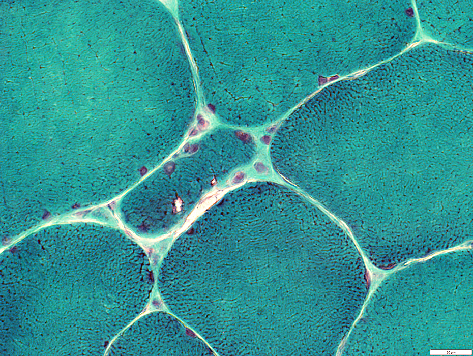
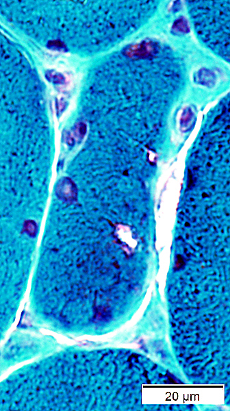
 VvG stain |
Vacuoles: Irregular shape; Gray border; Several in single muscle fiber (Right)
 VvG stain |
 VvG stain |
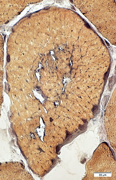 VvG stain |
 AMPDA stain |
 AMPDA stain |
 AMPDA stain |
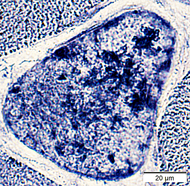 AMPDA stain |
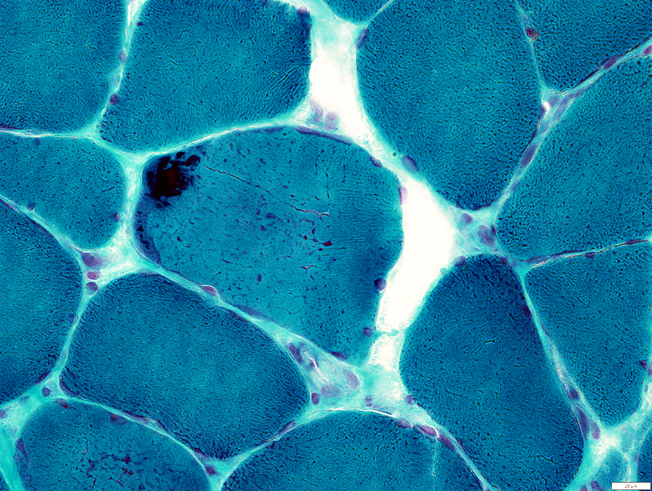
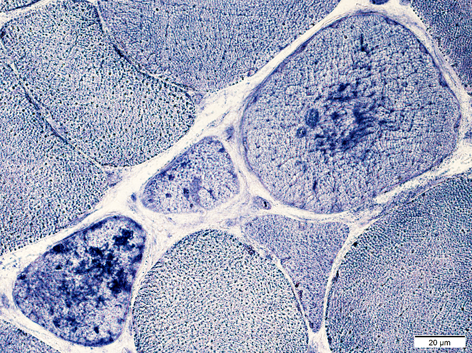
 Congo red stain |
 Congo red stain |
Vacuoles: Clear; Well defined border; May have basophilic debris (Below)
 Congo red stain |
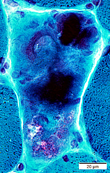
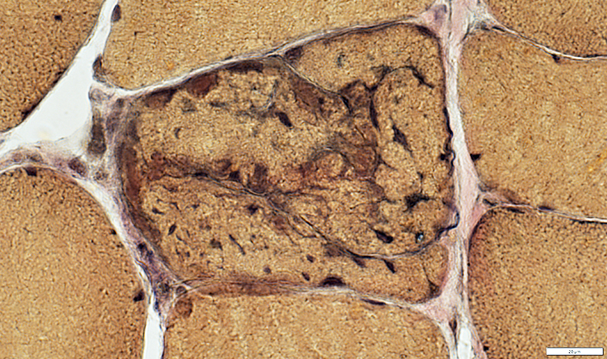
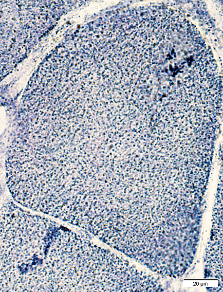
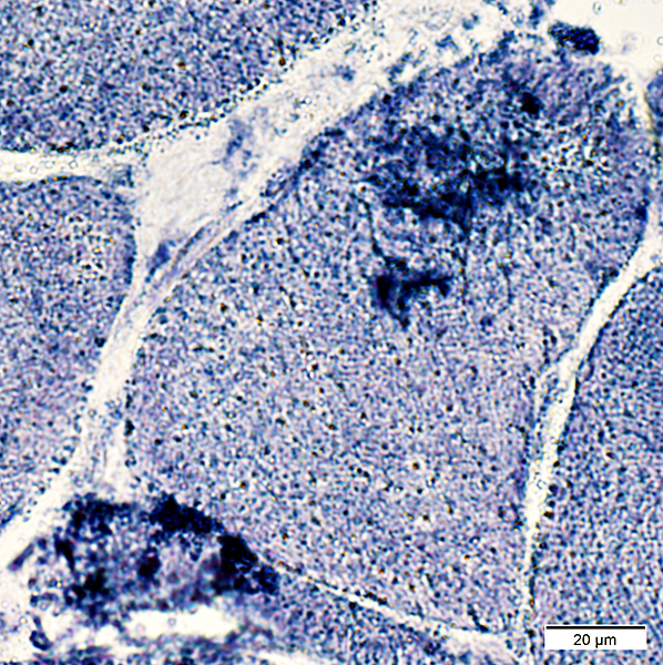
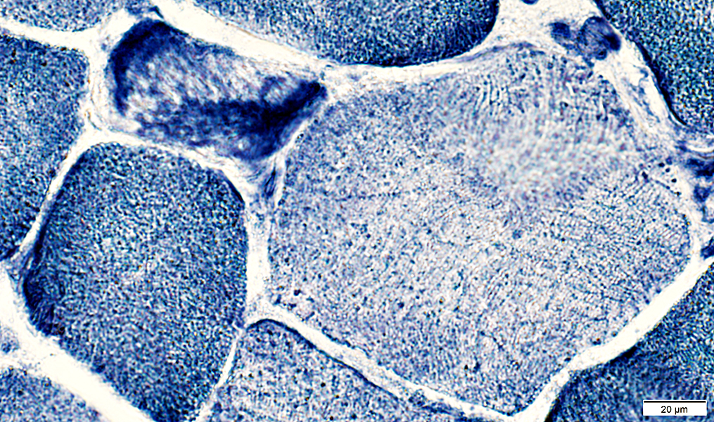
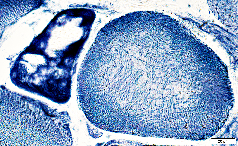 NADH stain |
Internal membranes: Clustered in some regions
 NADH stain |
Aggregates
Displace myofibrillar apparatus
Present in fiber types I & II
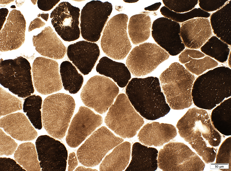 ATPase pH 9.4 stain |
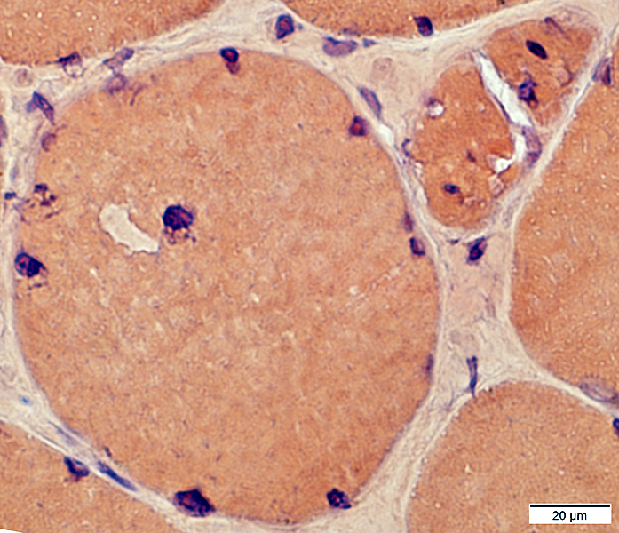
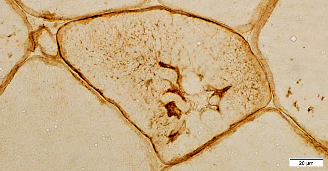
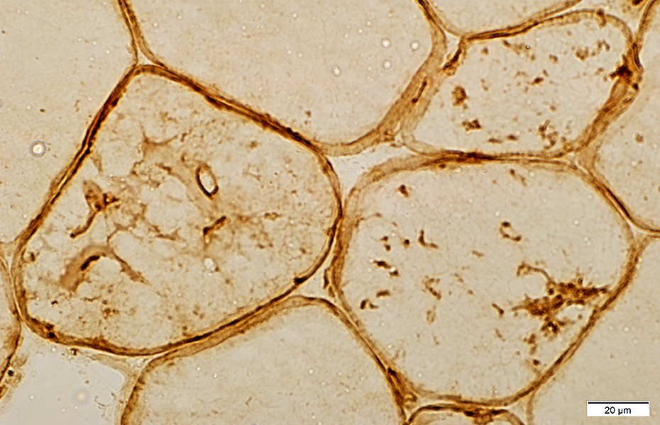
Desmin: Mildly irregular distribution in cytoplasm
 Desmin stain |
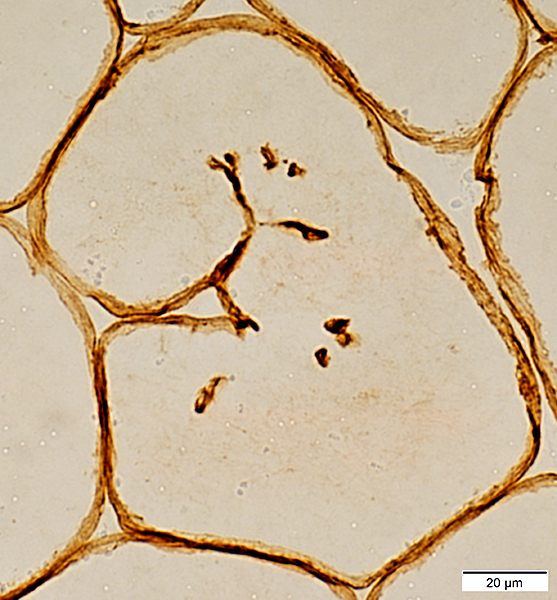
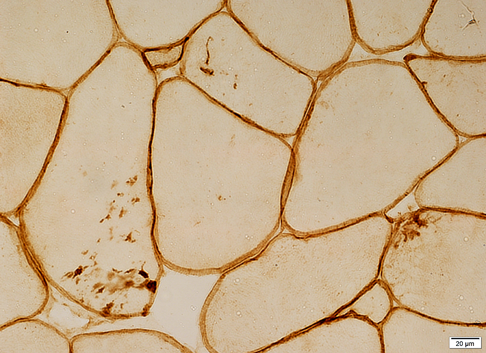 Caveolin-3 stain |
 Caveolin-3 stain |
 Caveolin-3 stain |
Dystrophin: Present in few splits & amp; aggregates
 Dystrophin (Dys2) stain |
Return to Neuromuscular Home Page
Return to Myopathies with wasting
5/10/2023