LGMD 2L (R12): Anoctamin 5 mutations
|
Amyloid Internal architecture Aggregates Partial fiber fusion (Splitting) Vacuoles & Blebs Myopathy Chronic Fiber sizes: Varied Necrosis Regeneration Ultrastructure |
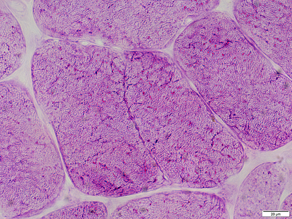
 H&E stain |
Fiber sizes: Varied
Clusters of small fibers
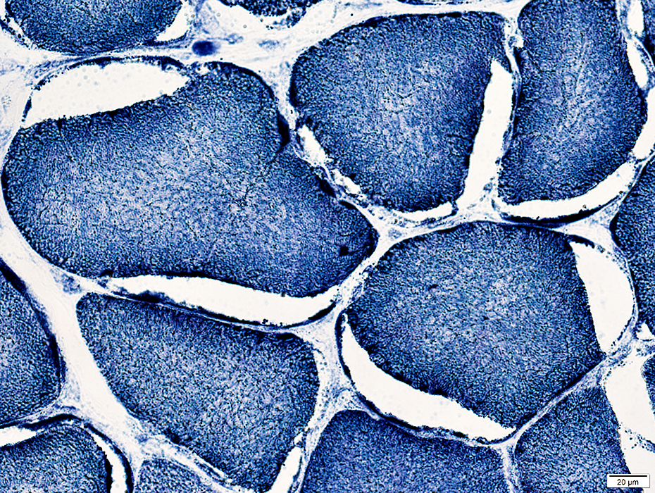
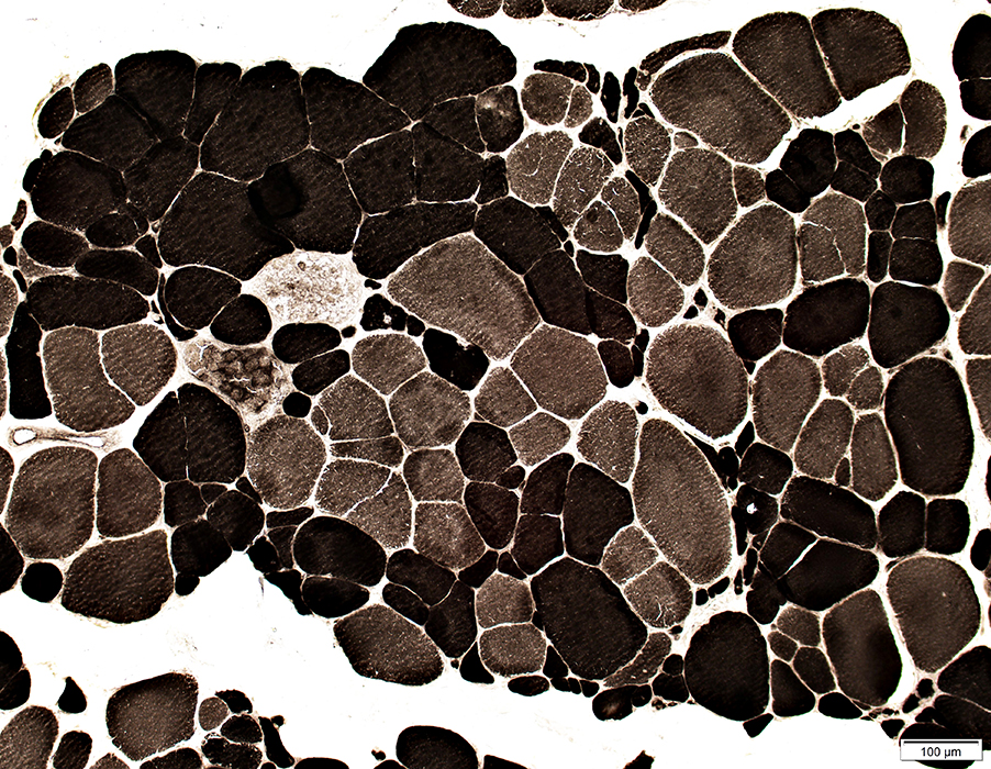 ATPase pH 9.4 stain |
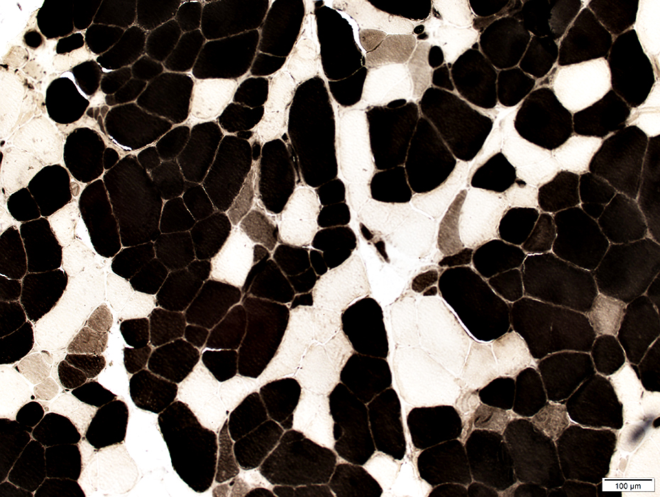 ATPase pH 4.3 stain |
Many immature type 2C muscle fibers
 H&E stain |
Fiber sizes
Varied; Clusters of small fibers
Necrosis & Regeneration
 H&E stain |
 |
Fiber size: Varied; Hypertrophy; Nuclear clumps
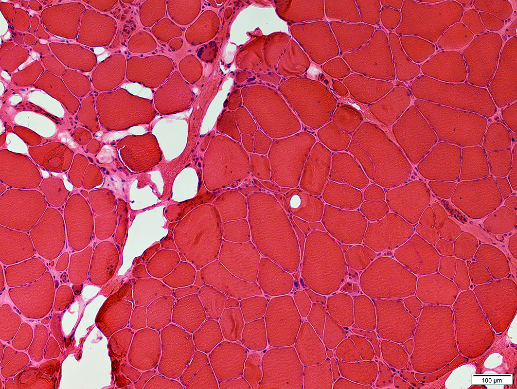
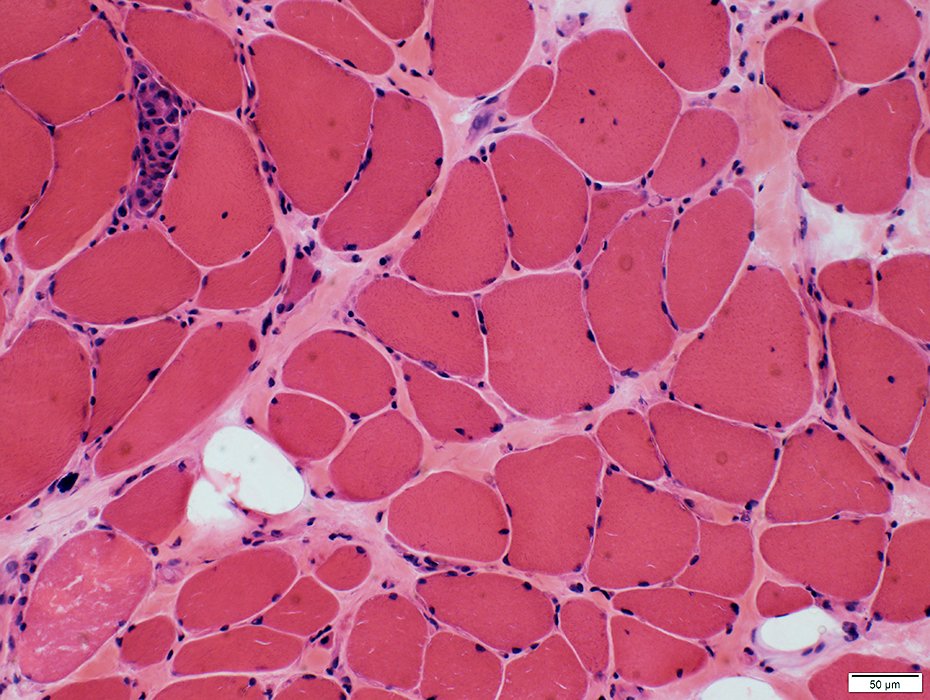
ANO5: Chronic
 H&E stain |
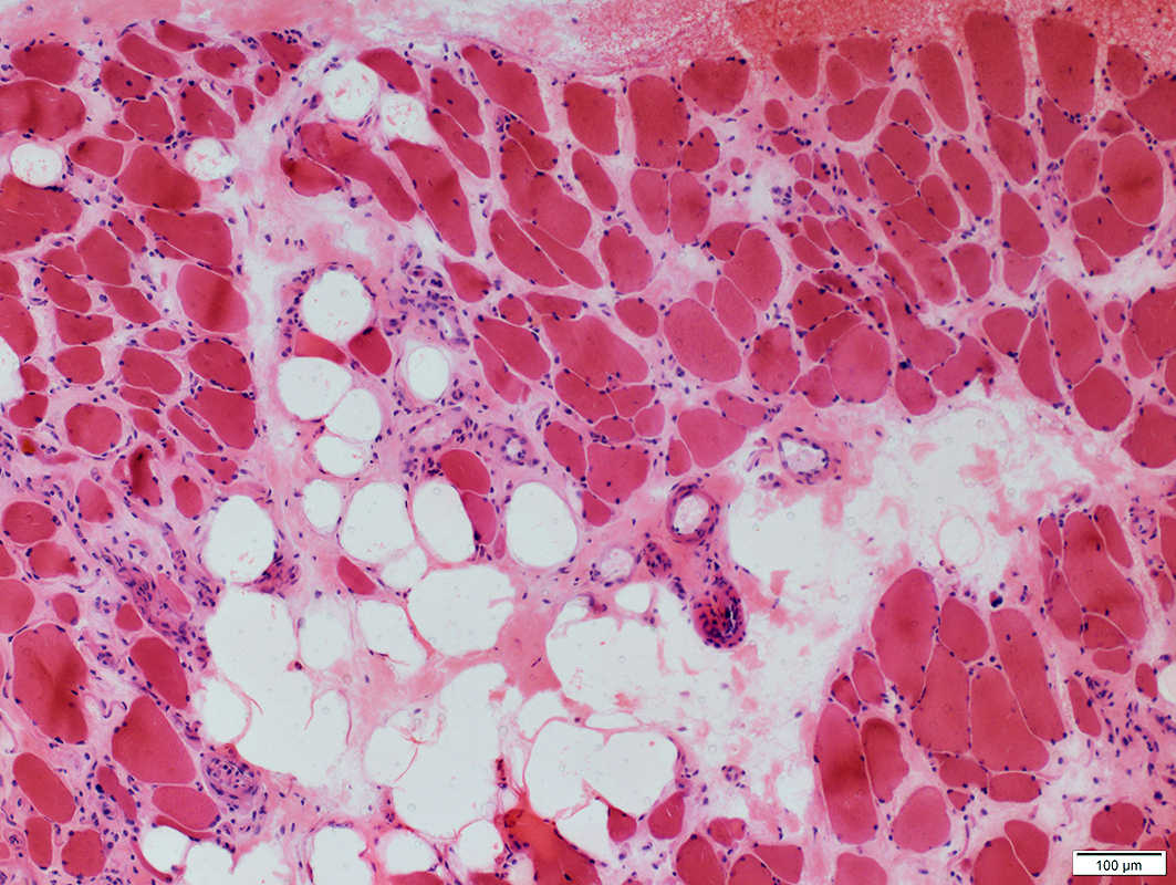
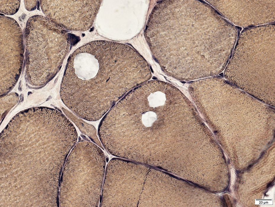
Fiber size: Varied
Endomysial connective tissue: Increased
Perimysium: Replaced by fat
 Congo red stain |
 Gomori trichrome stain |
Fiber size: Varied
Endomysial connective tissue: Increased
 H&E stain |
 AMPDA stain |
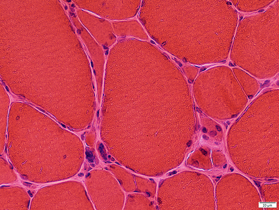
Fiber size: Varied
Internal architecture: Irregular
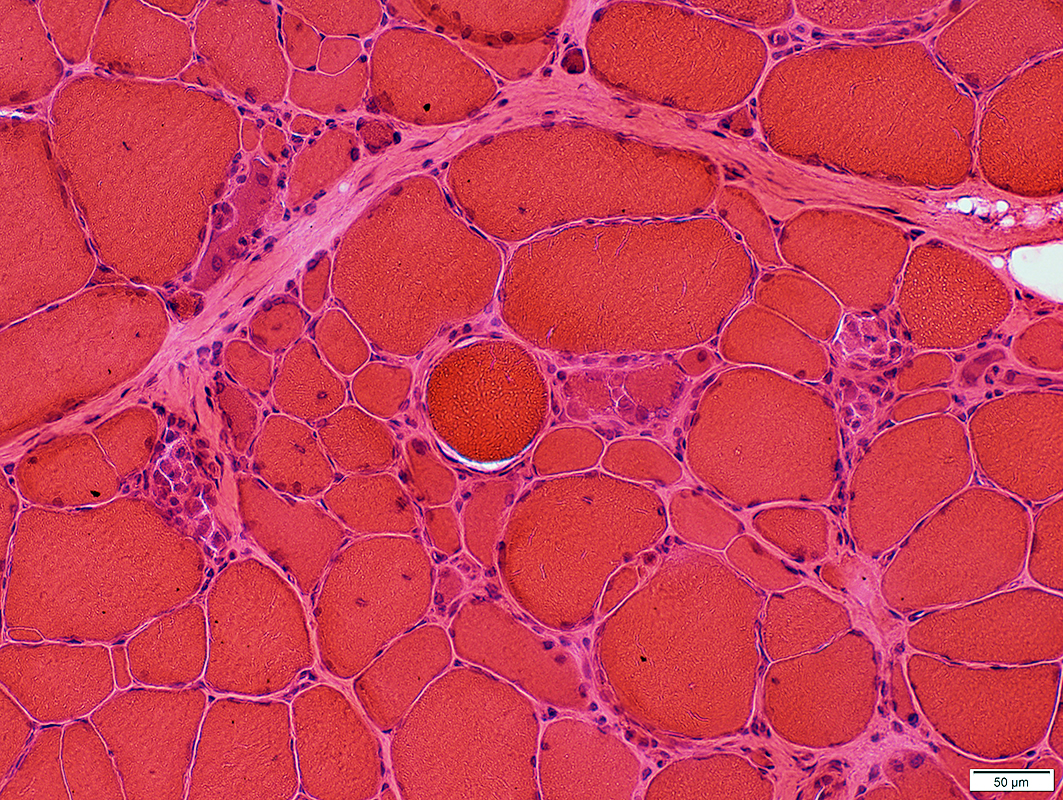
ANO5: Necrosis
 H&E stain |
Fiber size: Varied
Necrosis & Regeneration
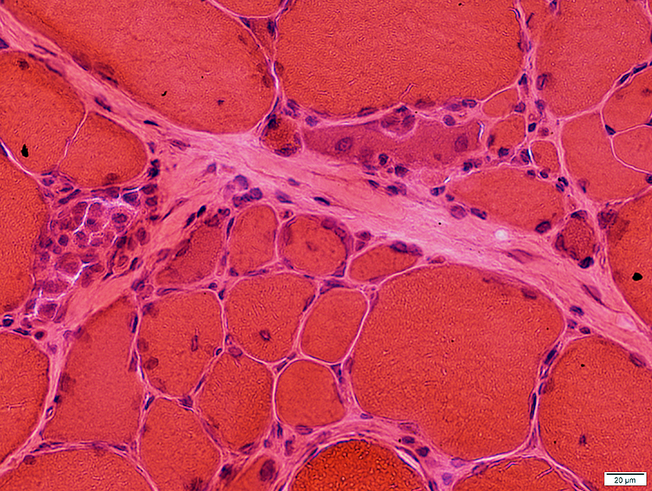 H&E stain |
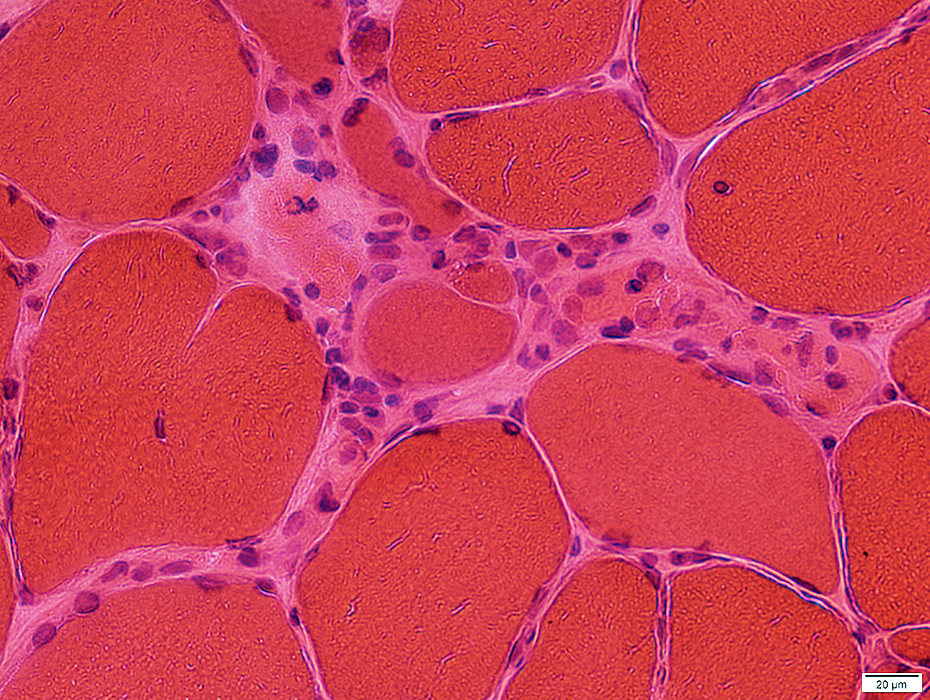 |
 H&E stain |
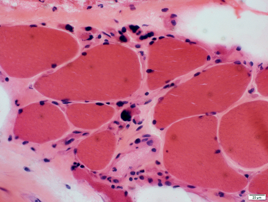
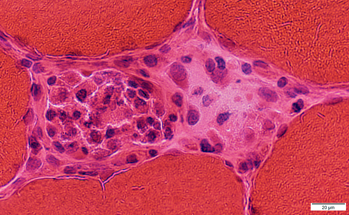
Early necrosis
Muscle fiber cytoplasm: Pale
Fibers invaded by few histiocytes
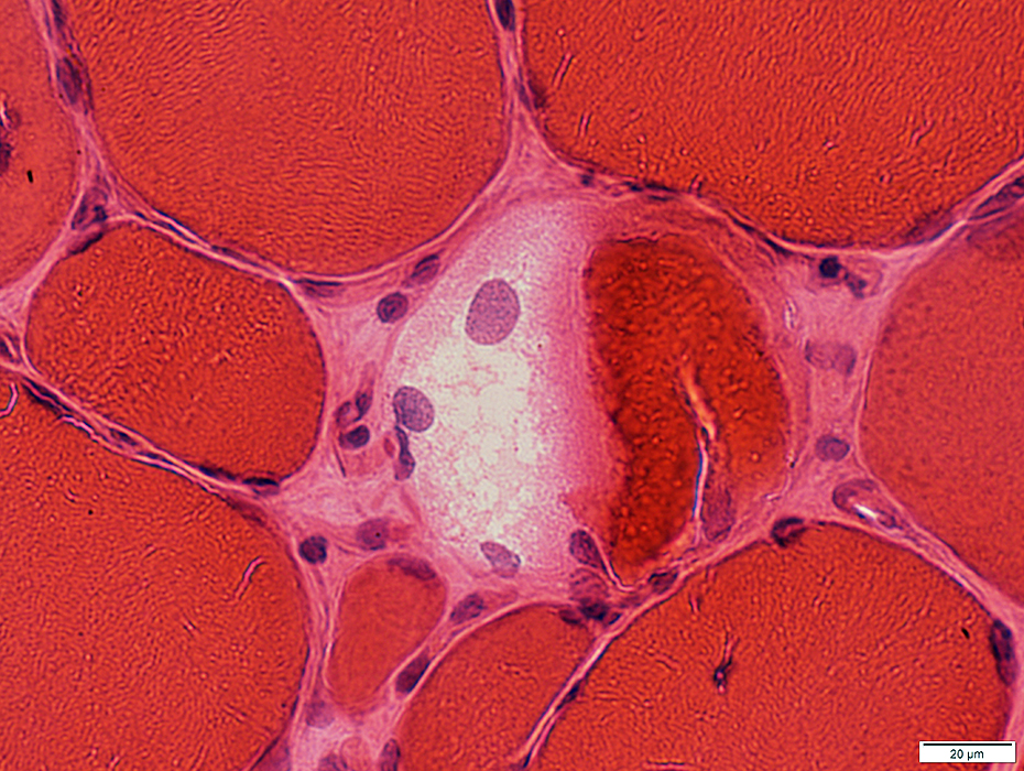
 H&E stain |
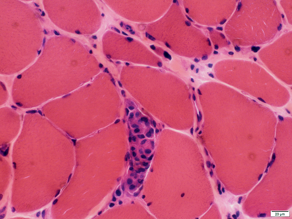
 H&E stain |
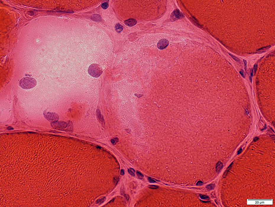
Necrosis
Muscle fibers: Replaced by histiocytic cells
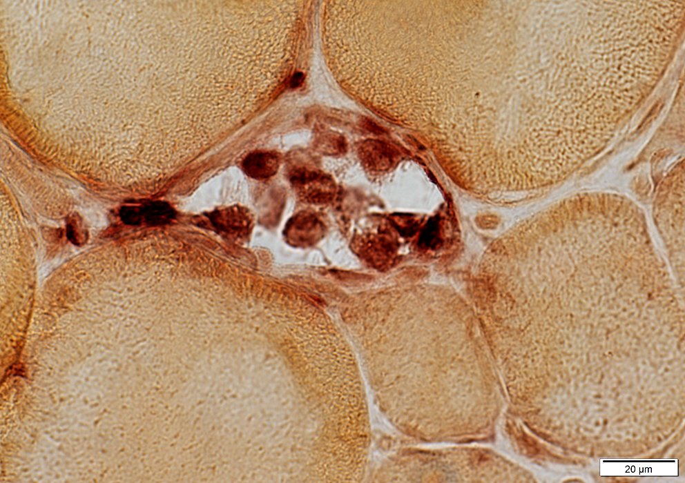 Acid phosphatase stain |
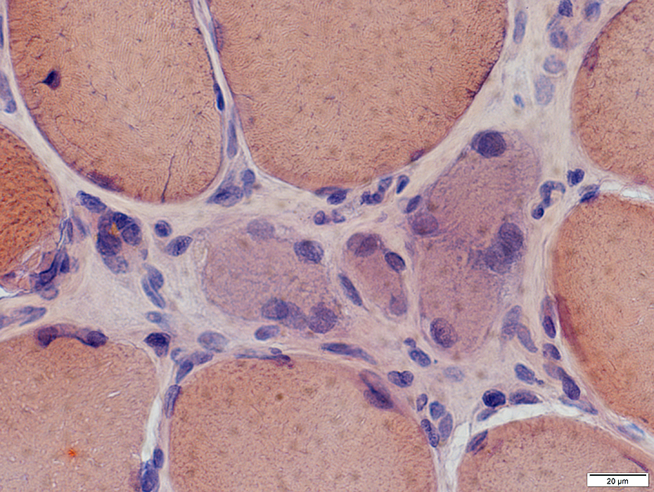
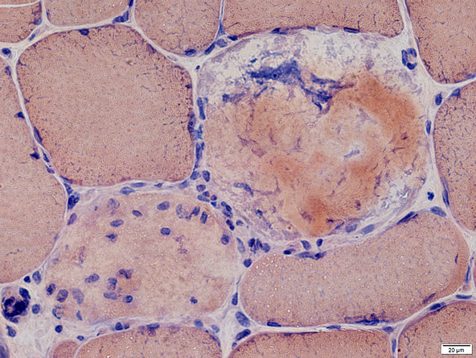
ANO5: Regeneration
 Congo red stain |
Regenerating Muscle fibers
Basophilic cytoplasm
Large nu;clei
 Congo red stain |
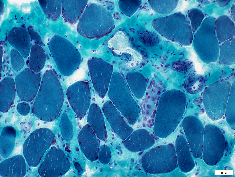
 Gomori trichrome stain |
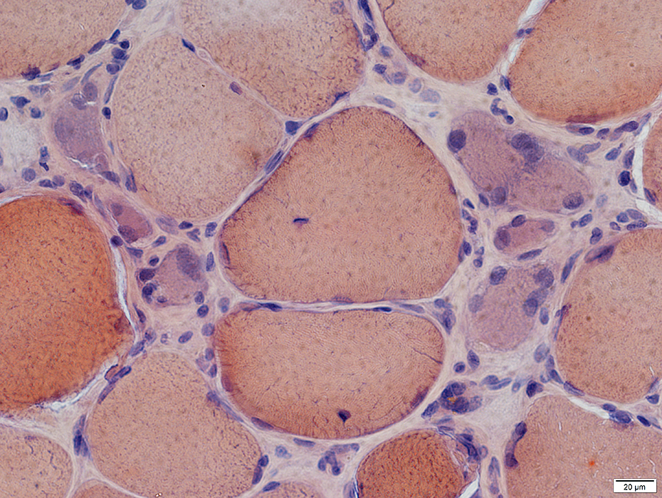
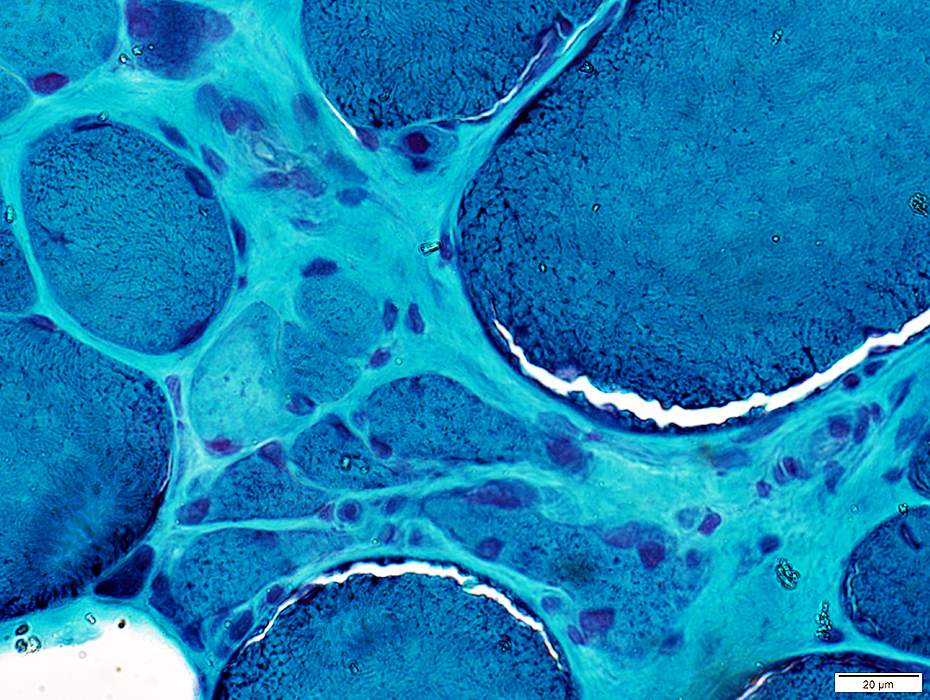
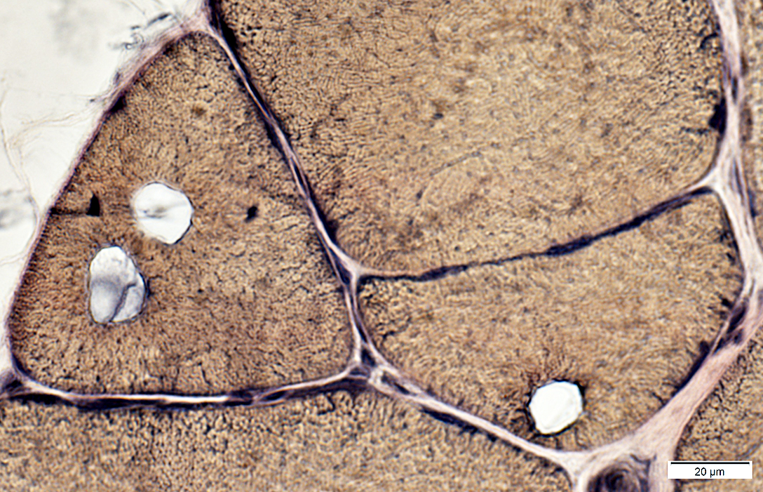
ANO5: Partial fusion of muscle fibers (Splitting; Branching)
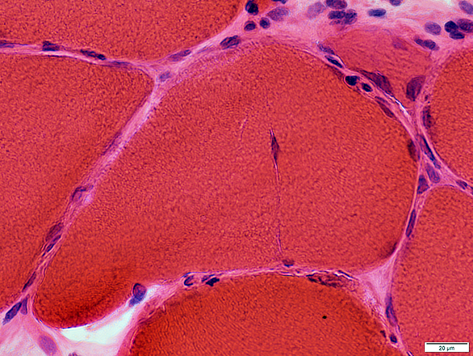 H&E stain |
Partially fused muscle fibers: Split across part of fiber
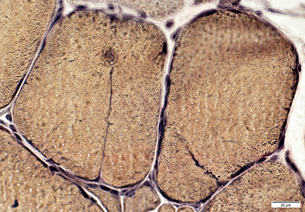 VvG stain |
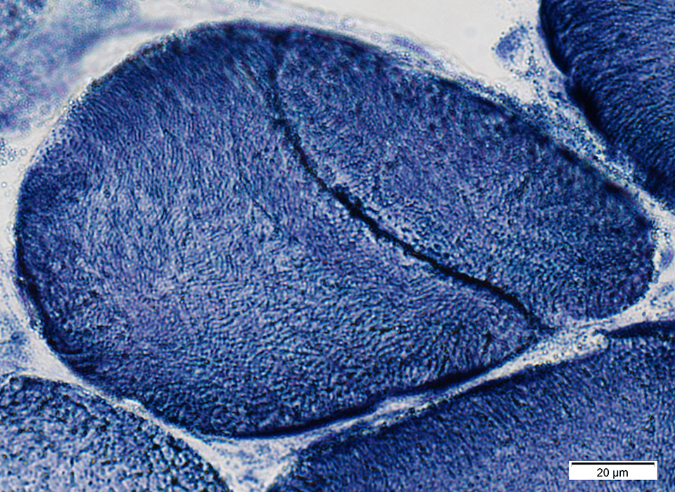 NADH stain |
Partially fused muscle fibers: Split across part of fiber
 PAS stain |
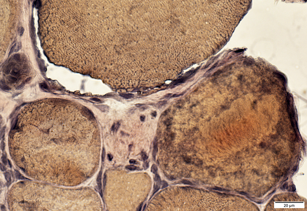
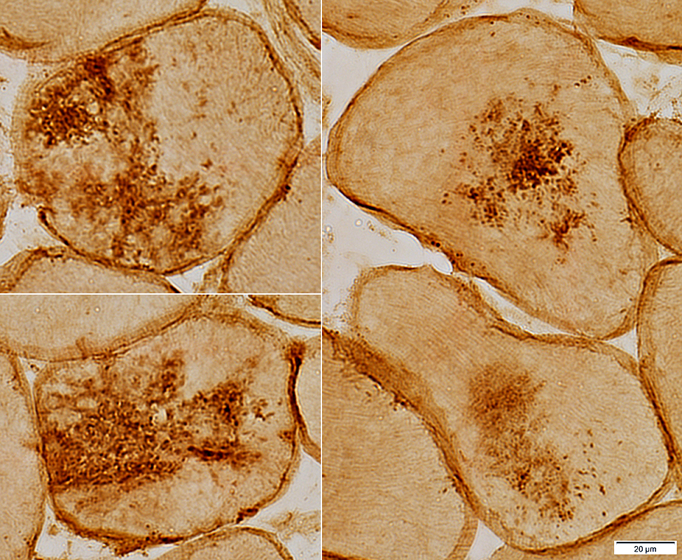
ANO5: Amyloid
 Congo red stain |
Amyloid: Birefringent; Deposited in vessel wall
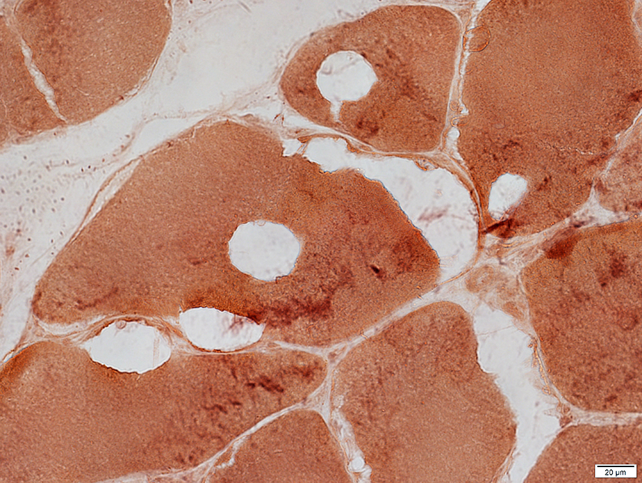
ANO5: Blebs & Vacuoles
 NADH stain |
Subsarcolemmal blebs
 VvG stain |
Cytoplasmic vacuoles
 VvG stain |
 Esterase stain |
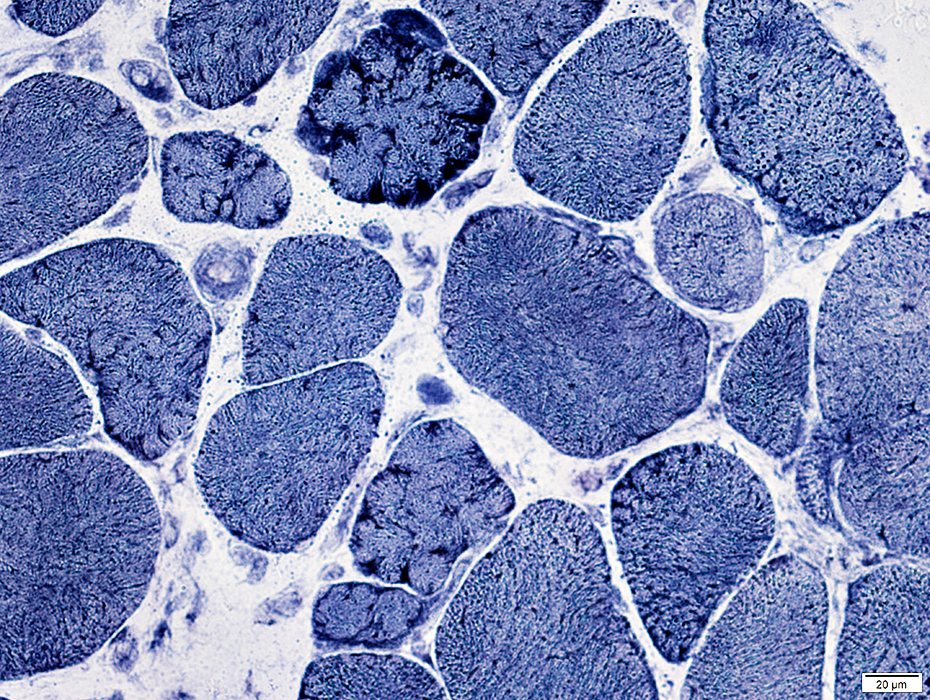
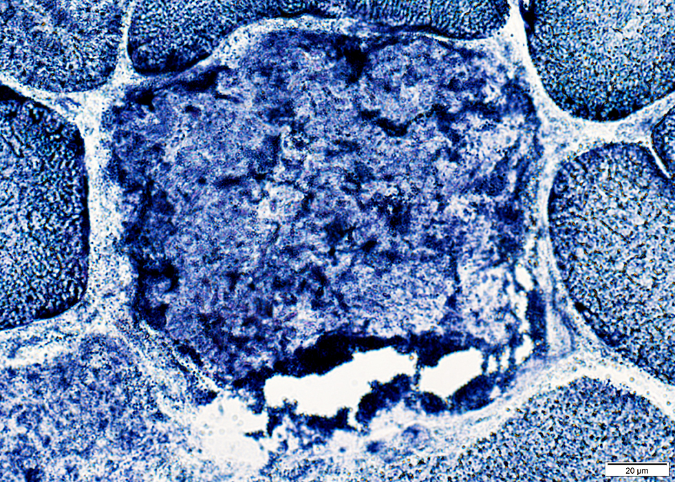
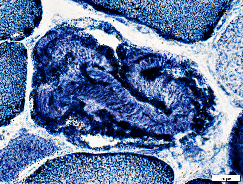
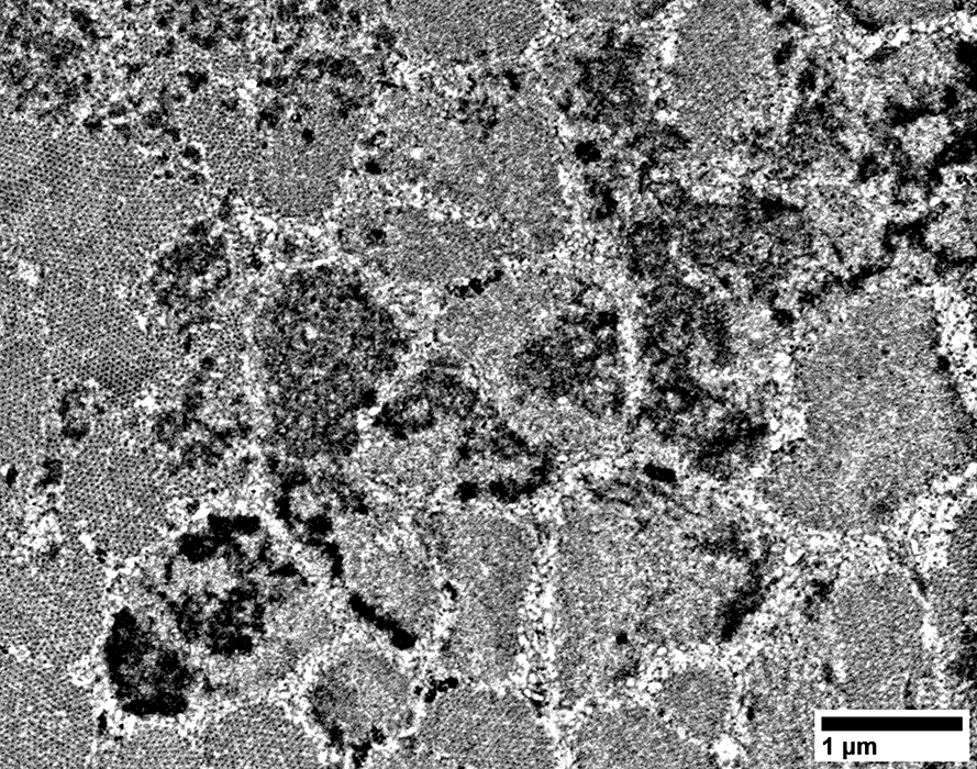
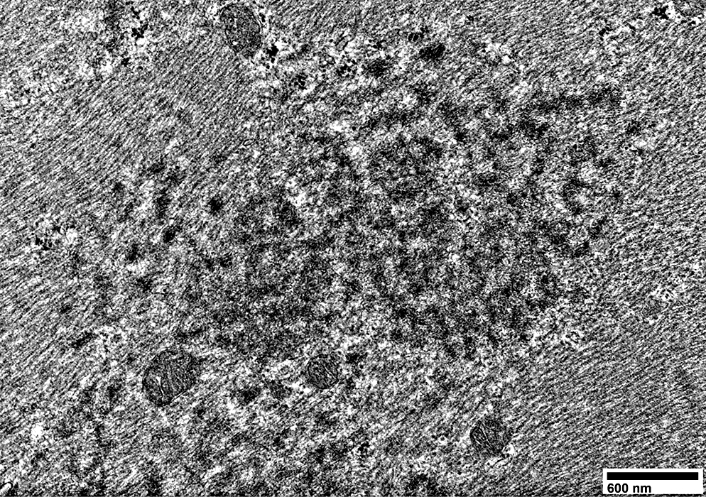
ANO5: Aggregates & Abnormal internal architecture
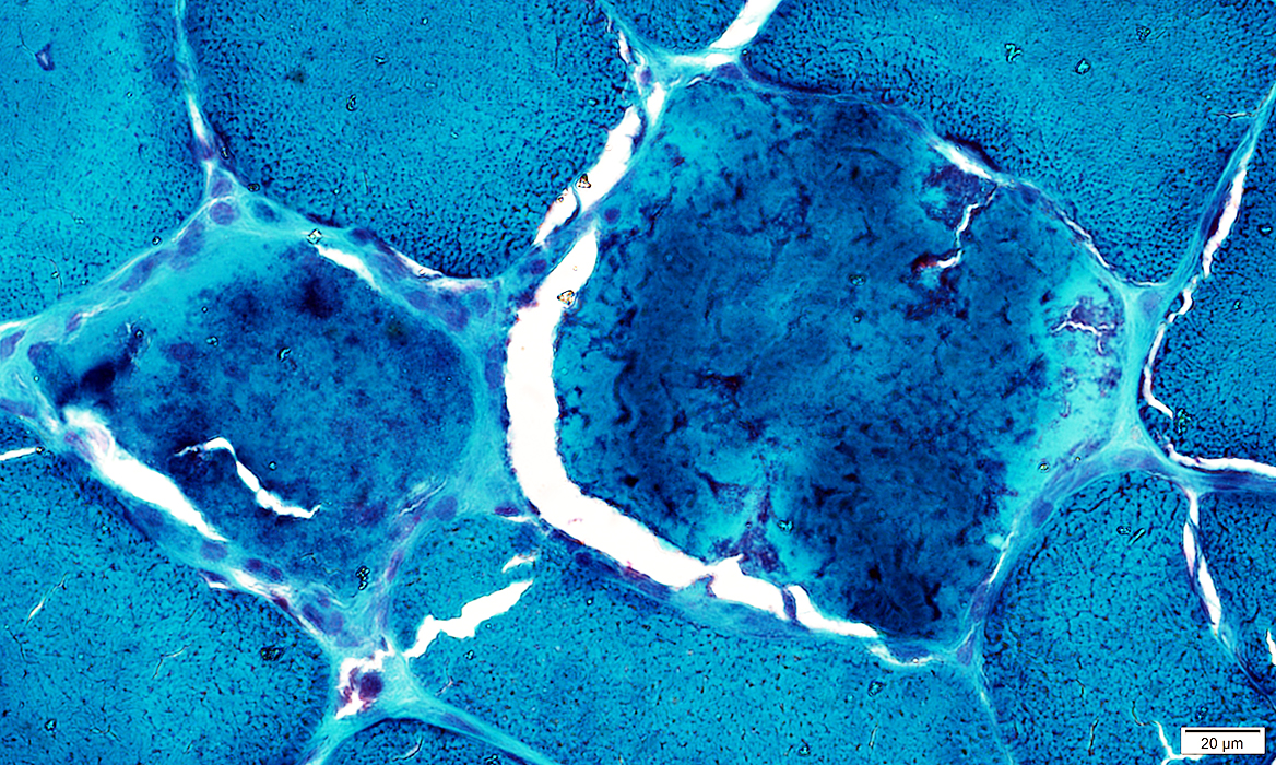 Gomori trichrome stain |
Fibers with clustered, irregular internal architecture
 Congo red stain |
 VvG stain |
Fibers with clustered, irregular internal architecture
 NADH stain |
 NADH stain |
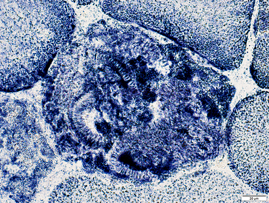 SDH stain |
Fibers with cytoplasmic aggregates
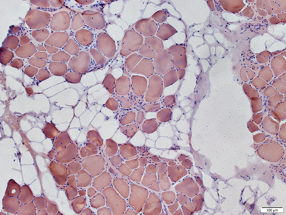
 Caveolin-3 stain |
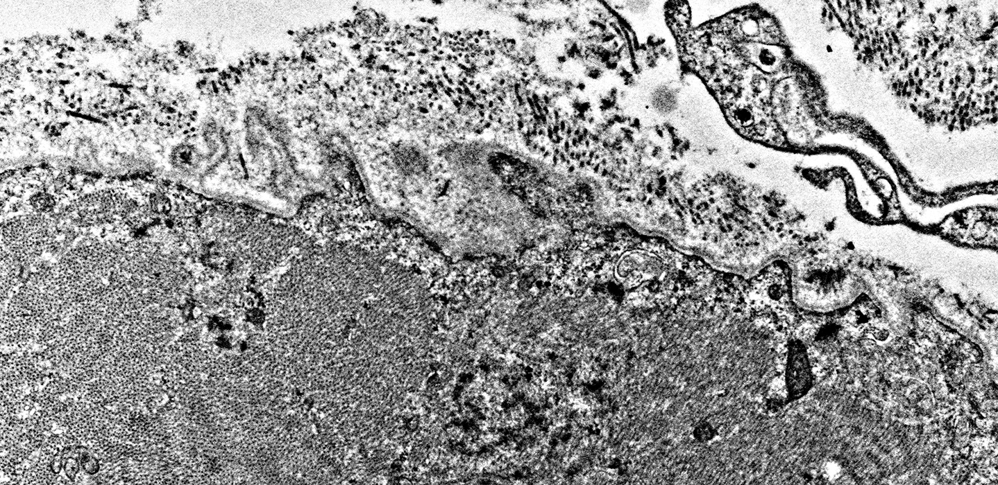
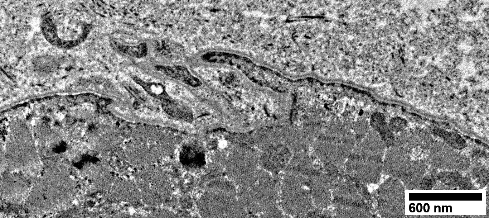
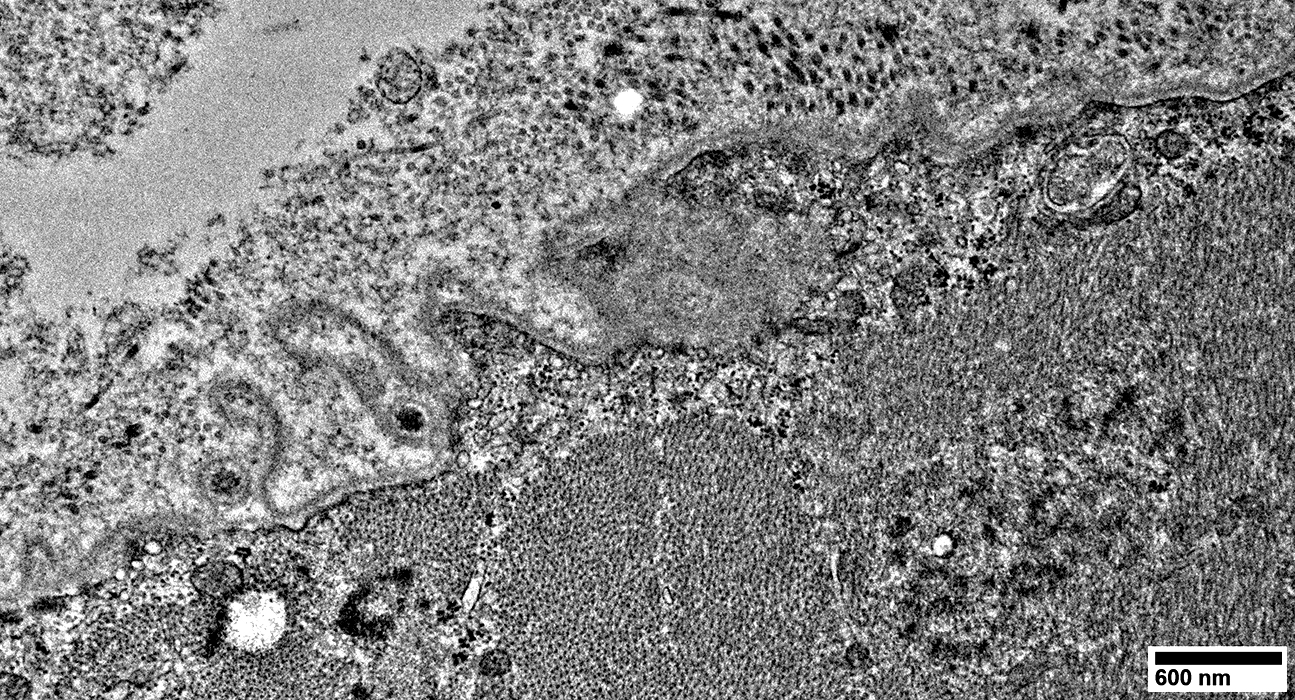
ANO5 mutations: Muscle fiber & Basal lamina Pathlogy
Basal lamina (Wide Black Arrow)
Irregular, wavy configuration
Sarcolemma beneath basal lamina: Interrupted
Muscle fiber under basal lamina: Sarcoplasm structure is disrupted; Dark, rod-like aggregates are present
Basal lamina, 2nd region (Blue arrow)
No muscle fiber cytoplasm underneath irregular region of basal lamina
Outside basal lamina: Connective tissue increased
 From: R Schmidt |
 From: R Schmidt |
 From: R Schmidt |
 From: R Schmidt |
Similar to: Dysferlin
 From: R Schmidt |
 From: R Schmidt |
Return to Neuromuscular Home Page
Return to Pathology index
9/9/2024