Batten Disease: Muscle & Nerve Pathology
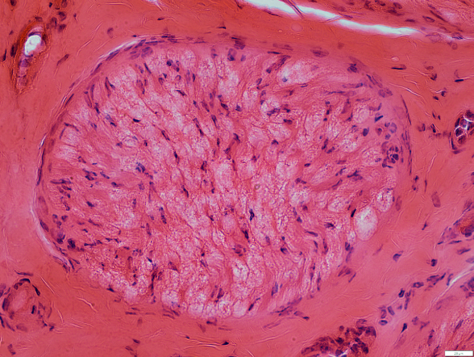
Muscle Pathology
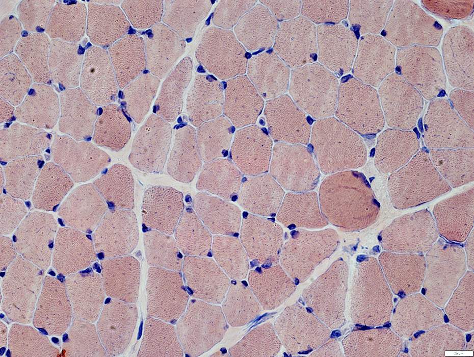
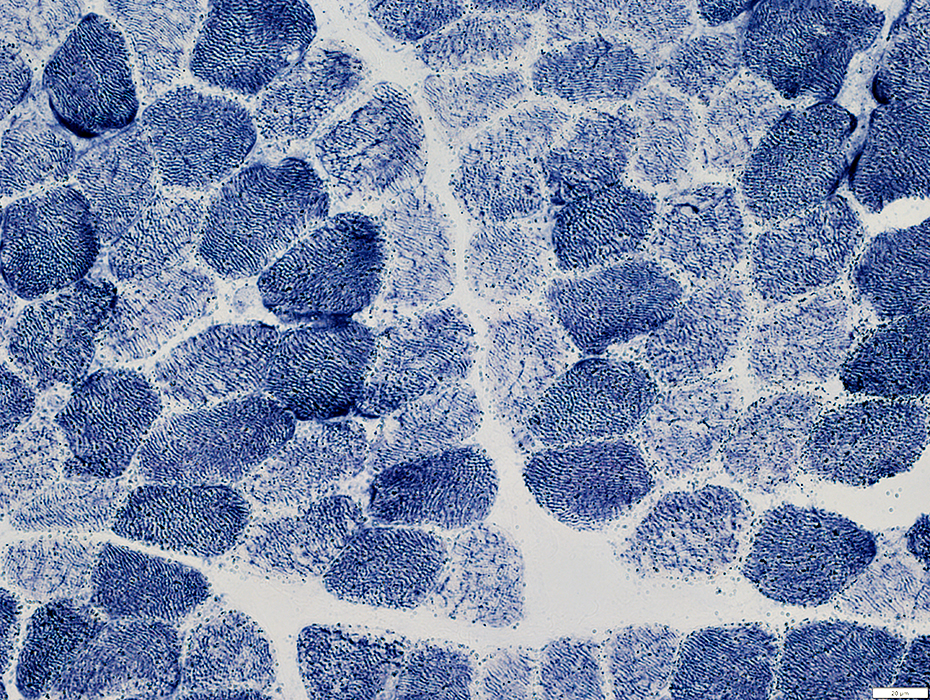
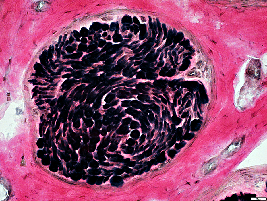
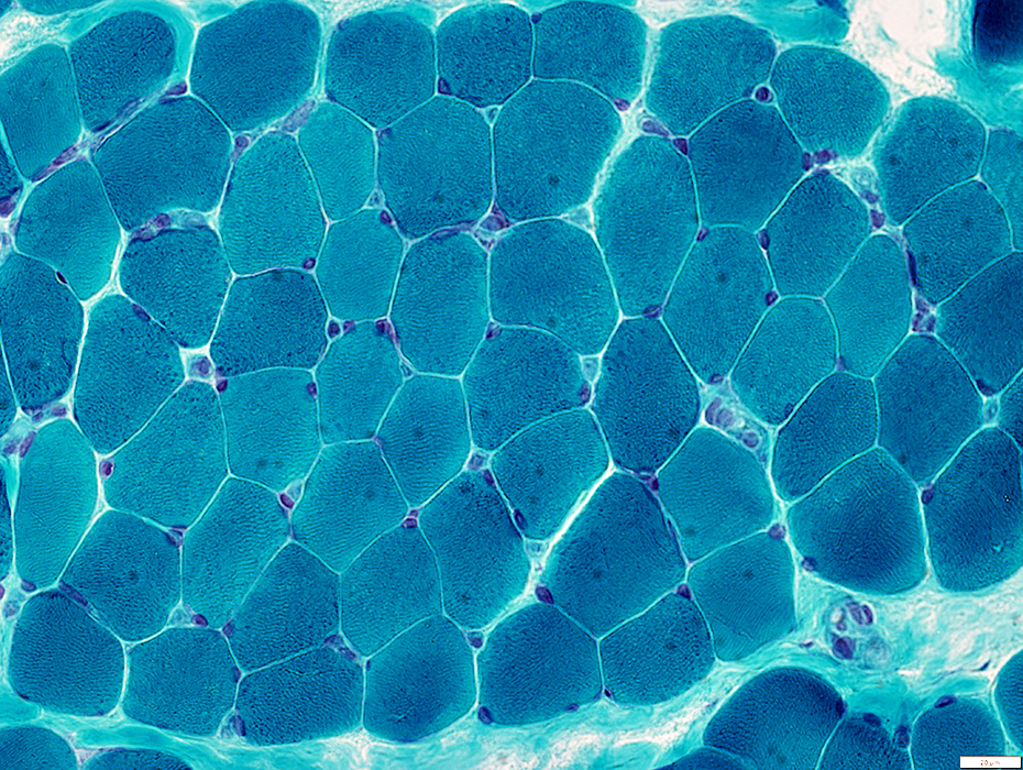 Gomori trichrome stain |
Mildly irregular internal architecture, especially in darker stained fibers
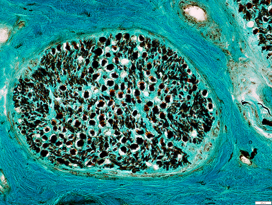
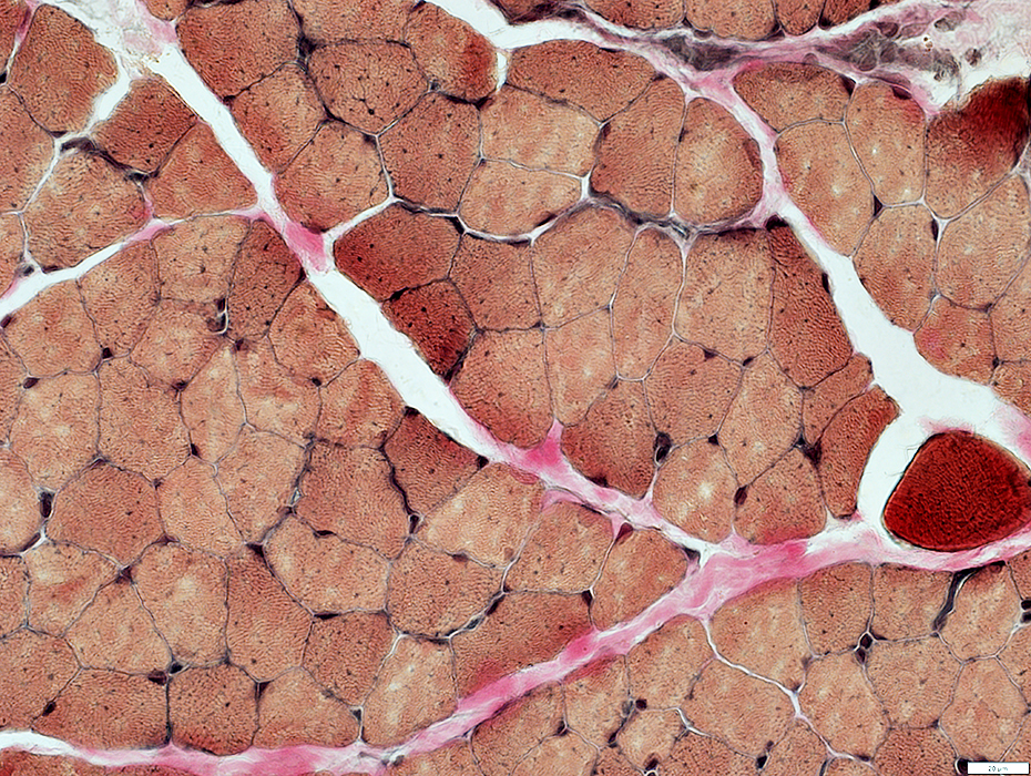 VvG stain |
Muscle Fiber Morphology
Mildly irregular internal architecture, with linearization in paler stained fibers
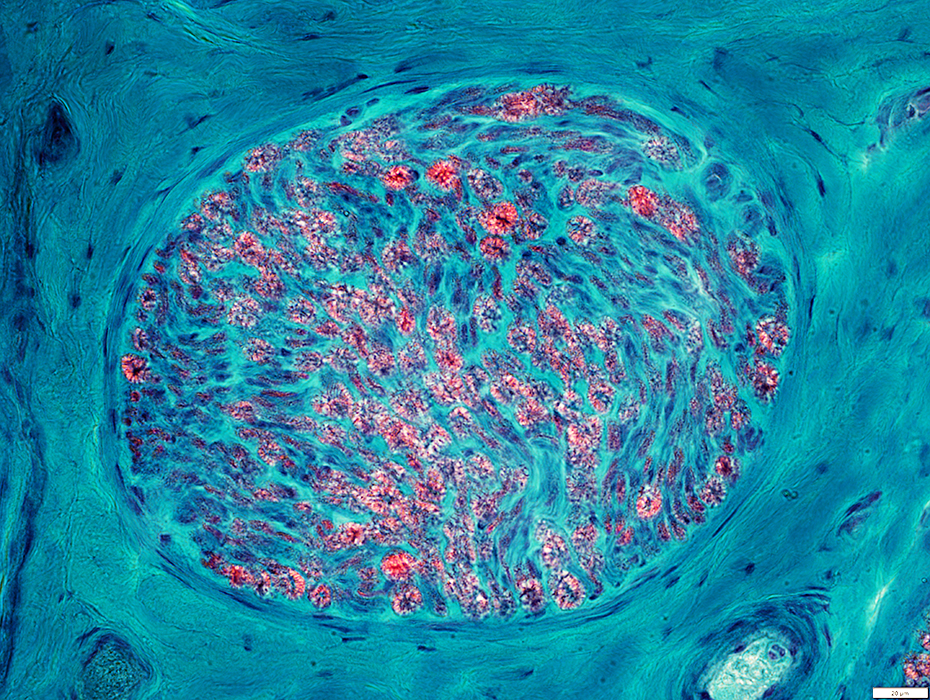
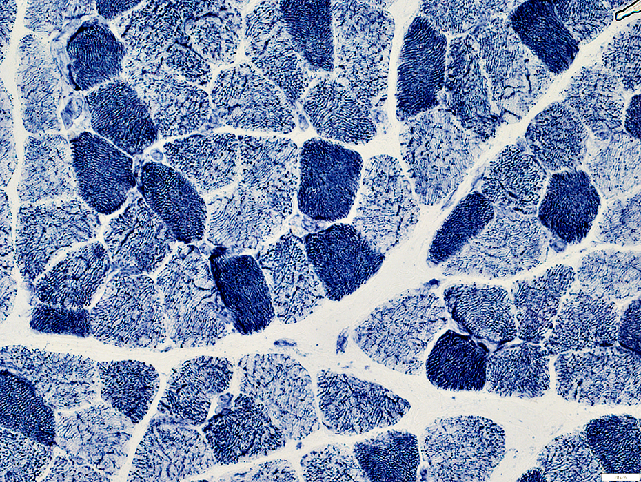 NADH stain |
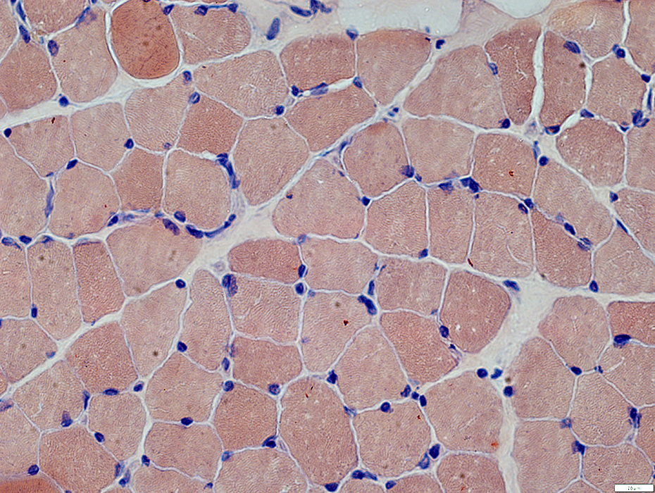 Congo red stain |
Size: Often large
Shapes: Irregular
 Congo red stain |
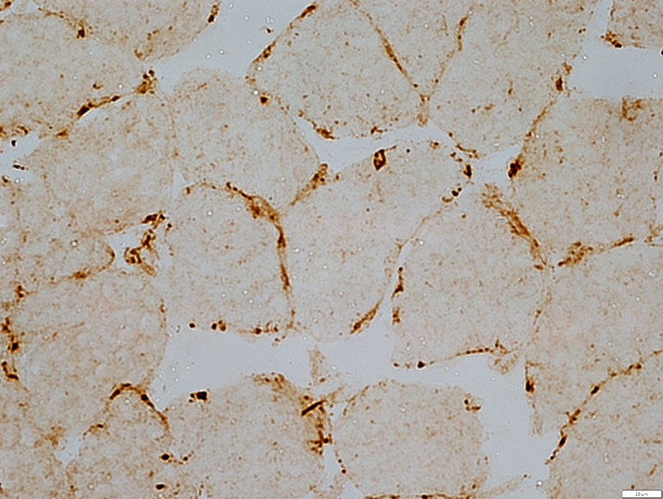
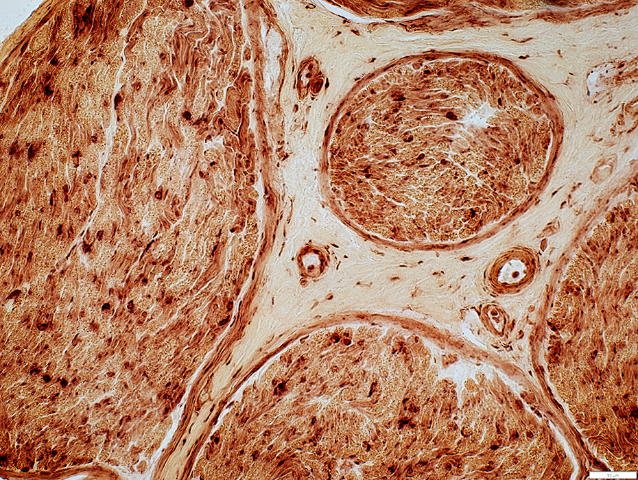
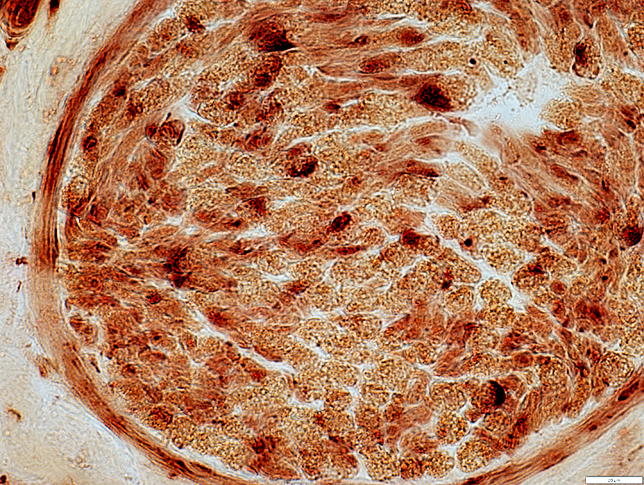
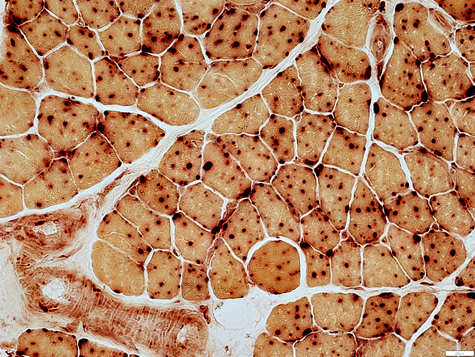 Acid phosphatase stain |
Several large, dark granules present in most muscle fibers
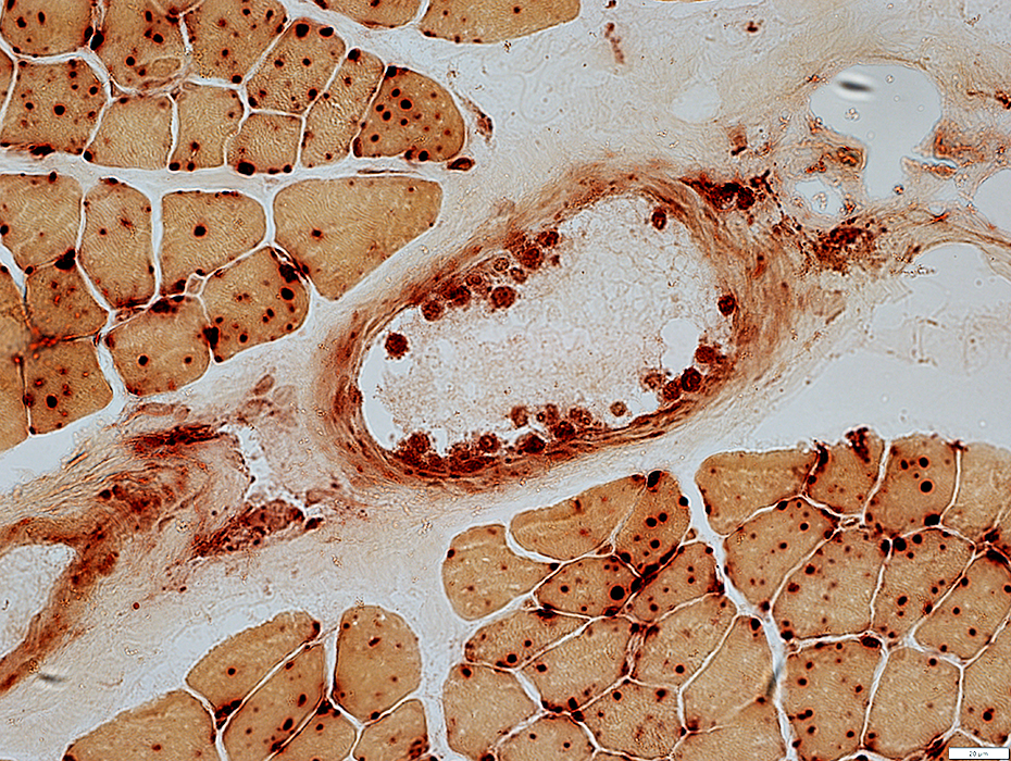
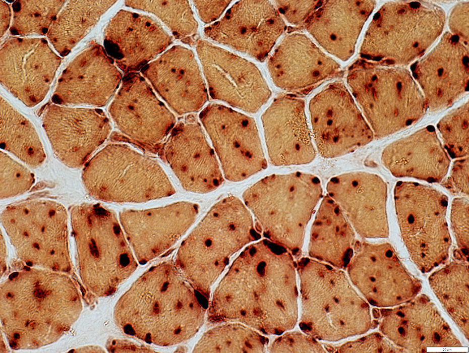 Acid phosphatase stain |
 Acid phosphatase stain |
Granules present in vessel walls
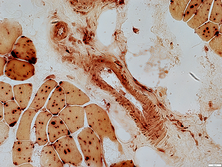
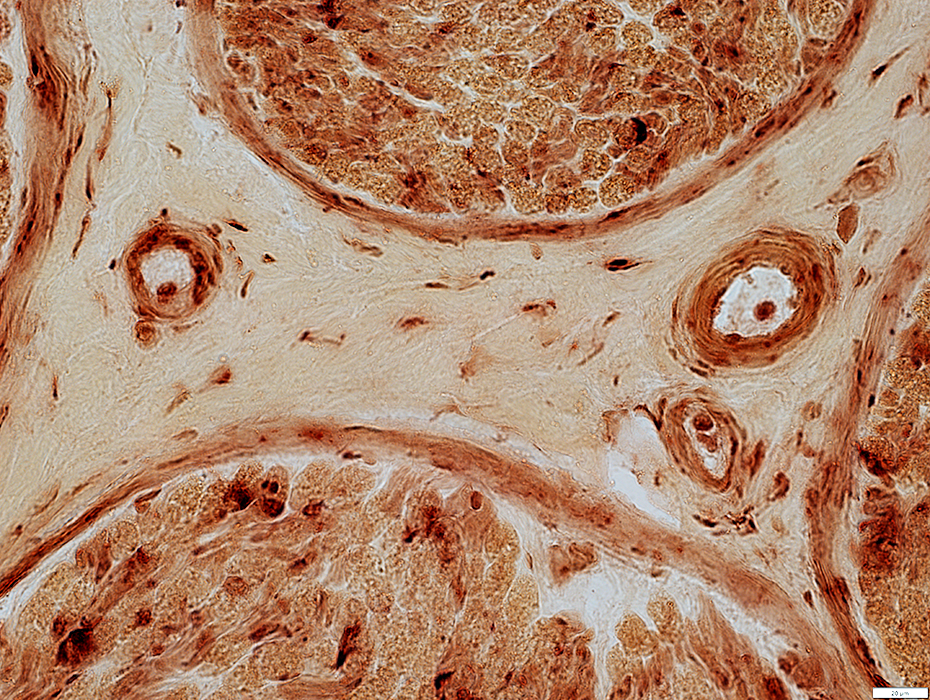
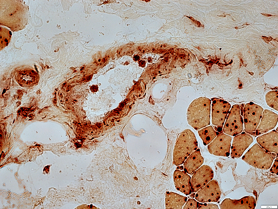 Acid phosphatase stain |
Granules present in vessel walls
Histiocytes adherent to vessel walls
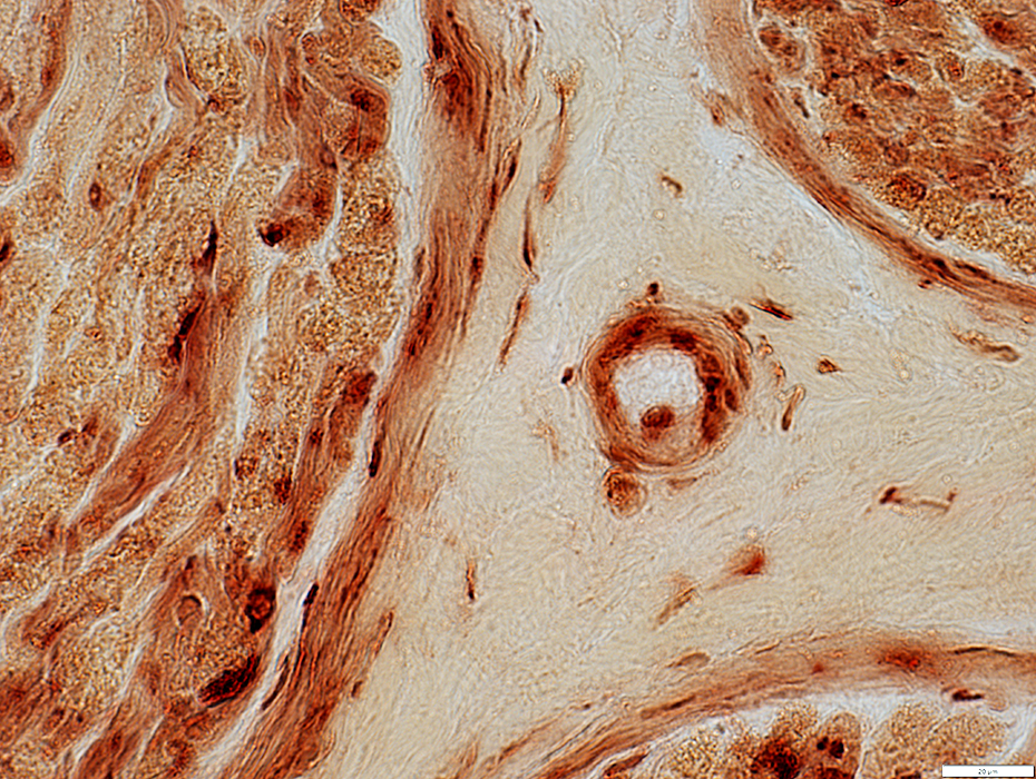
 Acid phosphatase stain |
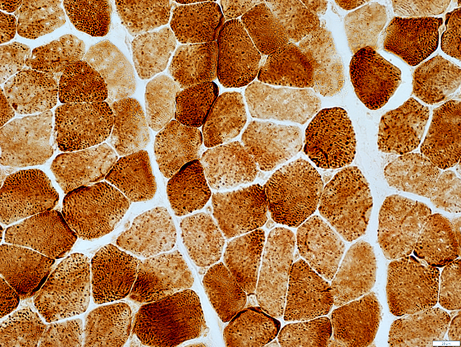
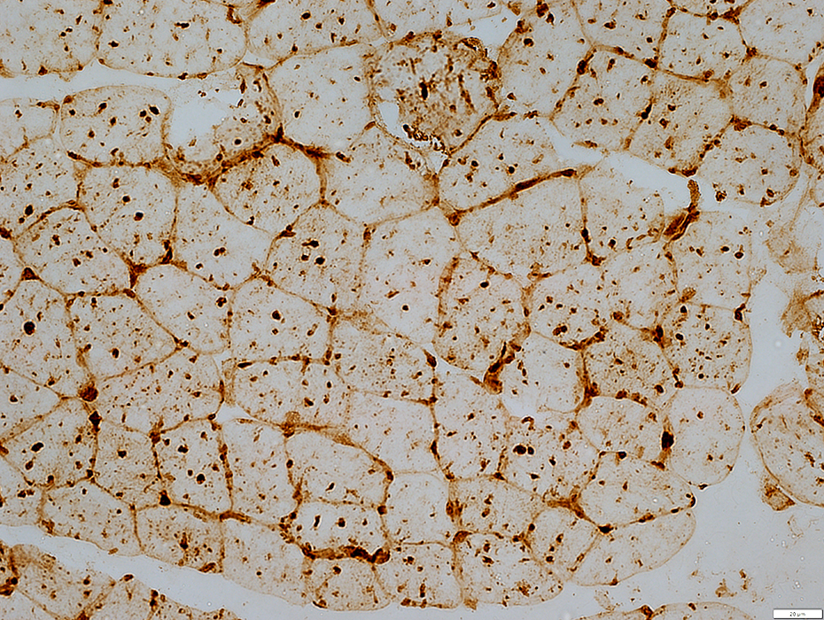 LAMP2 stain |
LAMP2 granules are prominent in muscle fiber cytoplasm
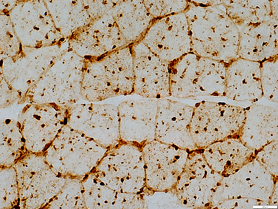 LAMP2 stain |
Control muscle
LAMP2 granules are not prominent in muscle fiber cytoplasm
 LAMP2 stain |
 COX stain |
No mitochondrial pathology
 SDH stain |
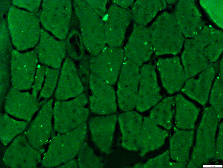
Batten Muscle: Autofluorescent Storage Material 1
 |
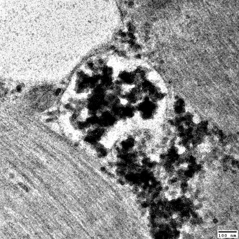
Batten Muscle: Aggregates, Ultrastructure
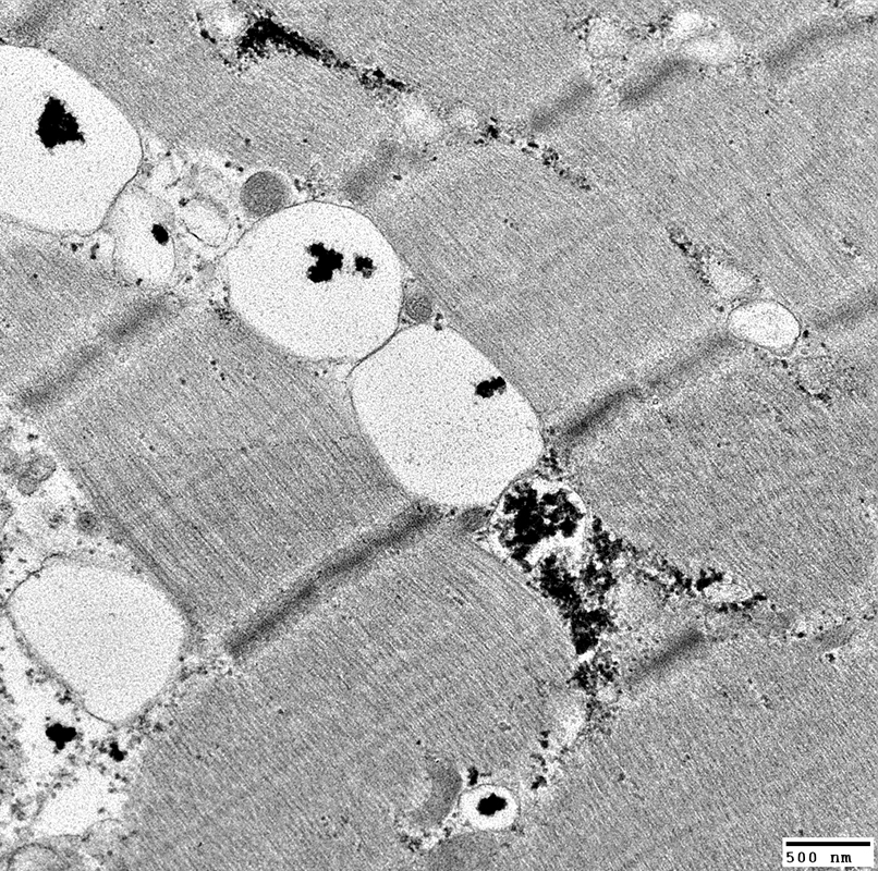 From: R Schmidt |
Dark
Shape: Irregular
Locations
Within membrane bounded vacuoles
Free in Cytoplasm
 From: R Schmidt |
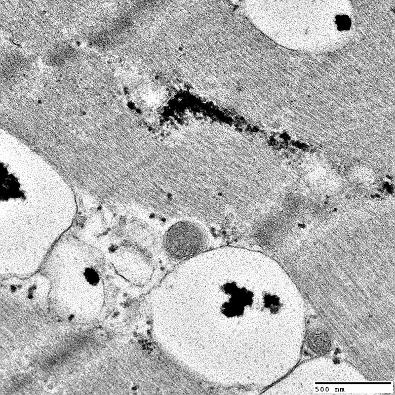
 From: R Schmidt |
Dark
Shape: Irregular
Locations
Within membrane bounded vacuoles
Free in Cytoplasm
 From: R Schmidt |
Batten disease: Nerve pathology
 H&E stain |
Normal endoneurium, perineurium & epineurium
 Gomori trichrome stain |
Mild loss
 VvG stain |
Normal numbers
 Neurofilament stain |
 Acid phosphatase stain |
Scattered in endoneurium
Present in walls of epineurial vessels
 Acid phosphatase stain |
 Acid phosphatase stain |
Present in walls of epineurial vessels
 Acid phosphatase stain |
Return to: Batten disease
Return to: Neuromuscular Home Page
References
1. Biochem Biophys Res Commun 2009;382:247-51
8/10/2024