Myopathy with DNAJB6 mutations (LGMD 1 D)
|
Aggregate stains Muscle fibers Fiber types Other changes Vacuoles |
DNAJB6: Myopathic changes
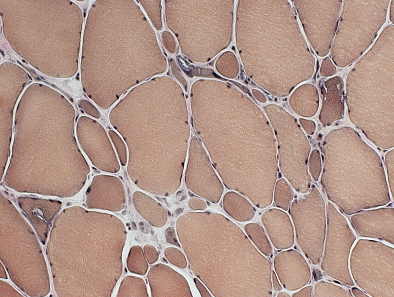
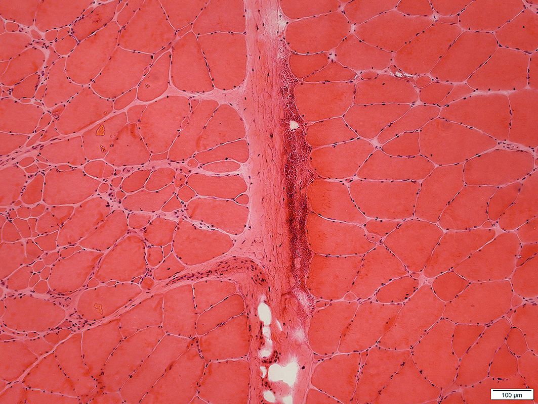 H&E stain |
 VvG stain |
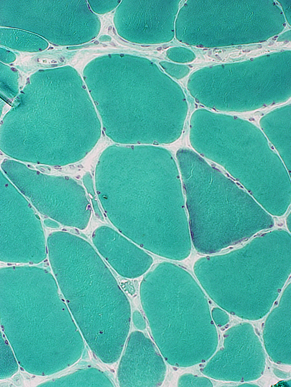
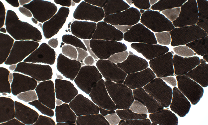
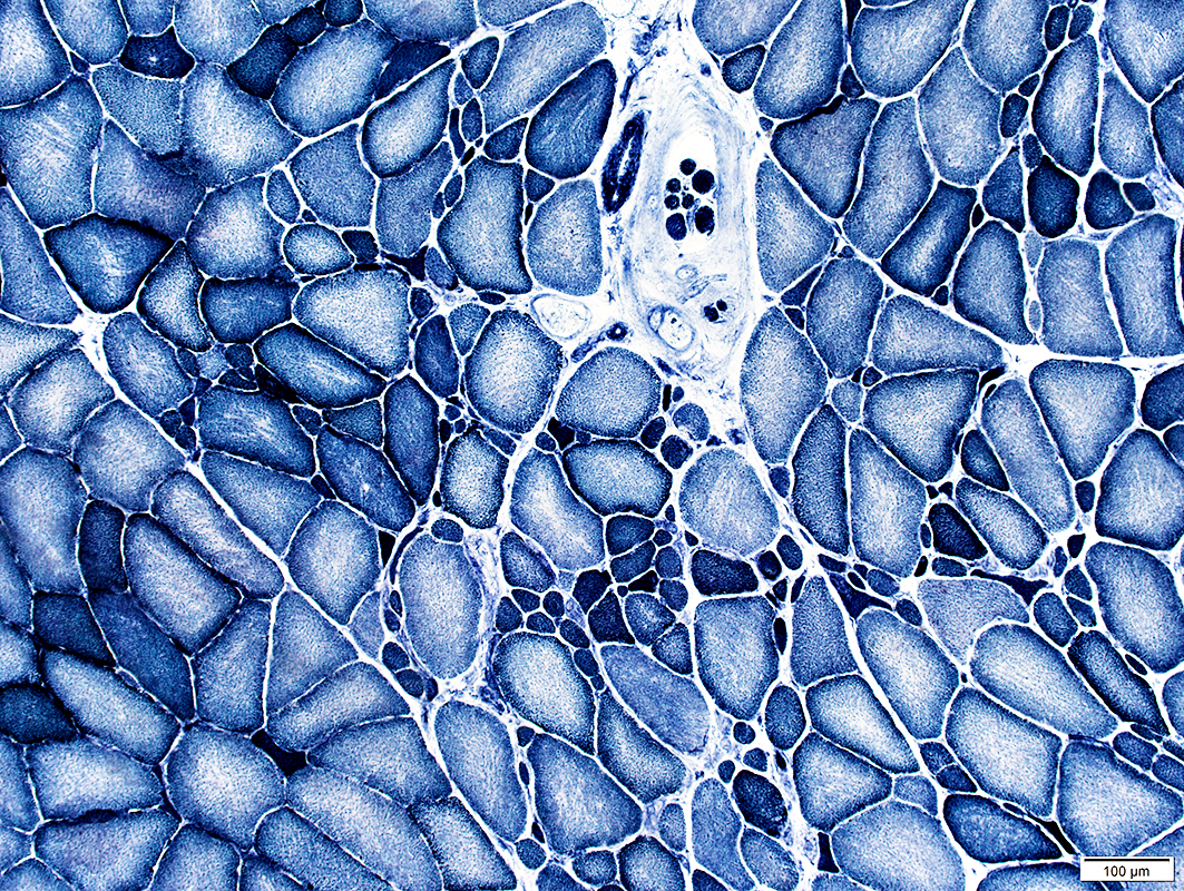 NADH stain |
Muscle fibers: Varied sizes
 H&E stain Large and small rounded muscle fibers. Fibers with vacuoles. Some internal nuclei, |
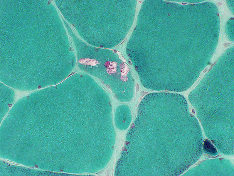
 Gomori trichrome stain Large and small rounded muscle fibers. Some fibers have vacuoles with red stained contents. |
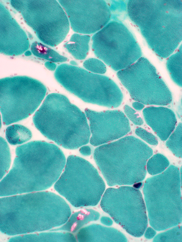 Gomori trichrome stain |
 Gomori trichrome stain |
|
 VvG stain Large and small rounded muscle fibers. Some fibers have vacuoles. |
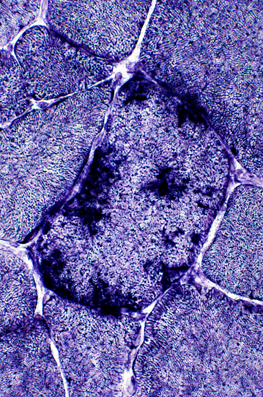
Muscle fiber types: Type 1 small; Type 2 predominant |
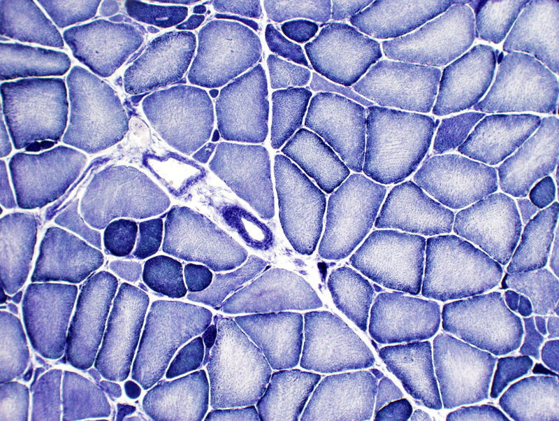
 ATPase pH 9.4 Varied fiber size: Type 1 & some Type 2 fibers are small. The largest fibers are Type 2. |
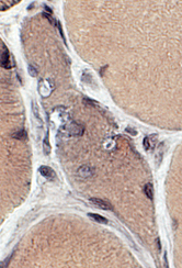
DNAJB6: Other |
|
 NADH stain Internal architecture is unremarkable. Small type 1 muscle fibers are dark stained. |
|
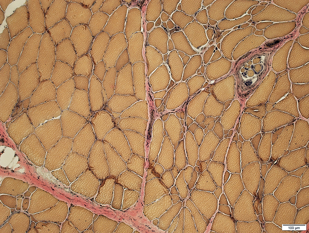
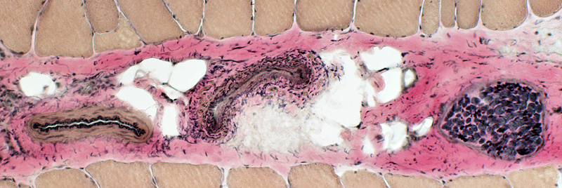 VvG stain Perimysial artery (left), vein (middle) & intramuscular nerve are normal. |
|
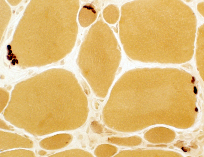 Esterase stain Neuromuscular junctions are multi-segmented. |
|
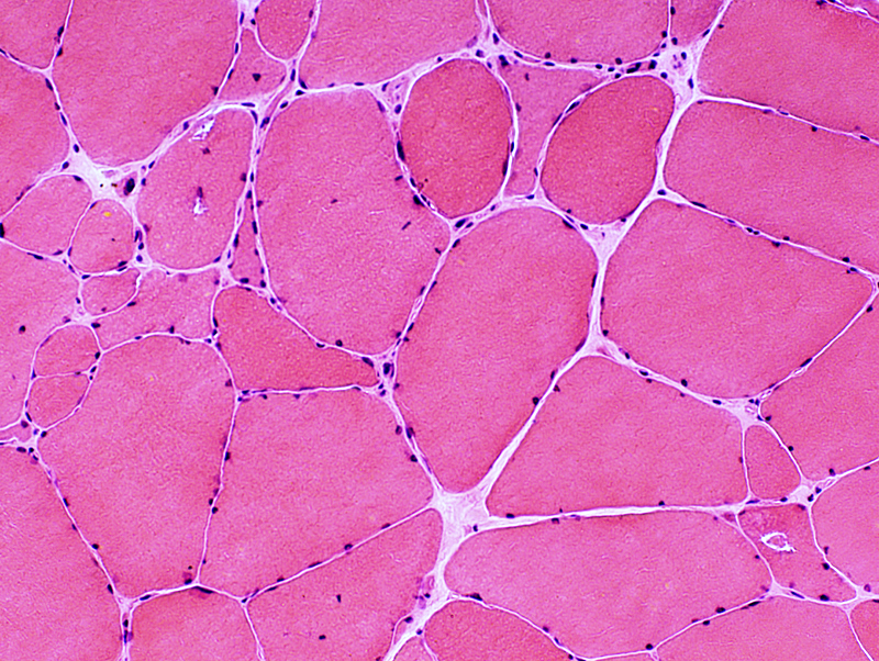
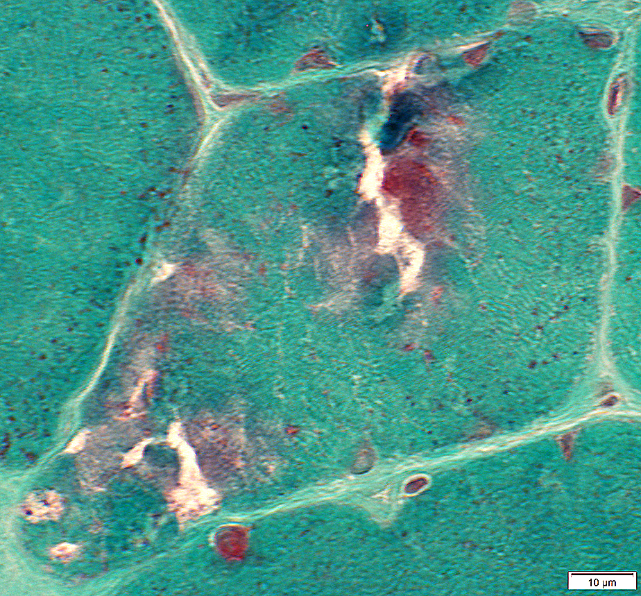
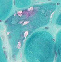
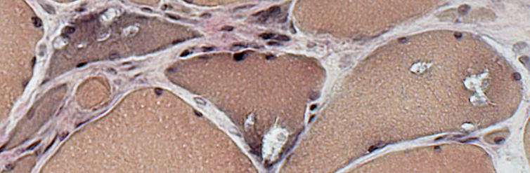
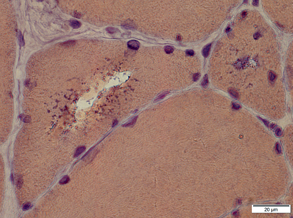
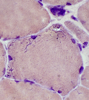
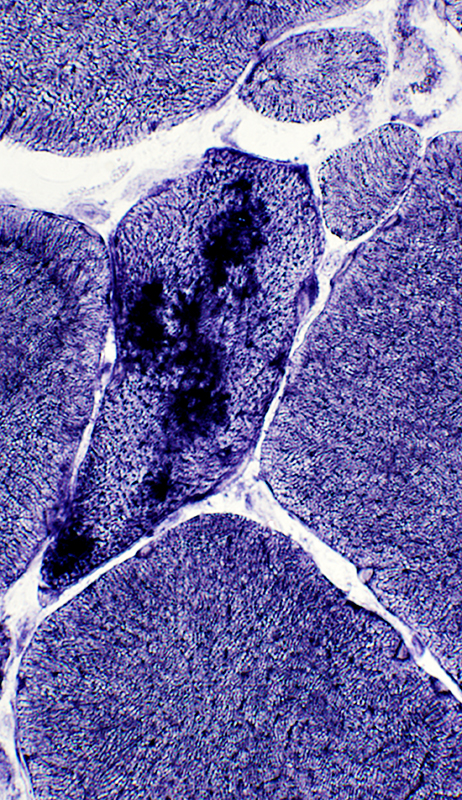
DNAJB6 myopathy: Vacuoles in large & small muscle fibers |
|
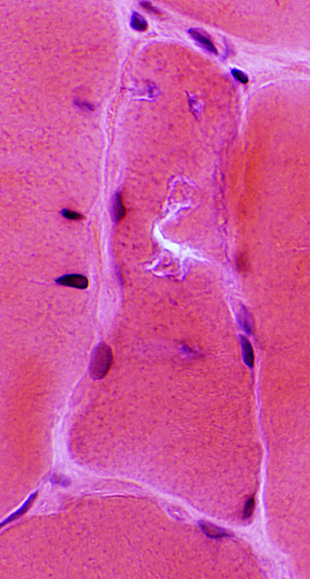 H&E stain |
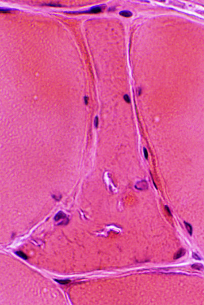 H&E stain |
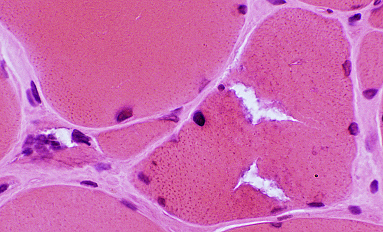 H&E stain |
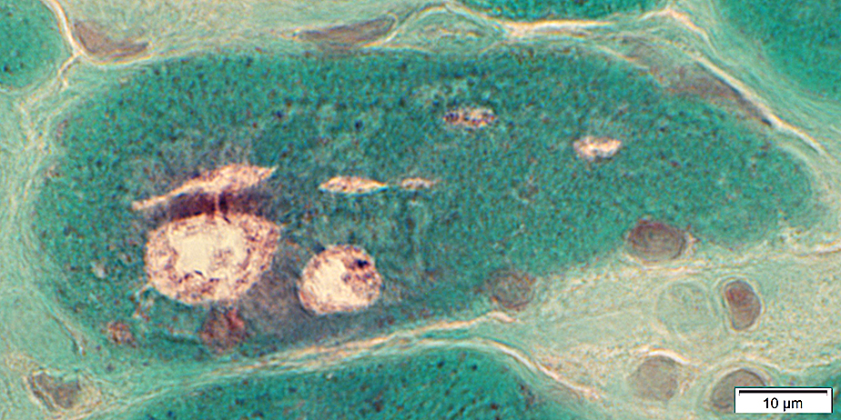 Gomori trichrome stain |
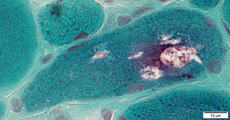 Gomori trichrome stain |
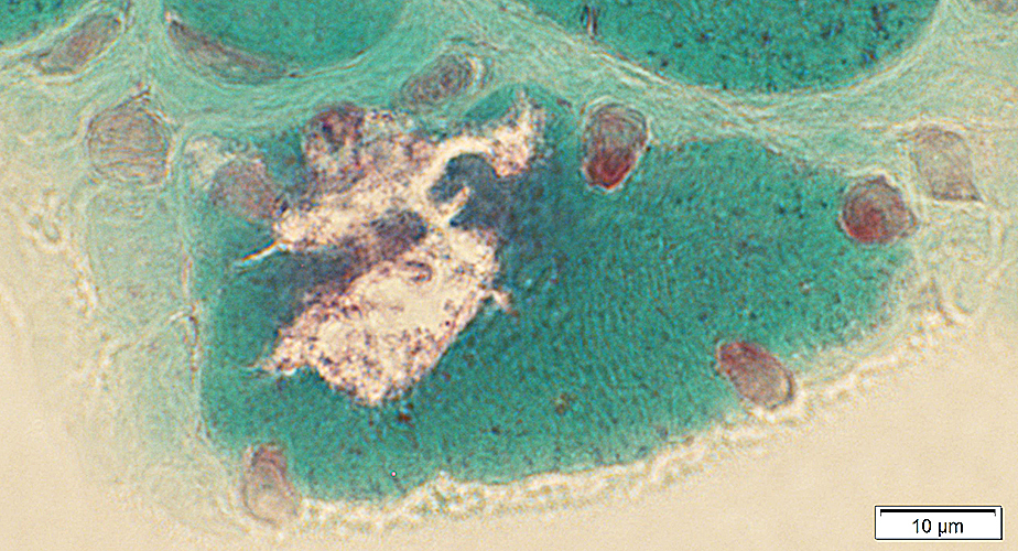 Gomori trichrome stain |
 Gomori trichrome stain |
|
 Gomori trichrome stain |
 VvG stain |
 Congo red stain |
|
Vacuoles associated with basophilic granular debris |
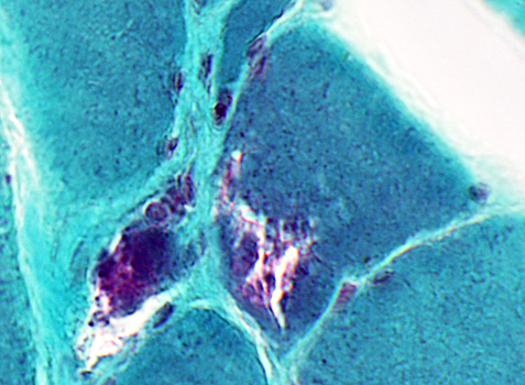
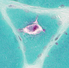
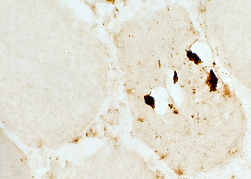
DNAJB6 myopathy: Aggregates in muscle fibers
 AMPDA stain Irregular aggregates: May be associated with vacuoles |
 AMPDA stain |
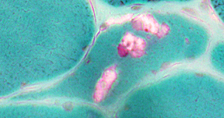
 SMI-31 Small & Large SMI-31+ aggregates in muscle fiber (Right) |
|
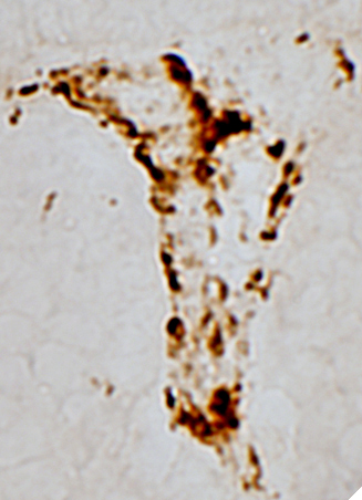 TDP-43 TDP-43+ aggregates in muscle fiber cytoplasm |
TDP-43 Desmin stains cytoplasm in some small muscle fibers but no aggregates. |
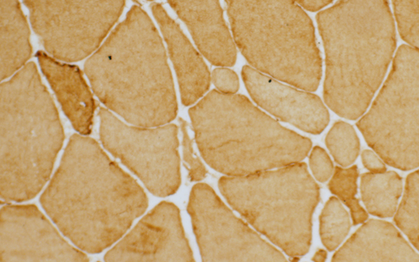
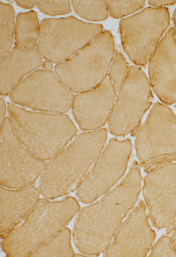 Caveolin-3 Caveolin-3 is reduced on sarcolemma. |
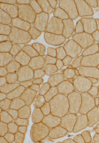 Caveolin-3 Control muscle: Normal caveolin-3 staining. |
Return to DNAJB6 myopathy
2/7/2015