FKRP Mutations
|
Muscular dystrophy Congenital (MDDGB5) Limb Girdle 2I (MDDGC5) |
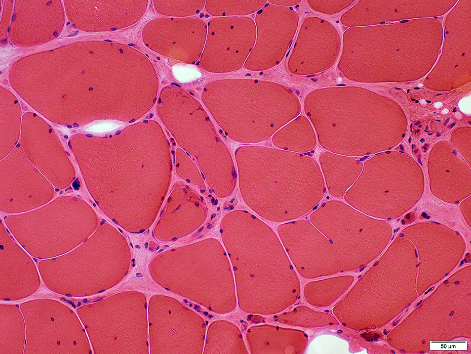
FKRP: Congenital Muscular Dystrophy (MDDGB5)
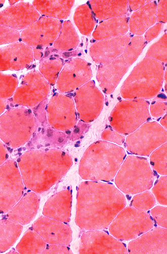 H & E stain |
 |
 |
|
Myopathy: Mild active & chronic changes Necrosis & Regeneration: Scattered Fiber size: Varied Small fibers: Rounded Endomysial connective tissue: Normal or Slightly increased Internal nuclei: Some fibers |
||
 H & E stain |
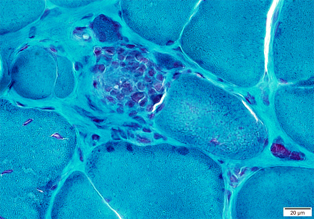
 Gomori trichrome stain |
 Acid phosphatase stain Necrotic muscle fibers: Phagocytosis |
 Esterase stain |
|
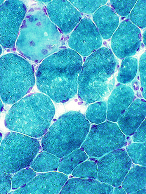
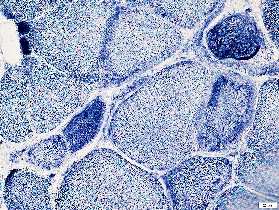
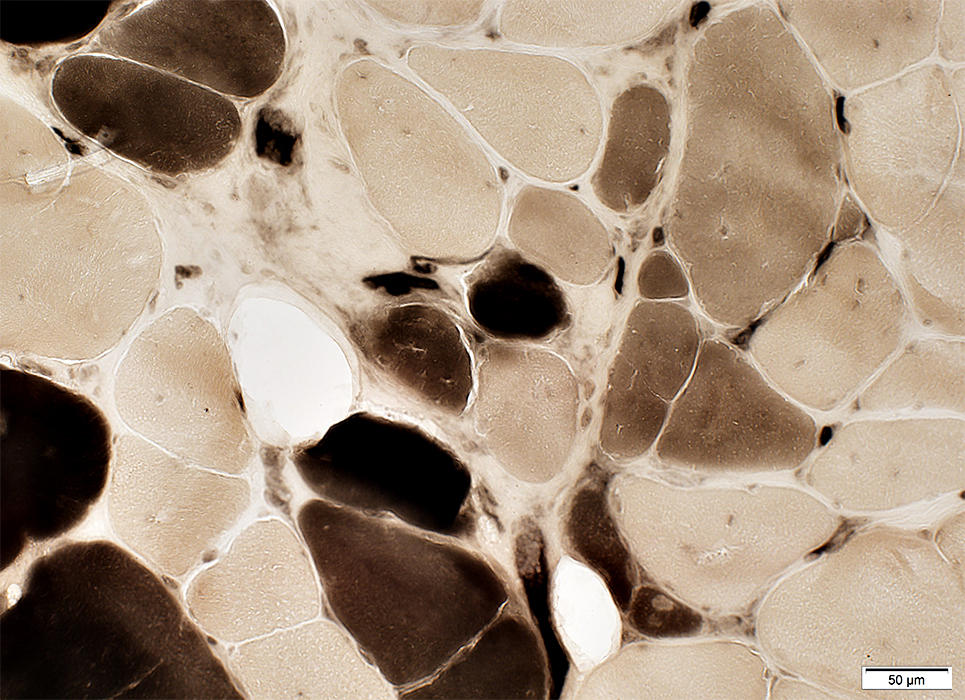
Necrotic (pale) muscle fiber (Dark arrow) Type I fiber predominance 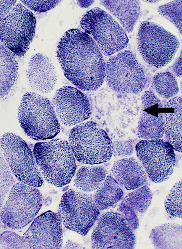 NADH stain |
Regenerating, immature muscle fibers Coarse internal architecture (White Arrow) 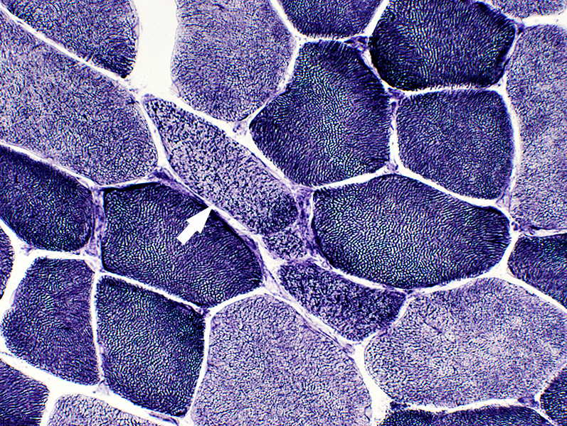 NADH stain |
Fiber types
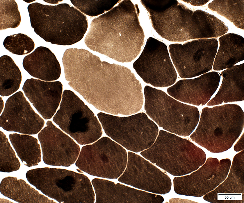
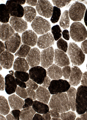 ATPase pH 9.4 Type I fiber predominance |
 ATPase pH 4.3 Scattered 2C fibers |
Neuromuscular Junctions: Normal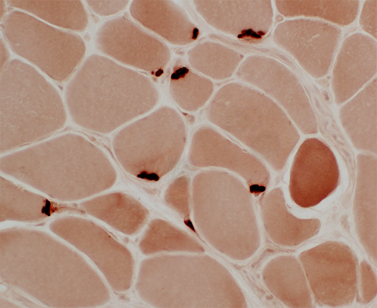 Esterase stain |
Intramuscular nerves: Normal VvG stain |
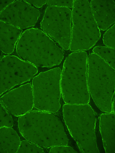
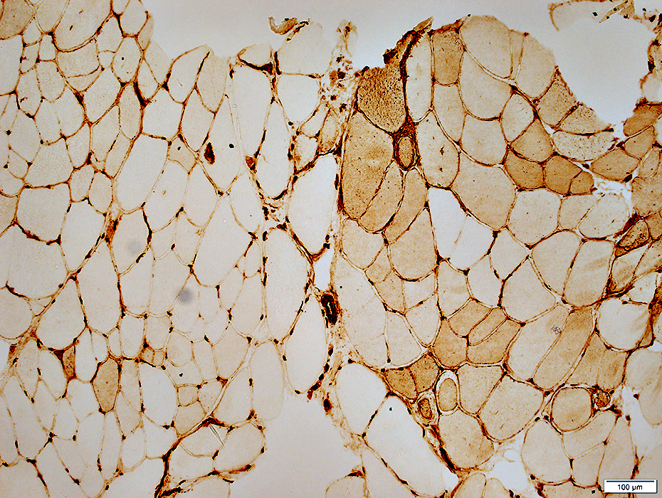
 α Dystroglycan FKRP mutations: Absent α-Dystroglycan around muscle fibers |
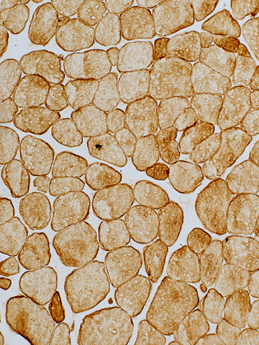
 α Dystroglycan Control muscle: Normal α-Dystroglycan around muscle fibers |
 Desmin stain Desmin: Stains cytoplasm in small regenerating fibers |
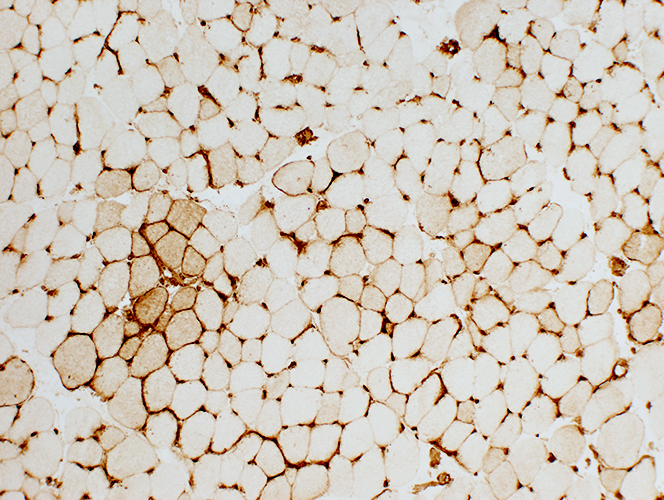 MHC Class I stain MHC-I is abnormally present on clusters of muscle fibers |
 MHC Class I stain |
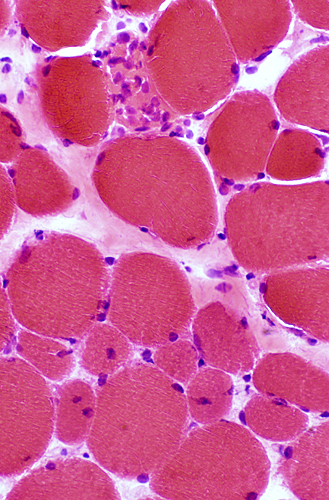
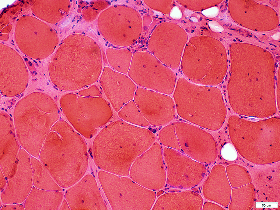
FKRP: Limb-Girdle Muscular Dystrophy 2I (LGMD 2I; MDDGC5)
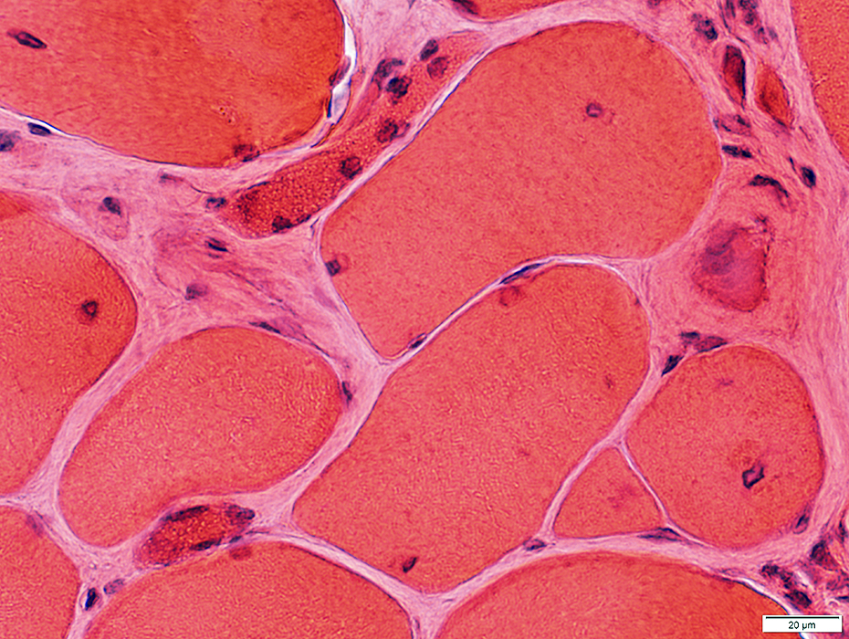
 H&E stain |
Fiber size: Varied; Hypertrophic & Small
Nuclei: Some are Internal or Large
Partially fused (or Split) fibers
Necrosis & Regeneration: Scattered
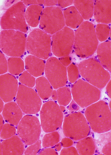
 H&E stain |
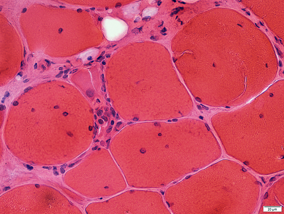 H&E stain |
Necrosis & Regeneration: Scattered
Immature muscle fibers
Internal architecture: Coarse
Nuclei: Large
Size Intermediate or Small
 H&E stain |
 Gomori trichrome stain |
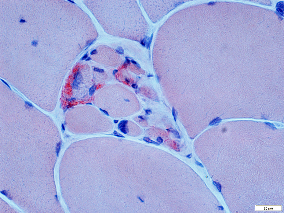
Necrosis: Muscle fibers replaced by phagocytic cells
Immature muscle fibers: Coarse internal architecture
Internal architecture: Coarse
Nuclei: Large
Size Intermediate or Small
 NADH stain |
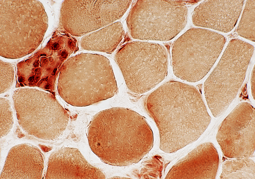
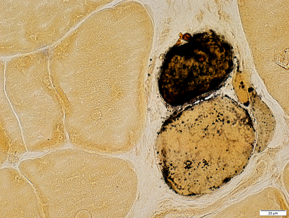 Alkaline phosphatase stain |
Immature muscle fibers: Cytoplasm stains for Alkaline phosphatase
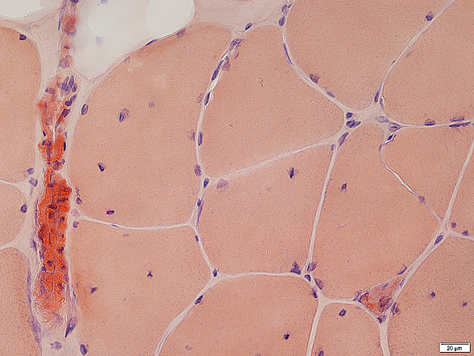
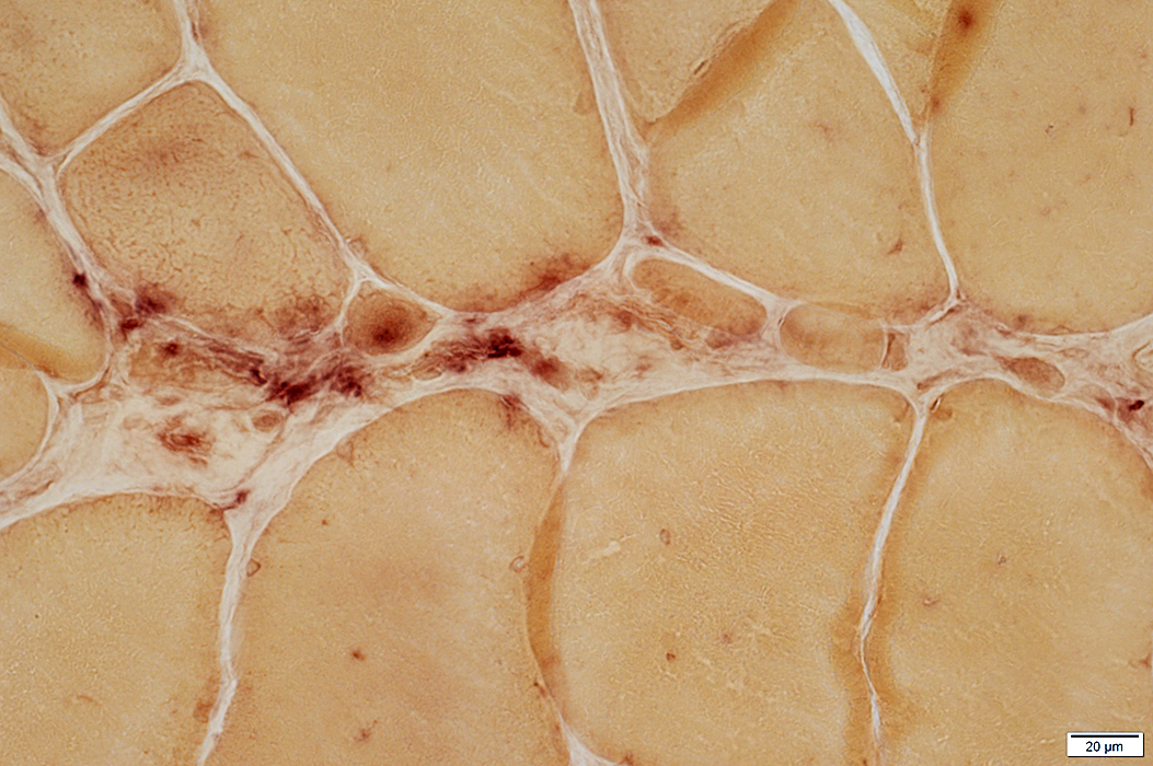
Necrotic fibers: Replaced by Acid phosphatase positive histiocytic cells
 Acid phosphatase stain |
FKRP: Ongoing Myopathy
Immature muscle fibers: Intermediate color with ATPase pH 4.3 stain
 ATPase pH 4.3 stain |
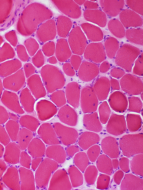
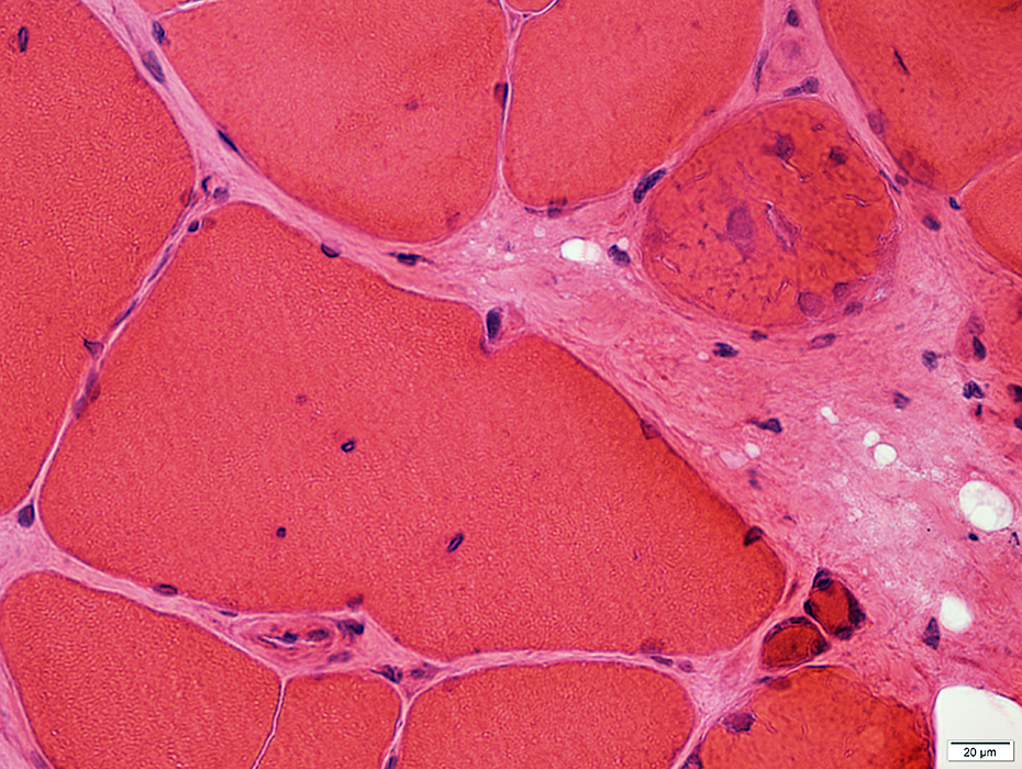
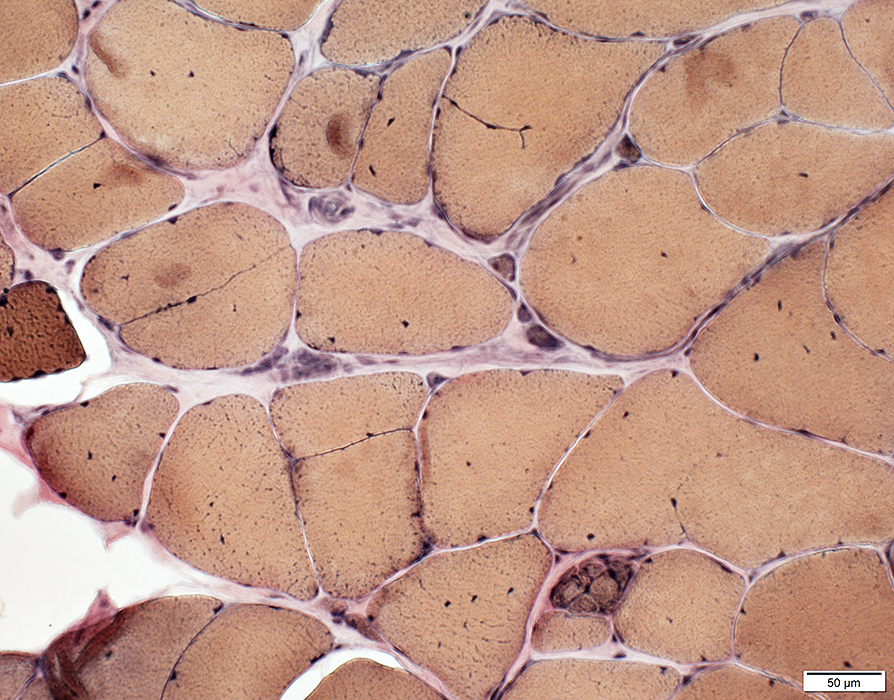
 H&E stain |
Endomysial connective tissue: Increased between muscle fibers
Partially fused (or Split) fibers
Nuclei: Some are Internal
Muscle fiber hypertrophy
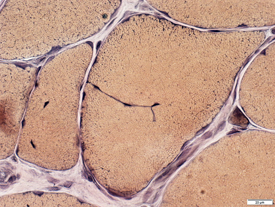
 VvG stain |
 Gomori trichrome stain |
Partially fused (or Split) fiber
 VvG stain |
 Congo Red stain |
Amyloid: Deposited in vessel walls & around small msucle fibers
 Congo Red stain |
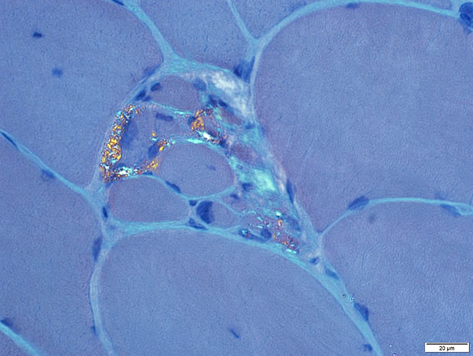 Congo Red stain |
FKRP: Other changes
 ATPase pH 9.4 stain |
FKRP: "Immune" changes
 MHC Class I stain |
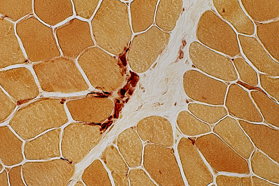
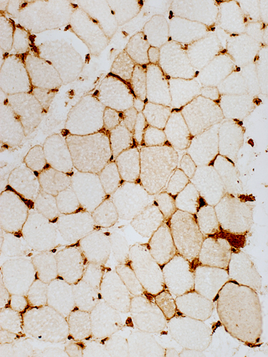
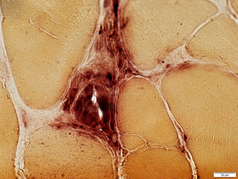
 Acid phosphatase stain |
Histiocytic cells (Acid phosphatase & CD4 positive) are present in Perimysial and Endomysial connective tissue
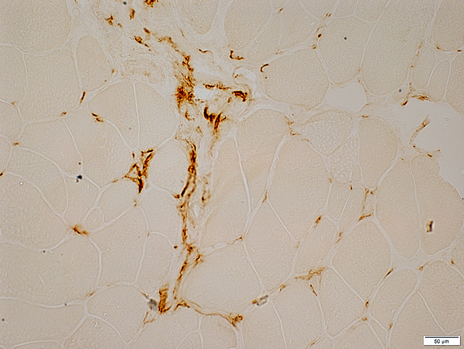 CD4 cell stain |
FKRP Myopathy
Reduced staining for carbohydrate moiety on surface of muscle fibers
Similar patterns seen with
Other hereditary disorders of α-Dystroglycan glycosylation
Graft vs Host myopathies
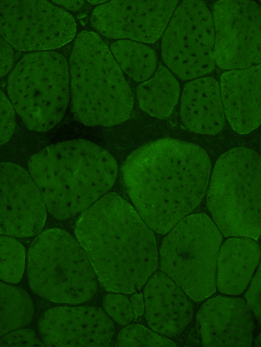
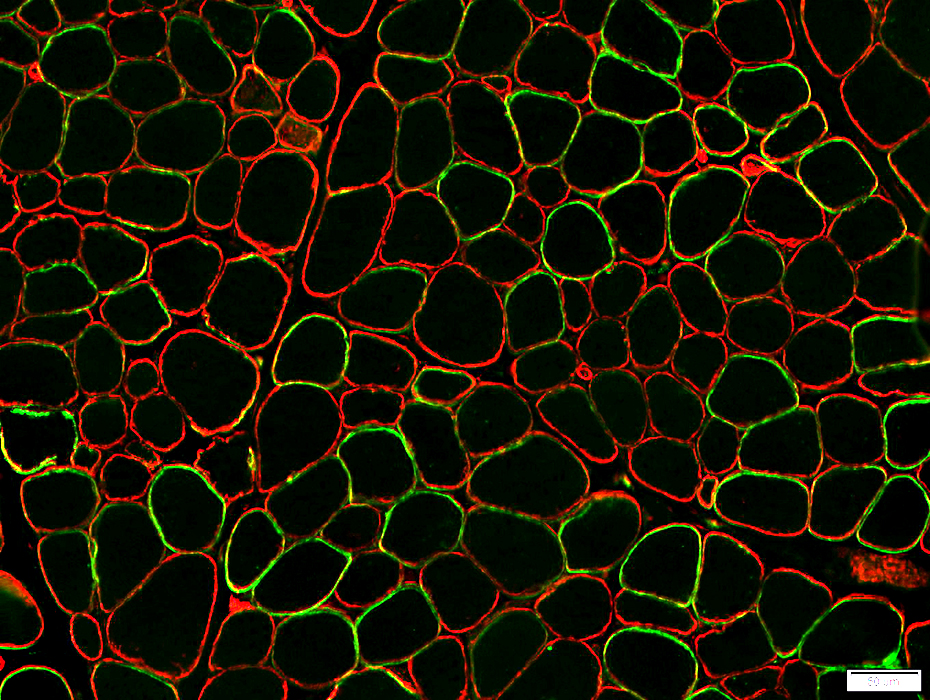 α-Dystroglycan - Green; Caveolin-3 - Red |
Reduced or Absent on the surface of some muscle fibers (Above)
Severely reduced on the surface of most fibers (Below)
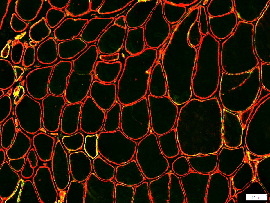 α-Dystroglycan - Green; Caveolin-3 - Red |
Return to Congenital Muscular Dystrophy.
Return to Neuromuscular syndromes
Return to Neuromuscular home page
10/17/2023