Graft vs Host Disease (GVHD)
|
General Myopathy Neuropathy |
Graft vs Host Disease: Neuromuscular, General
- Myopathy is more frequent than Neuropathy
- Tissue involvement
- No overall single pattern
- Individual patients often have disorders that involve more than one tissue
- May include
- Muscle
- Perimysium: Most common involvement
- Structure: Damaged
- Inflammation: Histiocytic cells
- C5b-9 deposits
- Endomysium
- C5b-9 deposits
- Cells: Histiocytic, scattered
- Endomysial capillaries
- Large
- Numbers: Reduced
- Glycosylation: Ulex stain reduced
- C5b-9 deposits
- Muscle fibers
- MHC I upregulation: Very common
- Glycosylation: Reduced α-Dystroglycan & Chondroitin-SO4
- Nuclei: Large; Abnormal distribution
- Aggregates: Lysosomal
- Perimysium: Most common involvement
- Nerve
- Epineurium
- Perineurium
- Vessels
- Axons
- Wallerian degeneration
- Axon loss: Differential fascicular
- Often secondary to involvemnt of another tissue type
- Myelin
- Muscle
- No overall single pattern
- Inflammatory cells: Usually histiocytic
GVH: Neuropathy
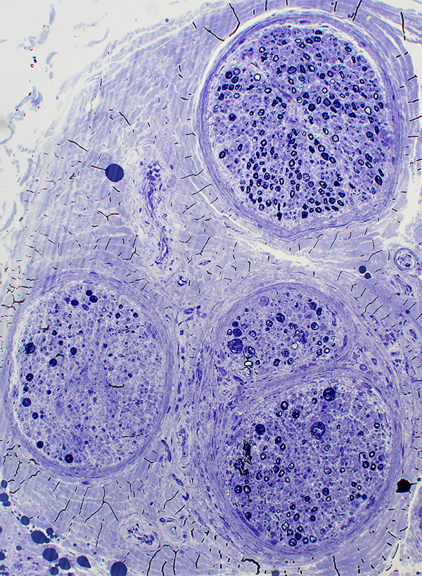 Toluidine blue stain Differential Fascicular Involvement: More axon loss in some regions than others |
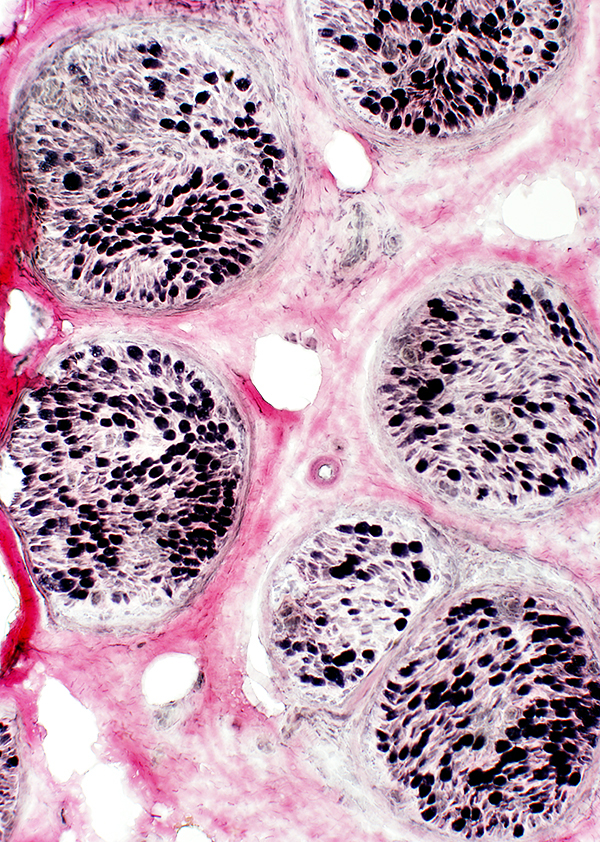 VvG stain |
 Neurofilament stain |
 ATPase pH 4.3 stain |
|
Wallerian Degeneration: Intrafascicular variation 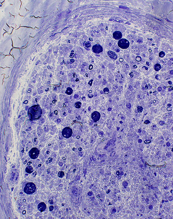 Toluidine blue stain |
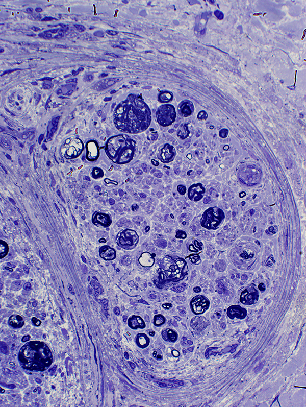 Toluidine blue stain |
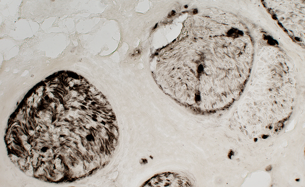
 Acid phosphatase stain |
|
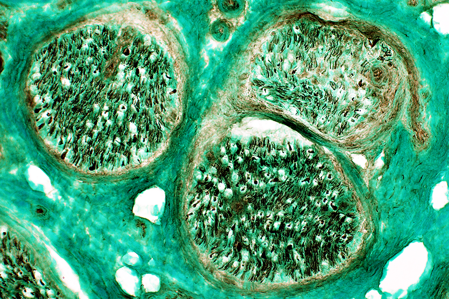
 Gomori trichrome stain |
|
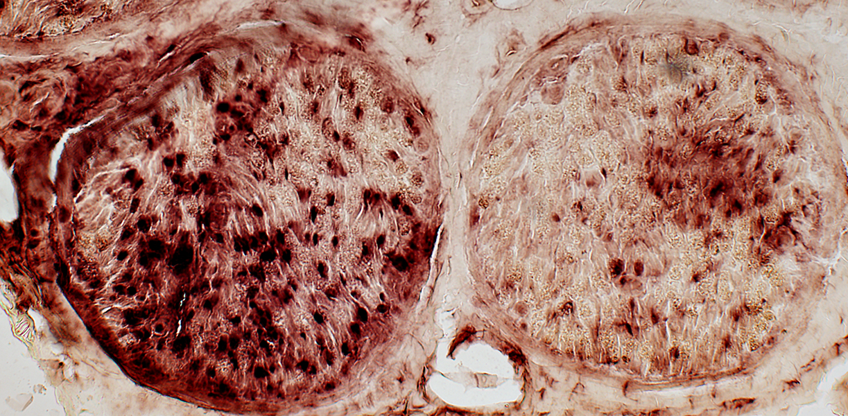
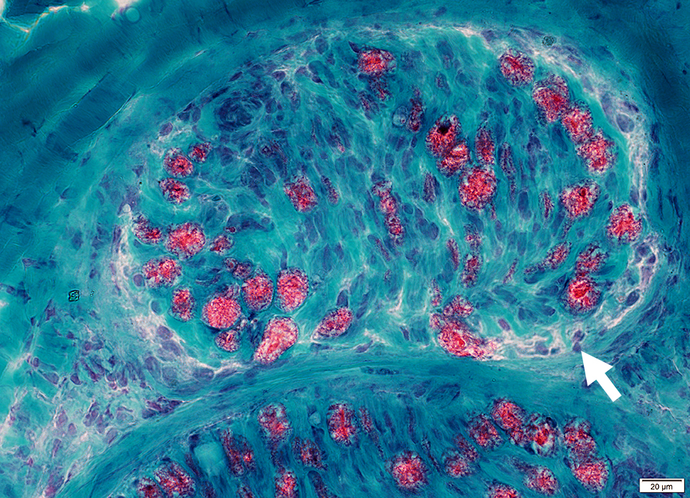
Immune damage: Connective tissue Perineurium (Arrow) & Epineurium: Damaged structure (Above) Epineurium: Alkaline phosphatase staining (Below) Perineurium: CD4 cells (Right) 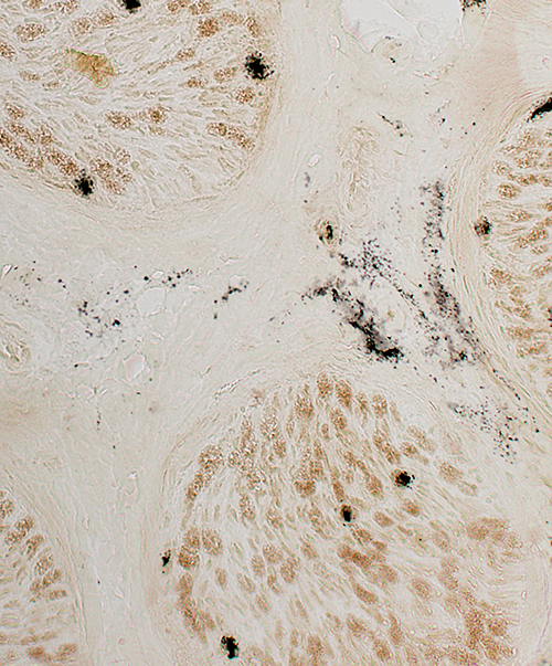 Alkaline phosphatase stain |
 CD4 stain |
 Gomori trichrome stain |
Pyknotic Nuclear Clumps
 NADH stain |
cGVH: Myopathy
|
Capillaries Muscle fibers Glycosylation Morphology Perifascicular Myonuclei MHC1 Perimysial connective tissue Histiocytes |
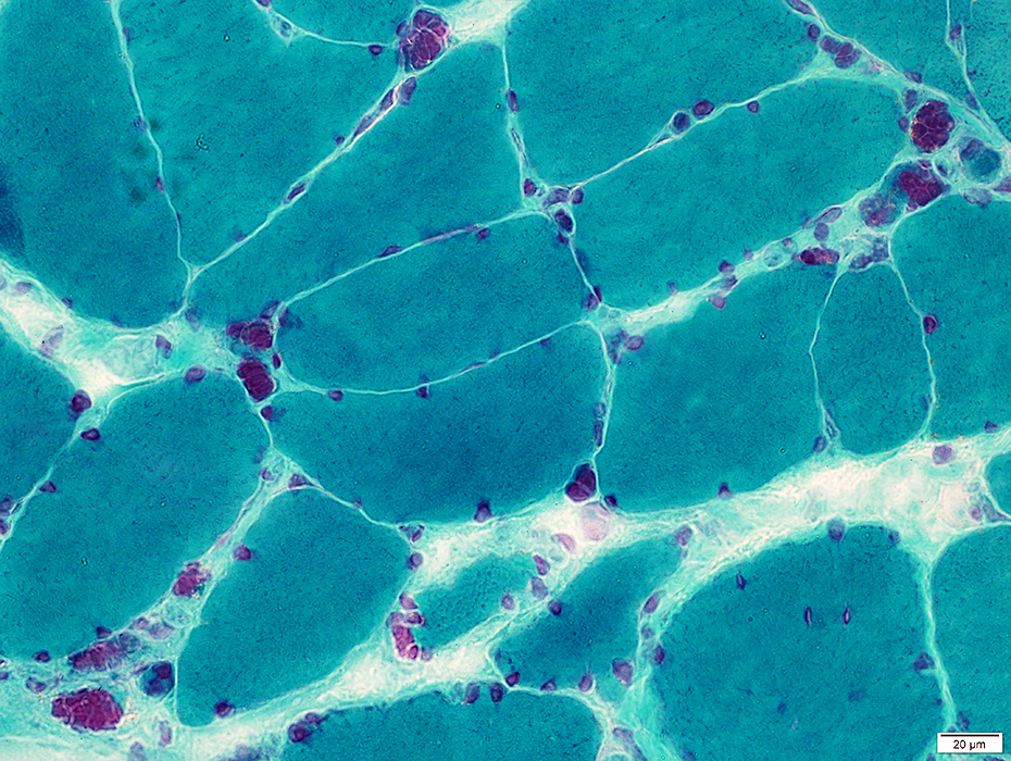
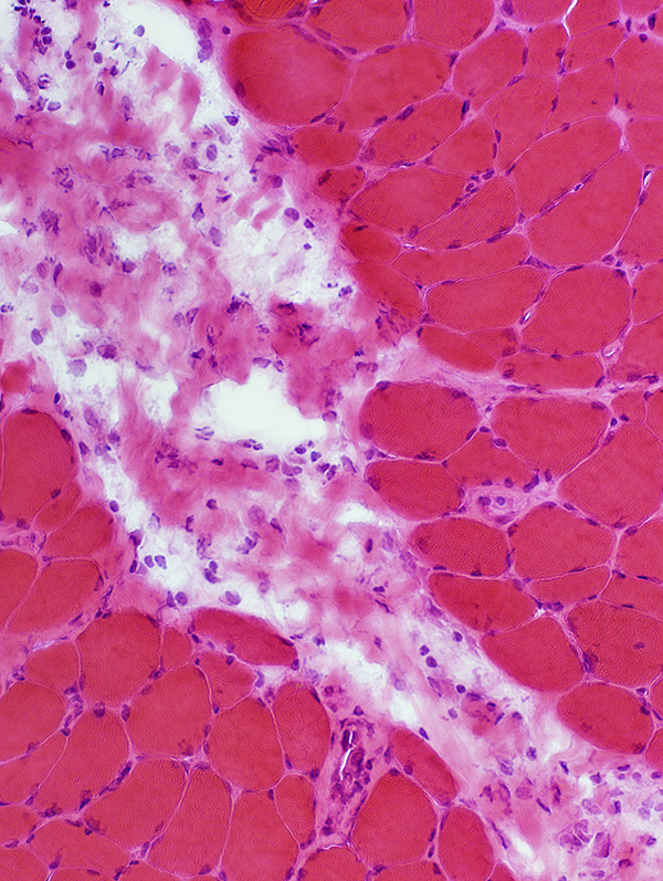
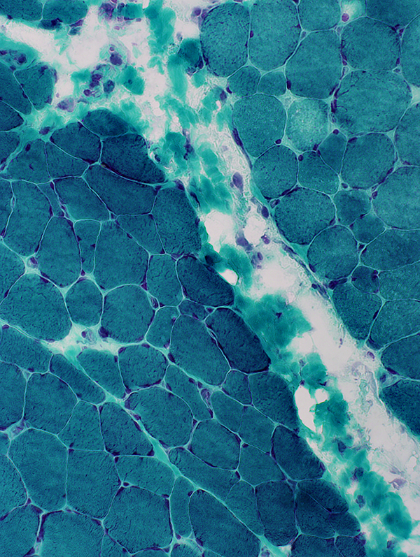
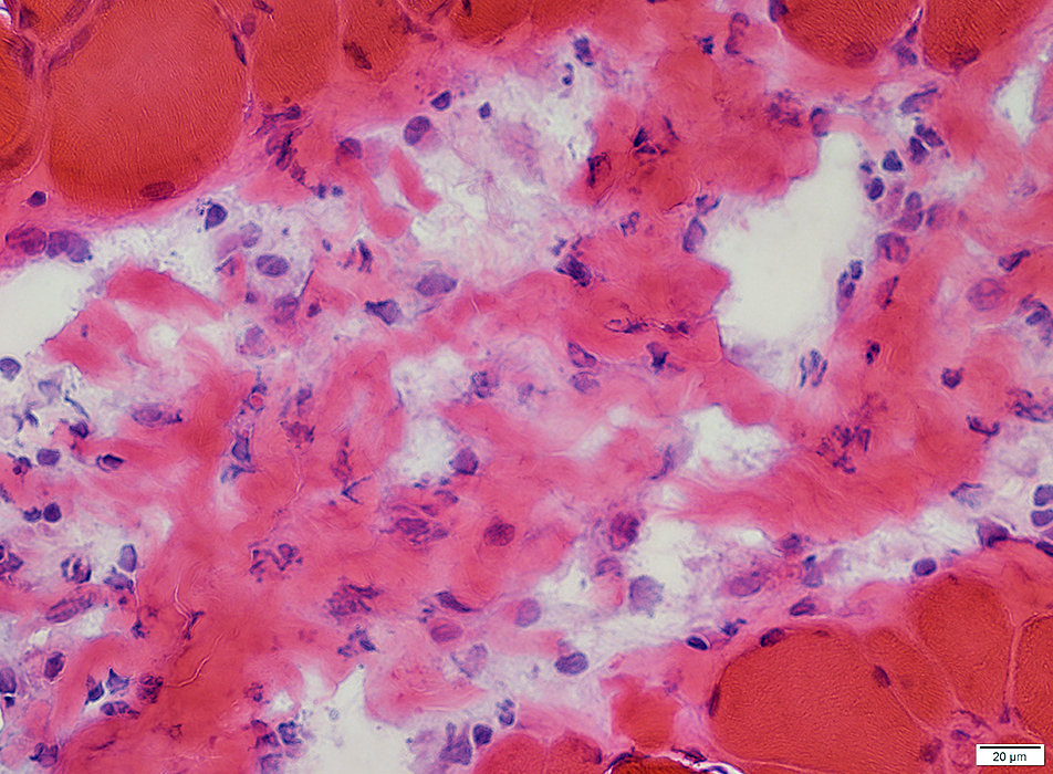
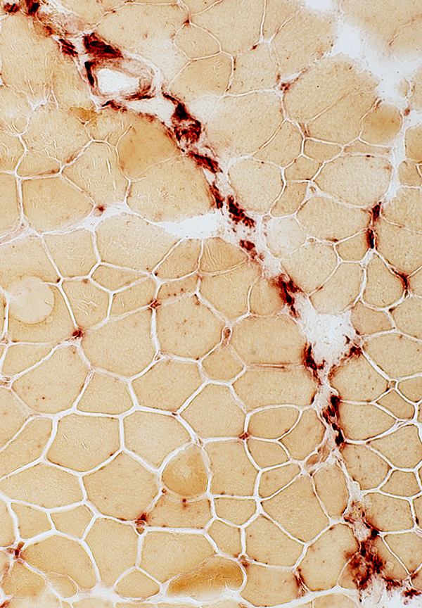
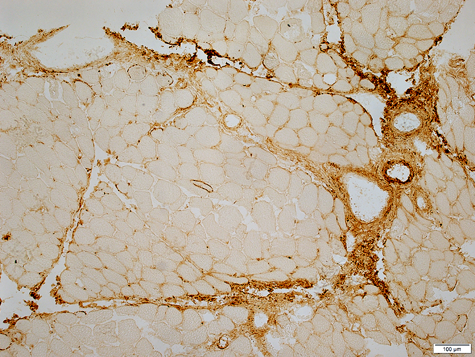
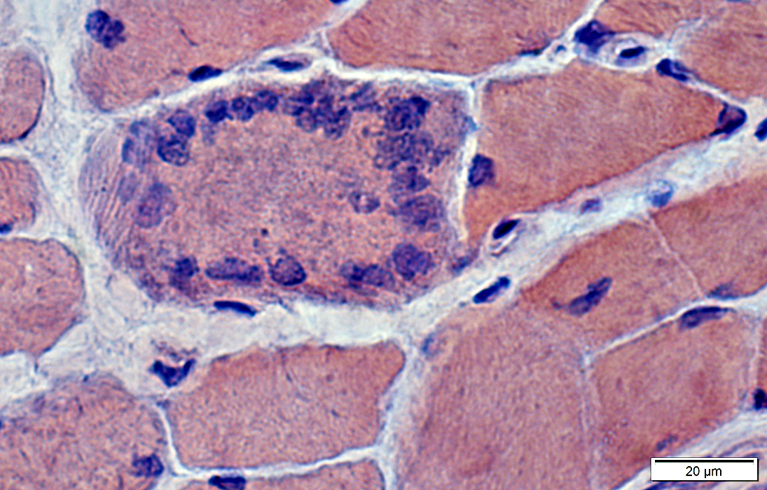
cGVH: Perimysial pathology
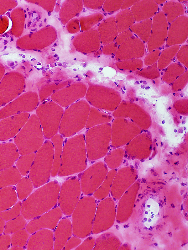 H&E stain |
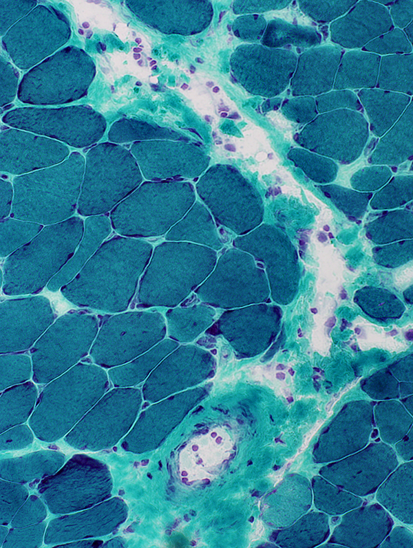 Gomori trichrome stain |
| Perimysium: Fragmented and damaged; Contains scattered large cells. Muscle fibers neighboring abnormal perimysium: Mildly small. | |
 H&E stain |
 Gomori trichrome stain |
 VvG stain |
 H&E stain |
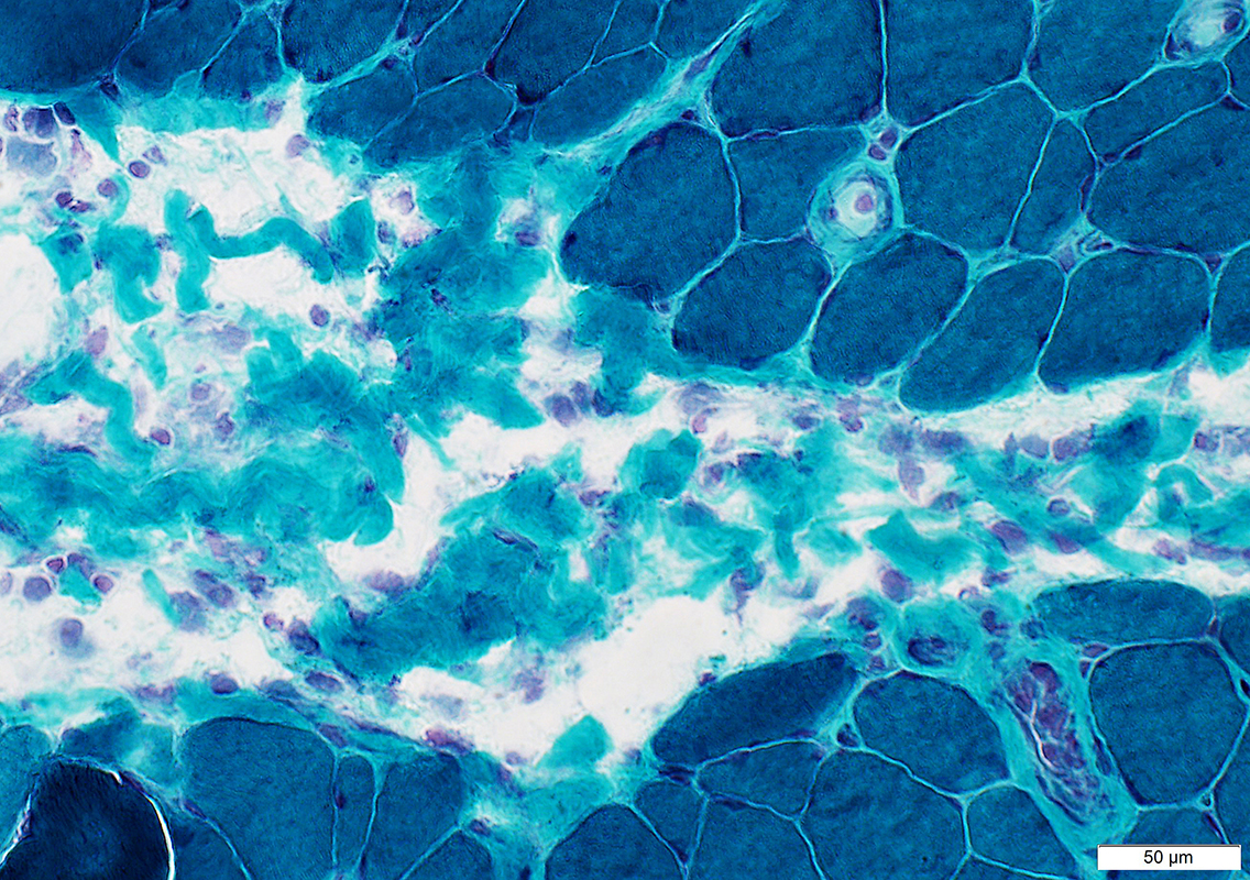 Gomori trichrome stain |
 Acid phosphatase stain |
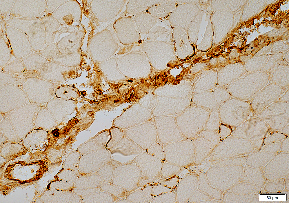
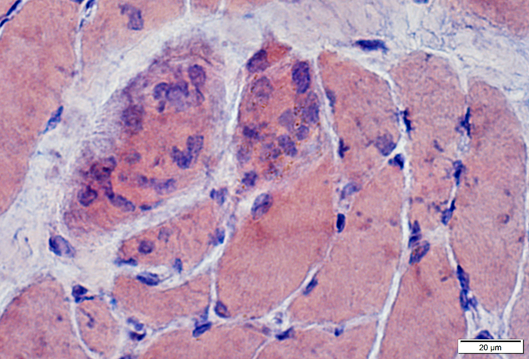
Muscle fibers at the edge of fascicles, near perimysium, tend to be smaller
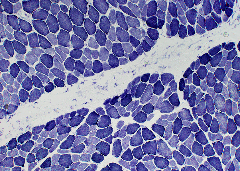 NADH stain |
 Acid phosphatase stain |
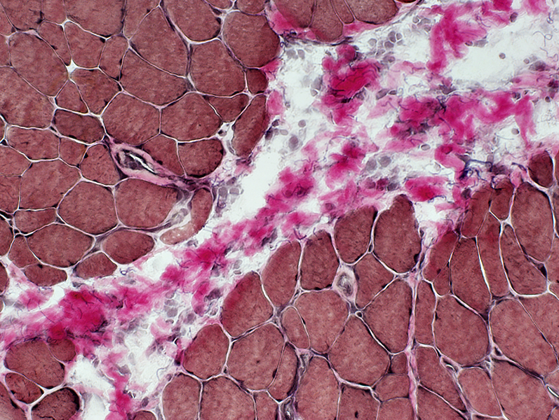
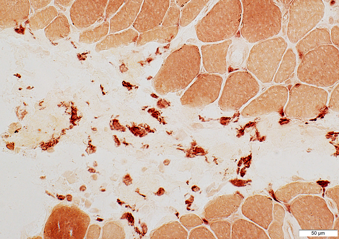
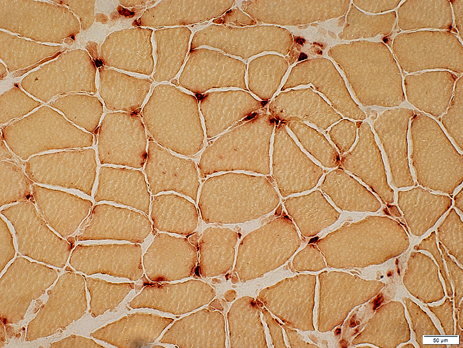
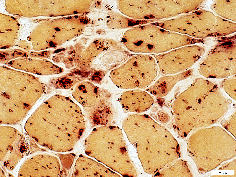
Perimysium: Acid phosphatase positive cells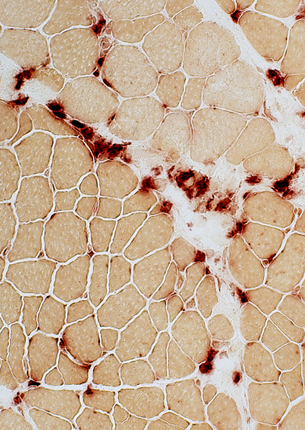 Acid phosphatase stain |
 Acid phosphatase stain |
 Acid phosphatase stain |
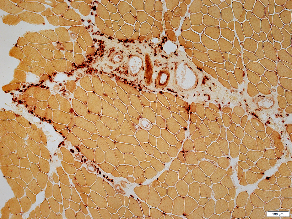
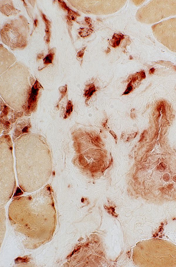
Histiocytic cells in perimysium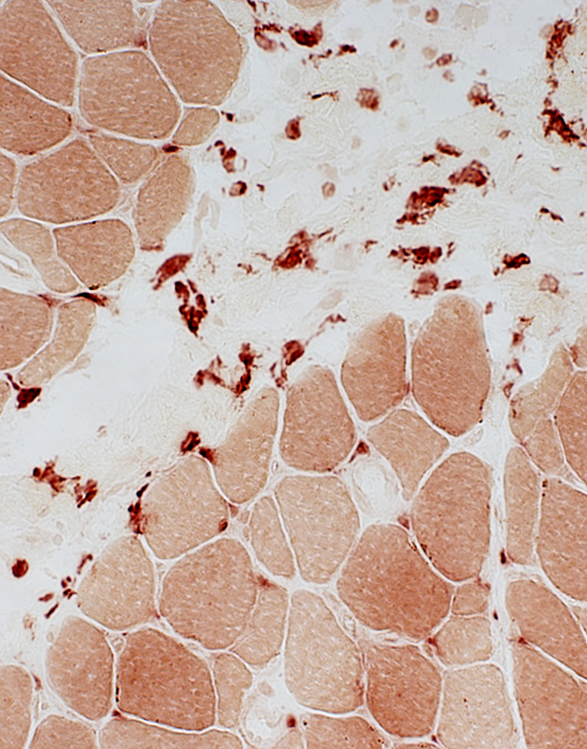 Esterase stain |
Histiocytic cells in endomysium
Often accompany upregulated MHC I by muscle fibers
 Acid phosphatase stain |
cGvHD: C5b-9 deposits, Locations
Connective tissue: Perimysium; May extend into endomysium
Capillaries: Endomysial
 C5b-9 stain |
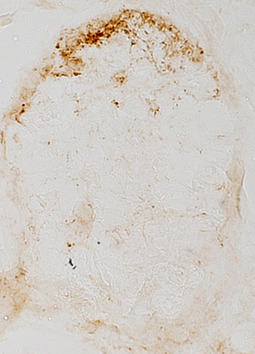
cGvHD: C5b-9 deposits, Locations
Perimysium
Surface of some muscle fibers, more often near perimysium
 C5b-9 stain |
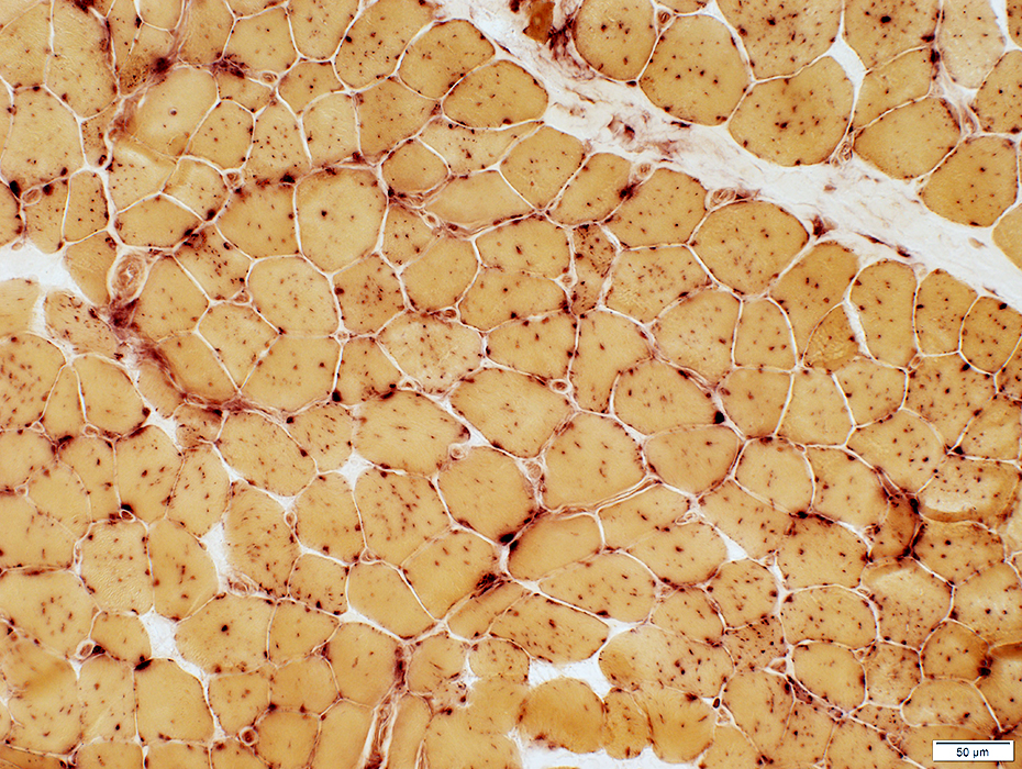
cGvHD: Muscle Fiber Morphology
Muscle Fibers
Sizes: Varied
Immature small fibers: Few
Necrosis: None or Rare
Endomysial Connective Tissue: Normal between fibers
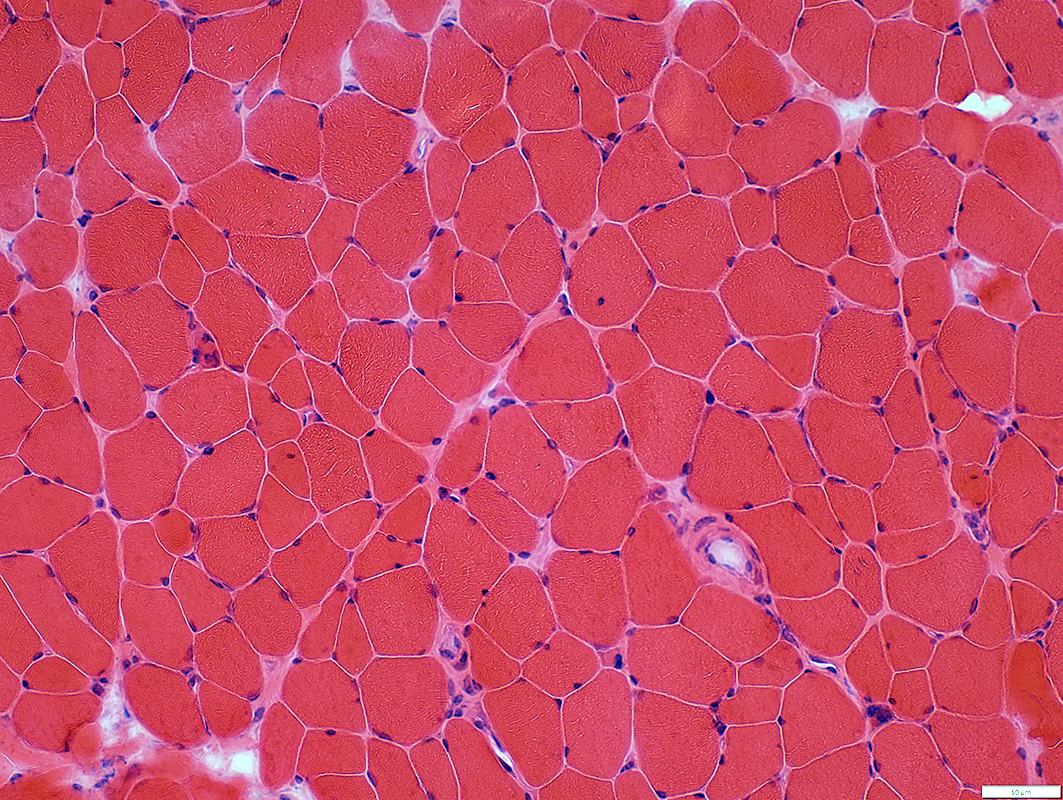 H&E stain |
cGvHD: Muscle Fibers
MHC Class I: Upregulated on all muscle fibers
 MHC Class I stain |
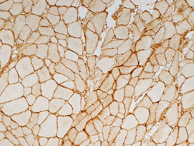
cGvHD Muscle Fibers: α-Dystroglycan
Reduced around surface of muscle fibers
Similar pathology to: Hereditary Muscle Glycosylation disorders
See FKRP
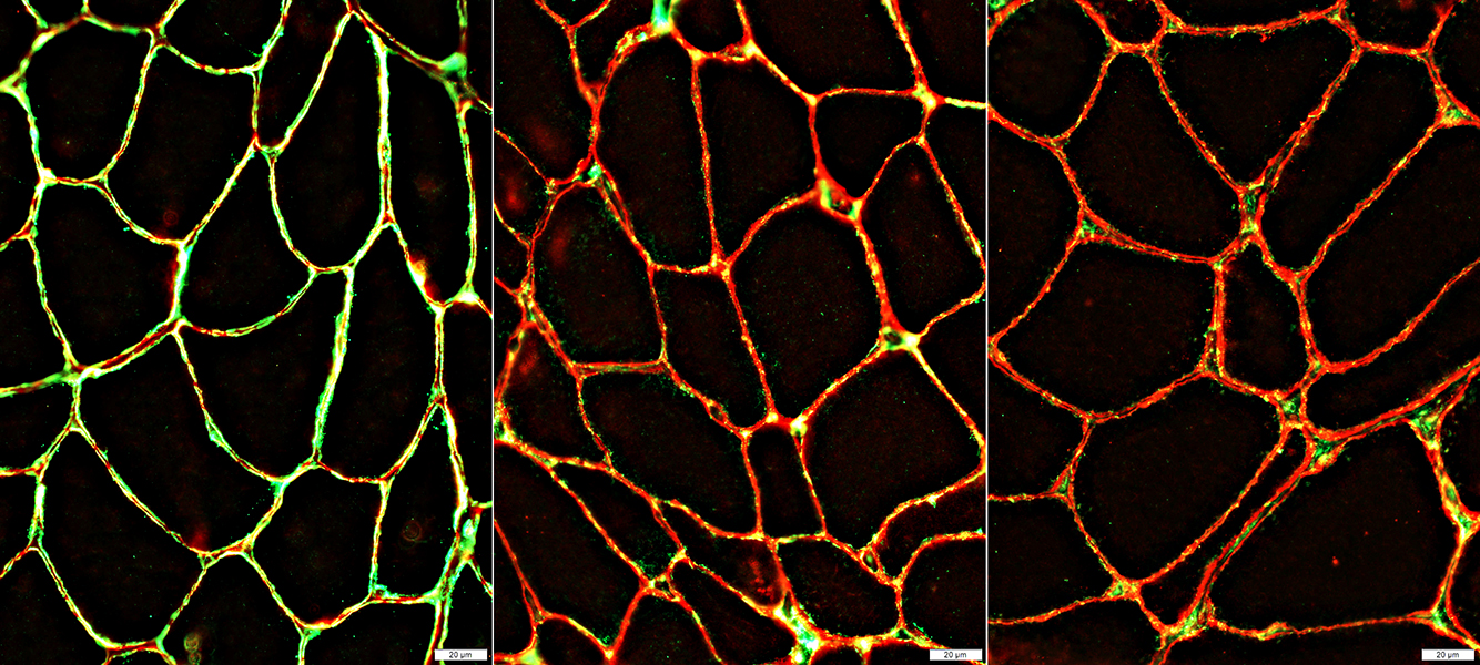 α-Dystroglycan stain: Glycosylation (Green); Protein (Red) |
|
| Normal Costaining of α-Dystroglycan Protein & Glycoslyation |
cGvHD α-Dystroglycan Glycoslyation is Reduced & Punctate |
 Chondroitin-SO4 stain |
|
| Normal Strong staining on the surface of muscle fibers |
cGvHD Reduced staining on surface of muscle fibers |
cGvHD: Muscle Fibers, Unusual features
Abnormal myonuclei
 Congo red stain |
 Congo red stain |
 Acid phosphatase stain |
 Acid phosphatase stain |
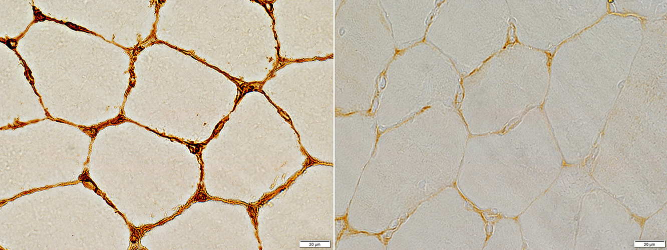
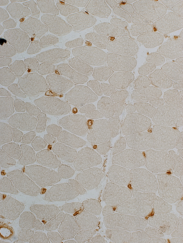
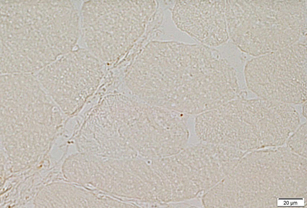
cGvHD: Capillary Pathology
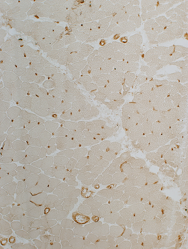 UEA I stain GVH: Capillaries Pale stained by UEA I Number: Reduced; Many muscle fibers with no neighboring capillary Size: Mildly large |
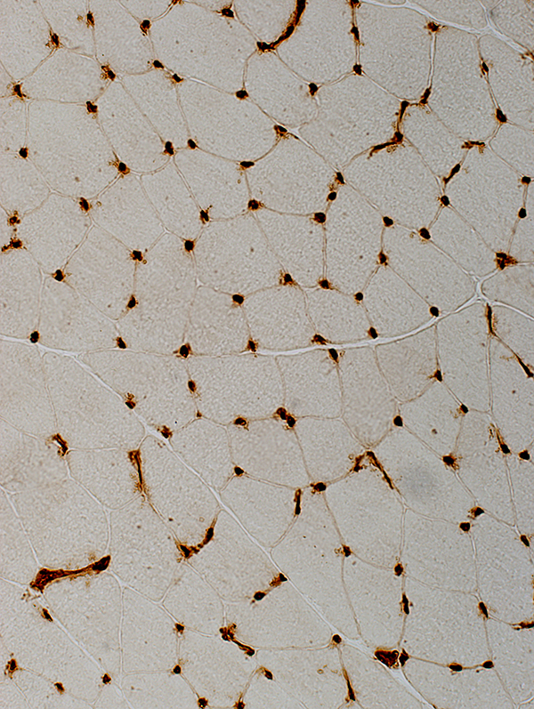
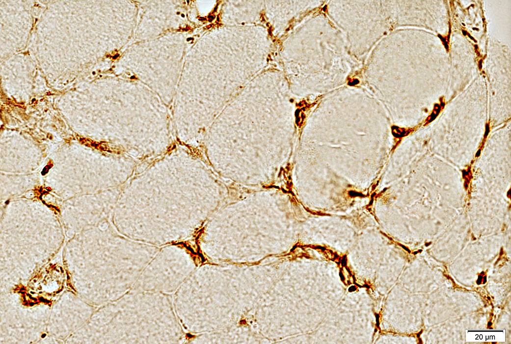
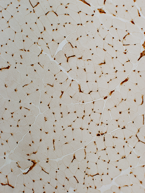 UEA I stain Control muscle: Capillaries Abundant: Capillaries neighboring most muscle fibers Dark stained by UEA I |
 UEA I stain |
 UEA I stain |
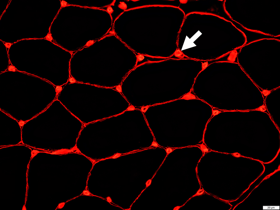 Collagen 4 stain (Red) |
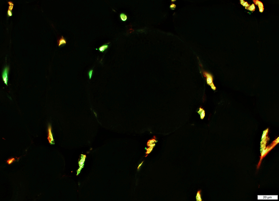
All muscle fibers have adjacent capillaries (Top; Arrow)
All Collagen 4 stained capillaries have associated endothelium (CD31; Yellow; Arrow)
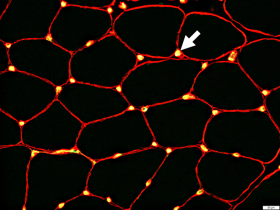 Collagen 4 stain (Red); CD31 stain (Green & Yellow) |
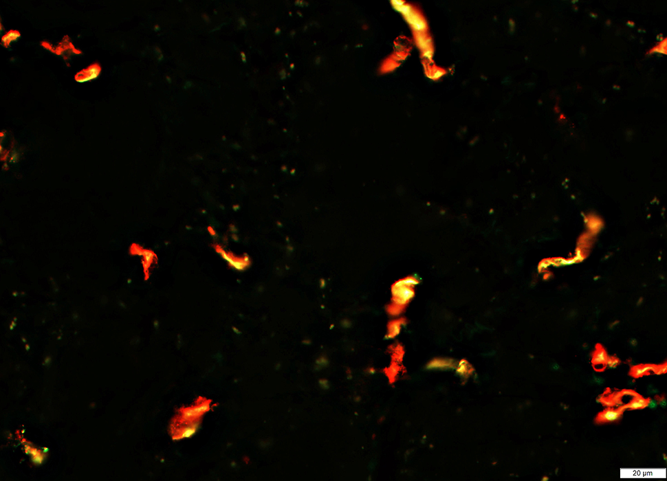
Capillaries: cGvHD
 Collagen 4 stain (Red); CD31 stain (Green & Yellow) |
Many Collagen 4+ capillary remnants with No CD31 endothelium (Red; Arrow)
Remaining intact capillaries (Yellow, Green & Red): Large; Reduced in number
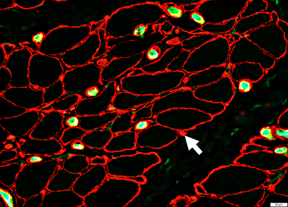
cGvHD Capillary Pathology
Capillaries are entirely lost: No Collagen 4+ capillary remnants
Remaining intact capillaries (Yellow, Green & Red): Large; Reduced in number
 Collagen 4 stain (Red); CD31 stain (Green & Yellow) |
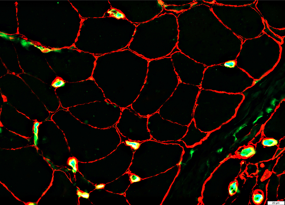
cGvHD Capillary Pathology
 UEA I (Ulex; Green & Yellow): CD31 Red) |
Costaining (Green & Yellow) of UEA I (Ulex) & CD31
Capillaries: cGvHD
UEA I (Ulex) is reduced in areas of CD31 stain (Red)
 UEA I (Ulex; Green & Yellow): CD31 Red) |
Capillaries: C5b-9 deposits
 C5b-9 stain |
cGvHD muscle: C5b-9 deposits on misoriented endomysial capillaries
 C5b-9 stain |
Return to Neuromuscular Home Page
Return to Graft vs Host
12/6/2019