LGMD 2A: Calpain-3 deficiency
LGMD 2A: Child
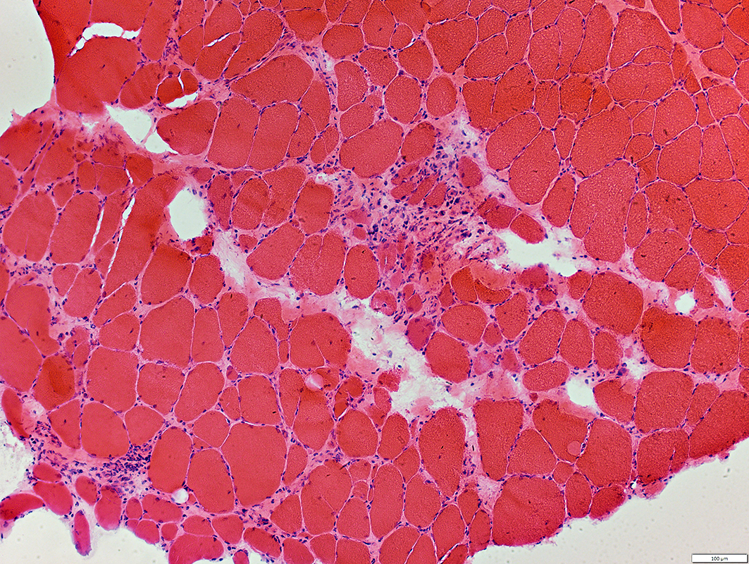
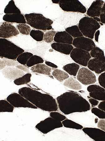
Myopathic changes
|
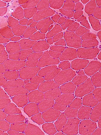
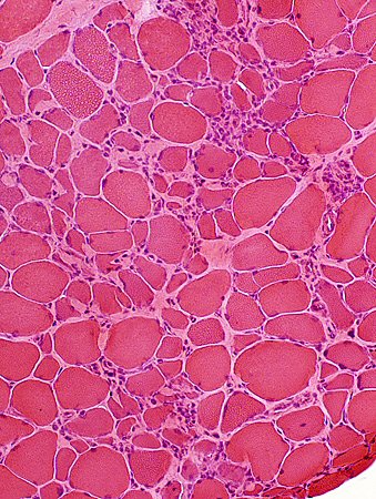
Muscle Fibers
Sizes: Varied
Large fibers: Hypertrophied
Small fibers: Round or Polygonal; Basophilic cytoplasm common
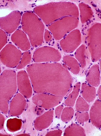
Myopathic groups
Endomysial connective tissue
Mildly increased in some regions
|
 H&E stain Myopathy Muscle fiber sizes: Varied Split muscle fibers Increased endomysial connective tissue |
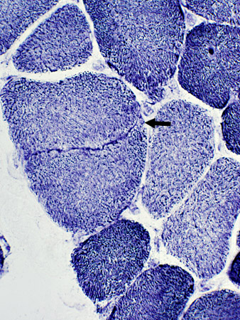
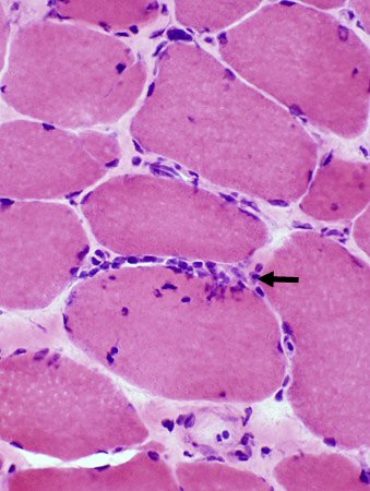
 H&E stain Myopathic groups Clustered basophilic fibers (Arrow) |
Muscle Fiber Splitting/Partial fusion
 H&E stain Split muscle fiber (Arrow) Small fibers in region of split fiber are clustered |
 NADH stain Split muscle fiber (Arrow) Split is highlighted by NADH stain "Splitting" is actually partial fusion |
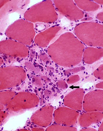
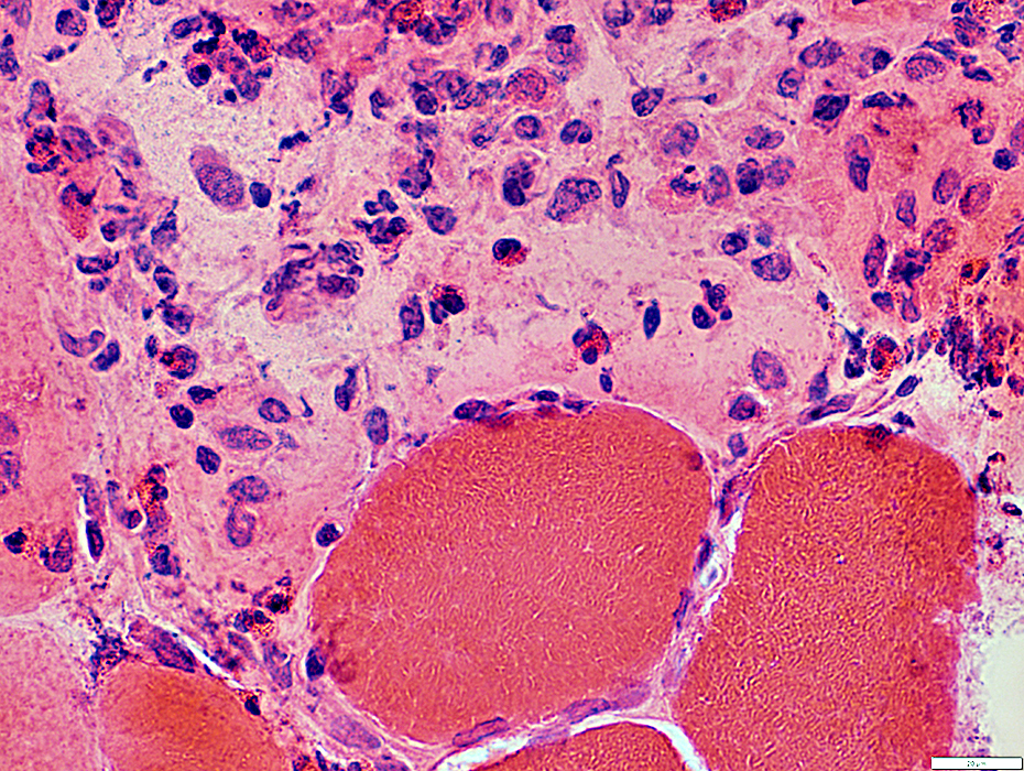
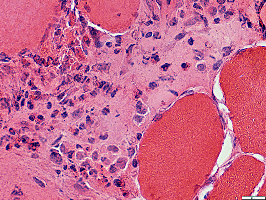
Muscle Fiber Necrosis: Myopathic Grouping
Clusters of Muscle fibers: All in early & similar stage of necrosis
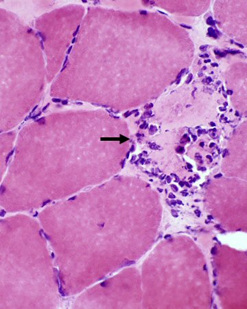 H&E stain Pale muscle fiber (Arrow)
invaded by cells |
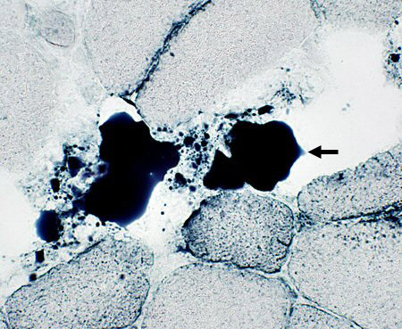
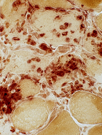 Acid phosphatase stain Acid phosphatase-stained cells
within pale necrotic muscle fibers |
Cluster of Muscle fibers: All in early & similar stage of necrosis
Muscle fibers are: Pale-stained (Arrow); Some invaded by cells
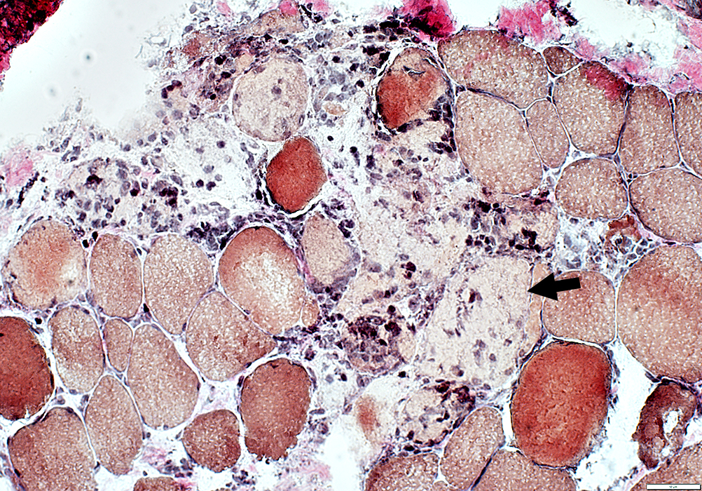 VvG stain |
Muscle Fiber Types
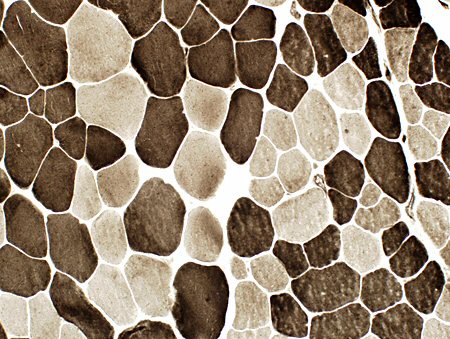
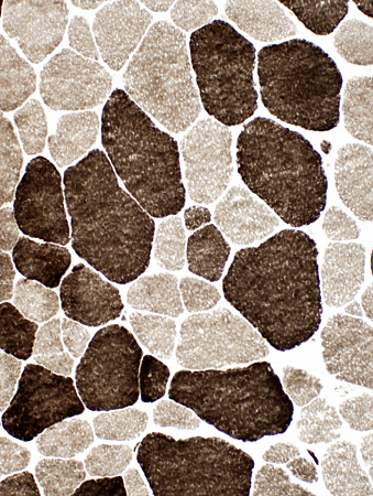 ATPase, pH 9.4 stain Most large fibers are Type 2
|
 ATPase, pH 4.3 stain Type 2C (intermediate staining) fibers are abundant
|
 ATPase, pH 9.4 stain |
Small fibers: Both types
Fiber type distribution: Non-random (Clusters of type 1 & type 2 fibers are present)
Immune features
|
Scattered in perimysium in regions of muscle fiber necrosis
|
 H&E stain Endomysial inflammation, minor
|
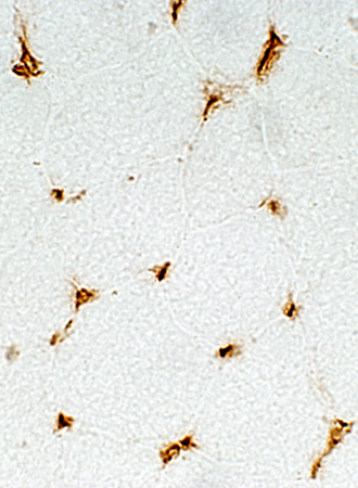 MHC Class I stain Normal distribution of MHC Class I: Only on capillaries
|
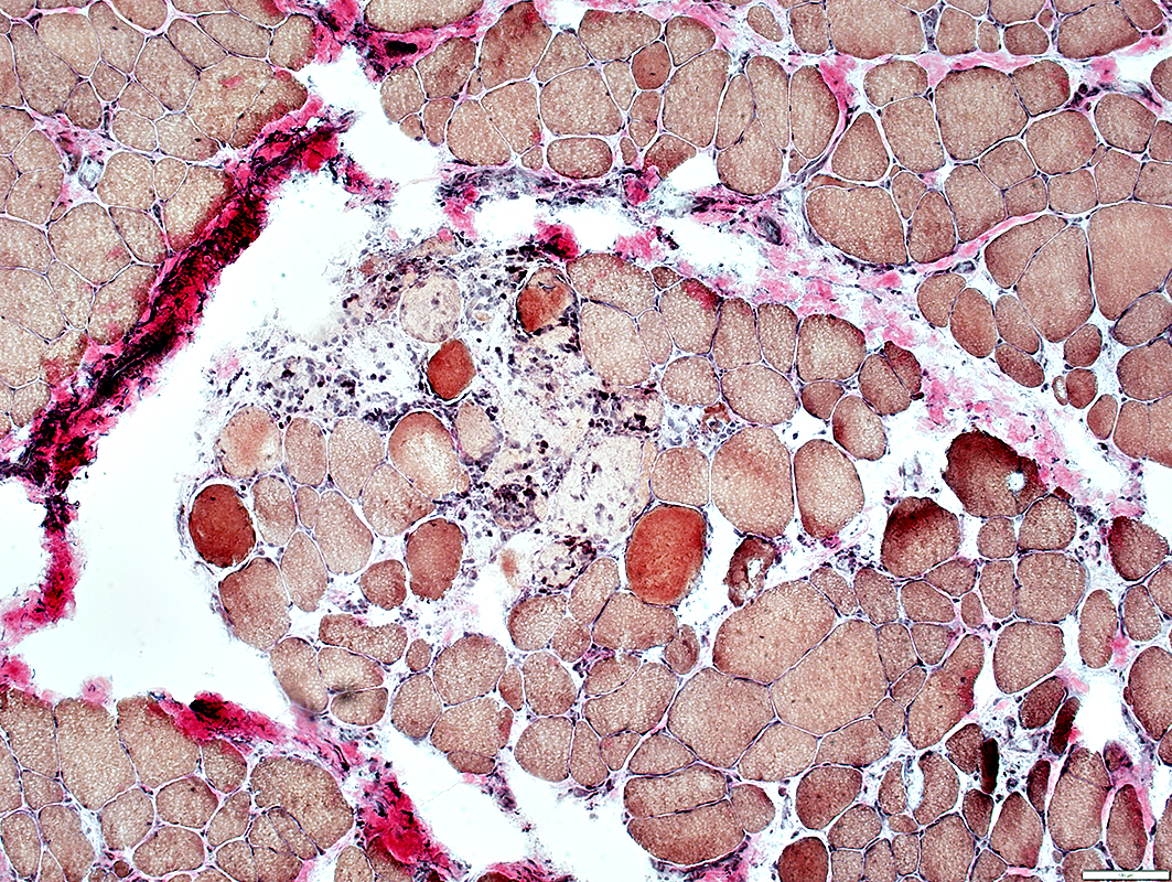
Fat accumulation in perimysium
 Sudan black stain |
LGMD 2A: Adult
Myopathic changes: Varied involvement in different fascicles
 H&E stain Varied fiber sizes
Little necrosis or regeneration |
 H&E stain Varied fiber size
Abundant necrosis and regeneration |
Myopathic Groups
|
Large group of small basophilic muscle fibers with large nuclei 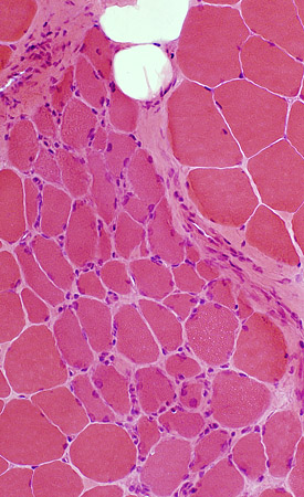 H&E stain |
Small muscle fibers have coarse internal architecture 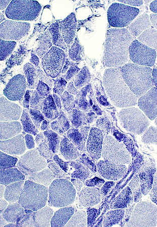 NADH stain |
|
Small muscle fibers are type 2C with intermediate staining 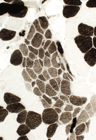 ATPase, pH 4.3 stain |
Small muscle fibers are immature with alkaline phosphatase staining 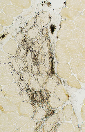 Alkaline phosphatase stain |
Fiber types
 ATPase, pH9.4 stain Small & large size fiber populations contain type I & type II
|
Return to Neuromuscular Home Page
Return to LGMD 2A
10/14/2022