Neuromuscular

Home, Search, Index, Links, Pathology, Molecules, Syndromes,
Muscle, NMJ, Nerve, Spinal, Ataxia, Antibody & Biopsy, Patient Info

NERVE BIOPSY

Diagnoses to look for
Pathology & Illustrations
Indications & Utility
Results: Differential diagnoses
Technical (Preparation)
  Freezing
Test form

Skin biopsy

Other
  Axons
  Myelin
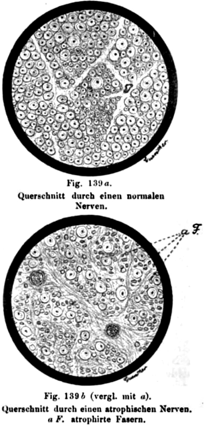
Nerve fascicle: Normal numbers of small and large axons

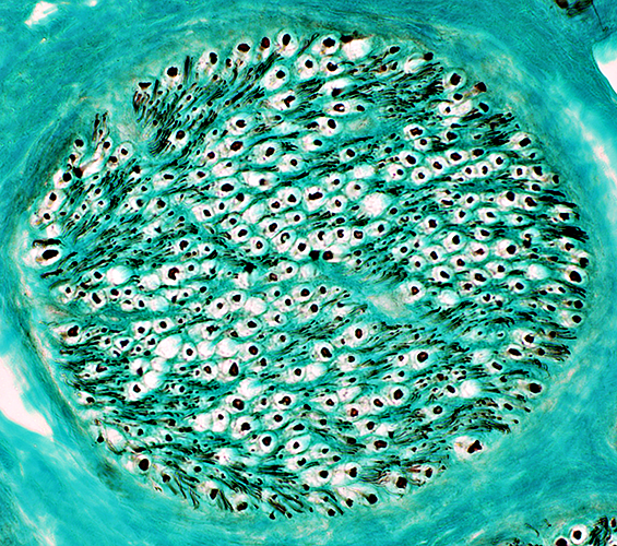
NFH + Peripherin stain
Myelinated axons: Surrounded by white halo
Unmyelinated axons: Present in clusters between myelinated axons


Useful biopsy results more likely
  • Clinical features
    • Clinical: Patient features
      • Significant functional disability
        • Weakness
      • Higher yield
        • Neuropathy is Asymmetric.
          • By history, examination, or electrodiagnosis
          • Nerve is biopsied
            • On more clinically affected side
            • On side of smaller sural action potential
        • Patient age > 65 years old
        • Laboratory: Abnormal nerve conduction studies
      • Neuropathy involves sensory loss
        • Biopsy location: Nerve in region with clear sensory loss
        • Sensory loss without weakness: Biopsy less likely to be useful
    • Evaluation strategy: Specific diagnoses are sought.
  • Surgery
    • An experienced surgeon should perform the biopsy
      • Sural nerves can be: Hard to find; Easy to damage
    • Biopsy whole nerve (rather than few fascicles): Allows evaluation of
      • Large vessels
      • Epineurial connective tissue
      • Comparative features of different fascicles
      • Immune features
    • Muscle: Biopsy at the same time
      • Increased yield of immune, vascular & amyloid pathology
  • Nerve biopsy specimens: Preparation
    • Size
      • Full thickness
      • Length: > 3 cm
    • Fresh: Transported in moist gauze on wood stick
    • Processed: 3 pieces of equal size
      • Frozen
      • Formalin fixed: 10% Buffered Formalin
      • Glutaraldehyde fixed
        • Modified Karnovsky's Fixative
          (3% Glutaraldehyde, 1% Paraformaldehyde in 0.1M sodium cacodylate)
  • Nerve biopsies: Staining
    • Stains
      • H&E
      • Gomori trichrome
      • Alkaline phosphatase
      • Acid phosphatase
      • Congo red
      • VvG
      • ATPase: Endoneurial cells & Epineurial vessels
      • Neurofilament (Axons)
      • NCAM (Schwann cells, non-myelinating)
      • Myelin
    • Normal
    • Immune features
  • Nerve analysis
    • Nerve is viewed in several preparations
      • Frozen sections
      • Paraffin embedded tissue
      • Plastic sections: Toluidine blue stained
    • Teased fibers
      • Main value: Distinguish axonal from demyelinating pathology.
      • Little diagnostic utility
    • Ultrastructural analysis is available

Oppenheim
Nerve biopsy: Diagnoses to look for
BIOPSY FINDINGS IN NEUROPATHY

Morphologic changes in nerve or muscle: Differential diagnoses Other nerve pathology Other pathology in neuropathy: Denervation of muscle
Neurofilament accumulations in neuropathy
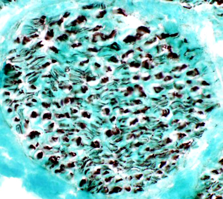
SMI 31 antibody
Sural nerve axons (Normal): Neurofilament stain

Mitochondrial abnormalities in Neuropathy
Polyglucosan bodies

Tomaculae 1

INFLAMMATORY OR IMMUNE NEUROPATHIES

Diseases Cellular infiltrates in nerve: Inferences from cell types & location Other pathological features of immune or inflammatory neuropathies

Skin Biopsy: Analysis of cutaneous nerves


Return to Neuromuscular Home Page
Return to Normal nerve
Return to Biopsy illustrations
Return to Polyneuropathy Index

References
1. JNNP 2000;68:483-488
2. J Neurosci 2013;33:10950–10961
3. Genes Dev 2014;28:290–303

7/5/2020