FASCIITIS
|
Immune Eosinophilic Toxic ICI IMPP |
Fasciitis: Immune
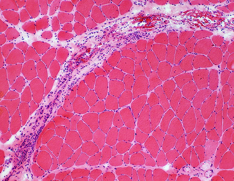
 H&E stain |
 H&E stain |
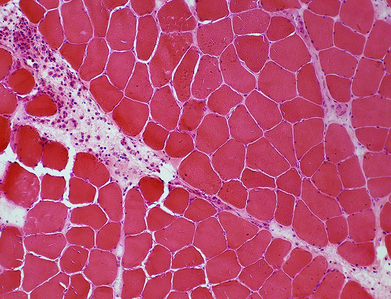
Muscle & Connective tissue pathology
|
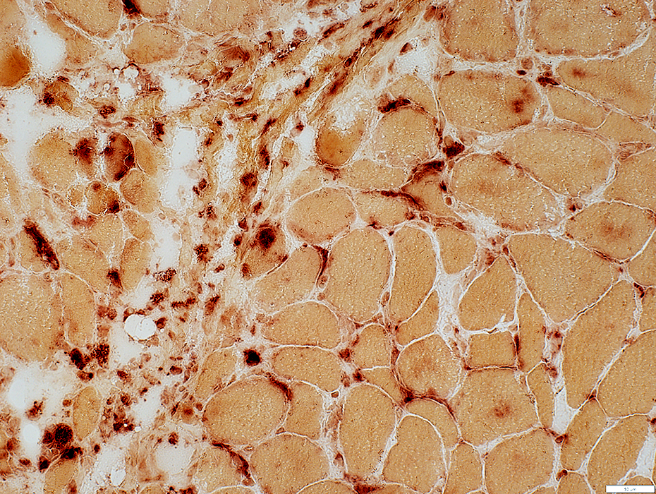
 Acid phosphatase stain |
|
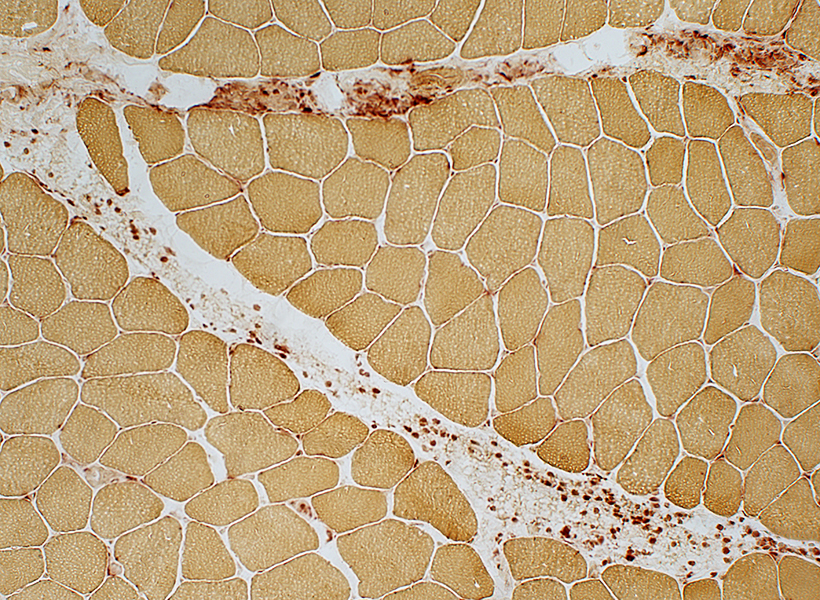
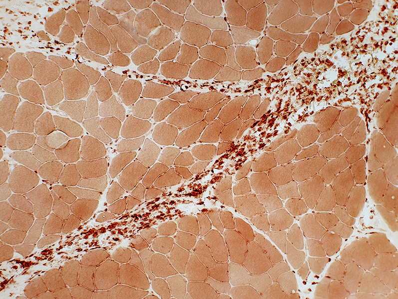
MHC Class I Perimysium: Mild staining Muscle fibers: Most are normal; Mild increase in some muscle fibers near perimysium Capillaries, endomysial: Normal staining 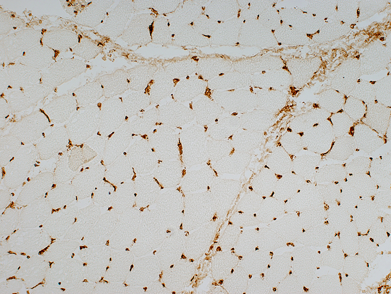 MHC Class I stain |
|
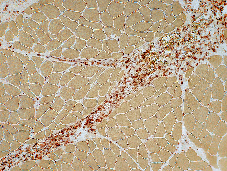
C5b-9 Deposition Perimysium: Moderate staining Endomysium: Diffuse deposition  MHC Class I stain |
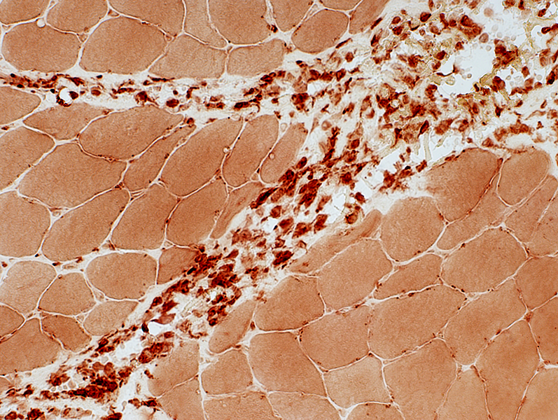
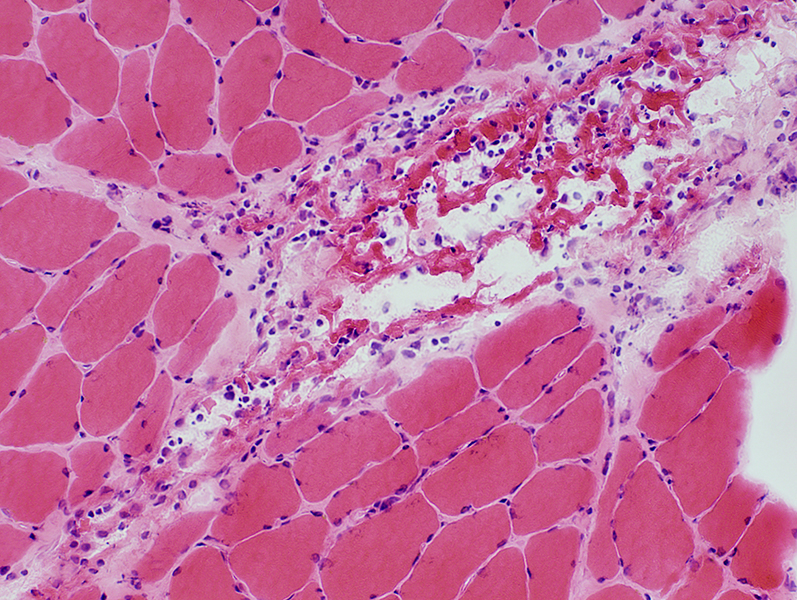
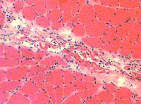
Fasciitis: Perimysial Histiocytic Inflammation
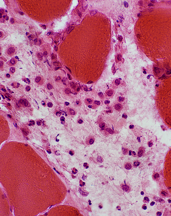 H&E stain |
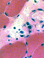
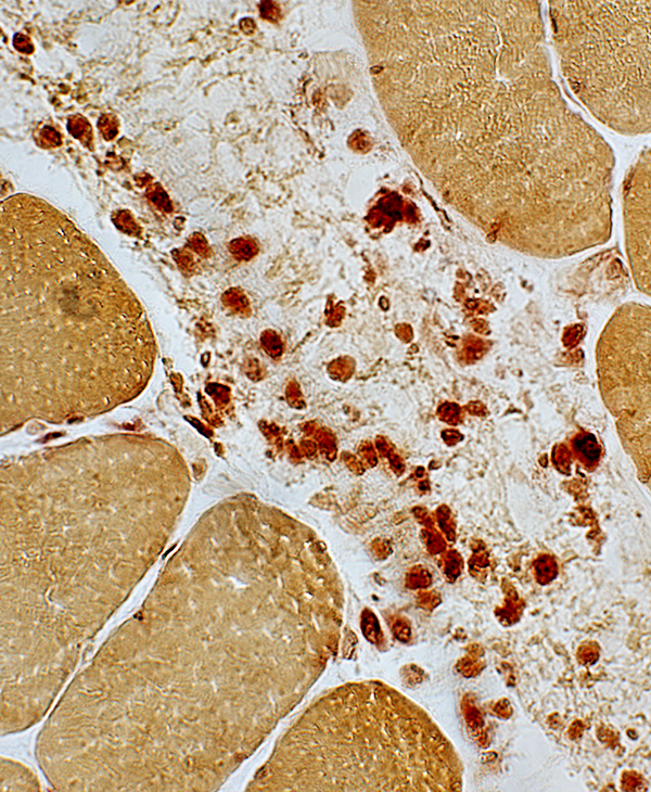 Acid phosphatase stain |
Inflammation
|
 H&E stain |
 H&E stain |
Fasciitis: MRI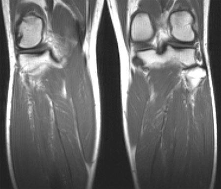 Multiple regions of increased T2 signal in leg muscles (L > R) |
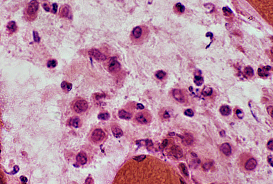
Fasciitis: Eosinophilic (Shulman syndrome) 1
- Pathology: Muscle
- Damage
- Perimysial connective tissue
- Epimysial connective tissue: Histiocytes
- Cells
- Types
- Histiocytes (CD206+; Anti-inflammatory)
- CD8 lymphocytes
- Eosinophils: Eosinophil major basic protein (EMBP)+, Some patients
- Location: Perimysium
- Types
- Muscle fibers
- Upregulation of MHC1 & MHC2: Especially perifascicular
- Atrophy: Some fibers
- Damage
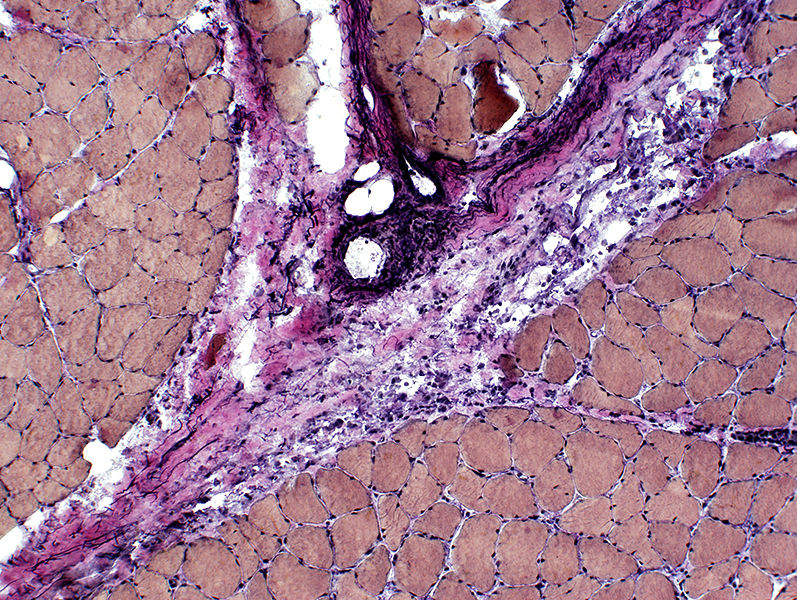
Fasciitis Perimysium: Fragmentation; Condensation
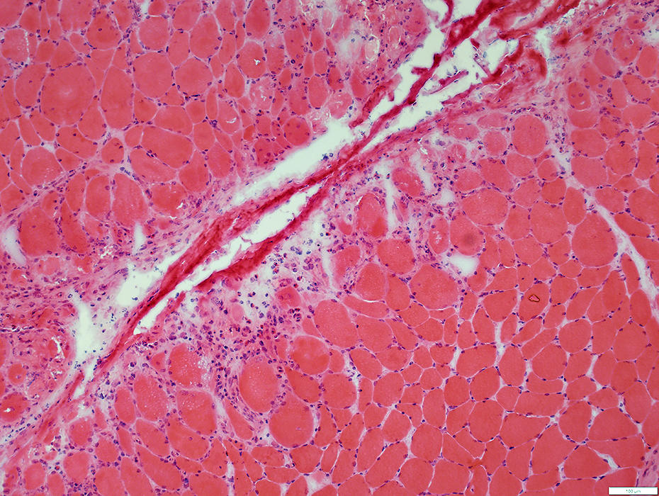 H&E stain |
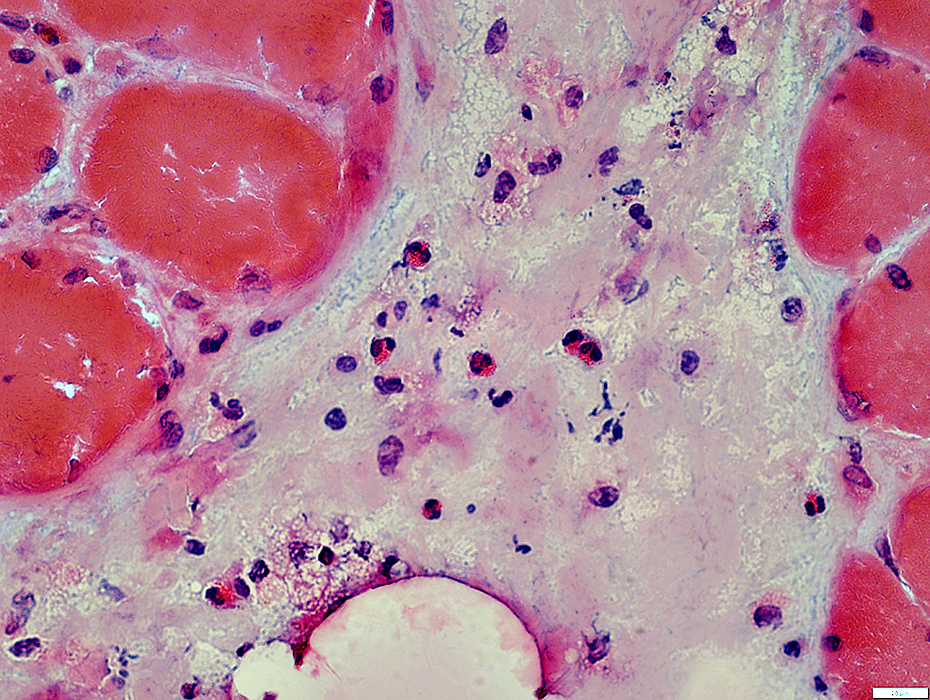
Fasciitis Perimysium: Palor
 H&E stain |
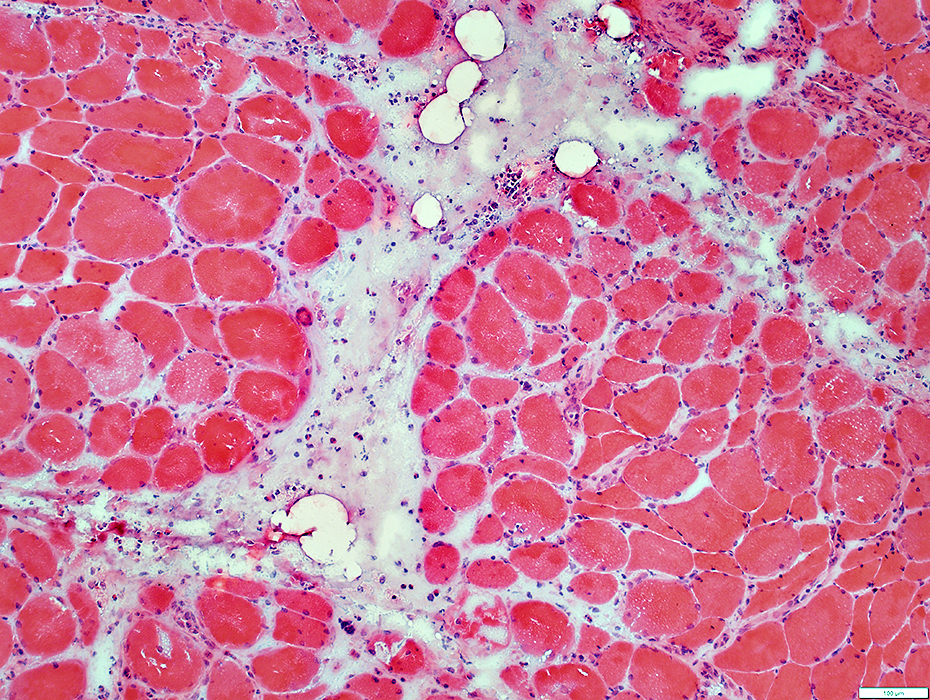
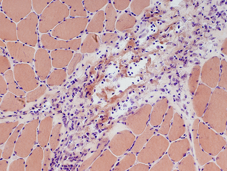
Fasciitis Perimysium: Eosinophils; Histiocytes
 H&E stain |
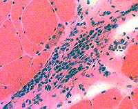
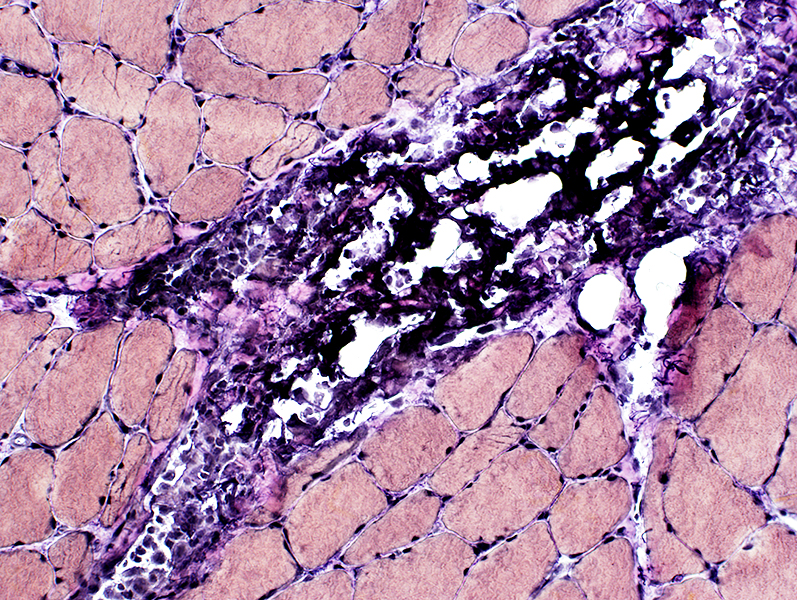
Fasciitis Perimysium: Histiocytes
 Acid phosphatase stain |
Fasciitis Perimysium: Alkaline phosphatase positive
 Alkaline phosphatase stain |
Fasciitis: Toxic
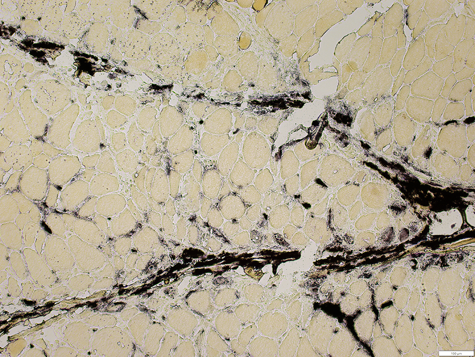
 H&E stain |
 VvG stain |
 Acid phosphatase stain |
 Esterase stain |
 Esterase stain |
 H&E stain |
 H&E stain 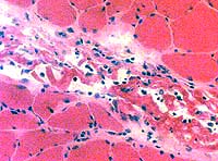 H&E stain |
 VvG stain |
 Congo red stain |
Return to Neuromuscular Home Page
Return to Inflammatory myopathies
References 1. Rheumatology (Oxford) 2023;62:2005-2014, Autoimmun Rev 2014;13:379-82, Rheumatology (Oxford) 2023;62:2005-2014
2/5/2024