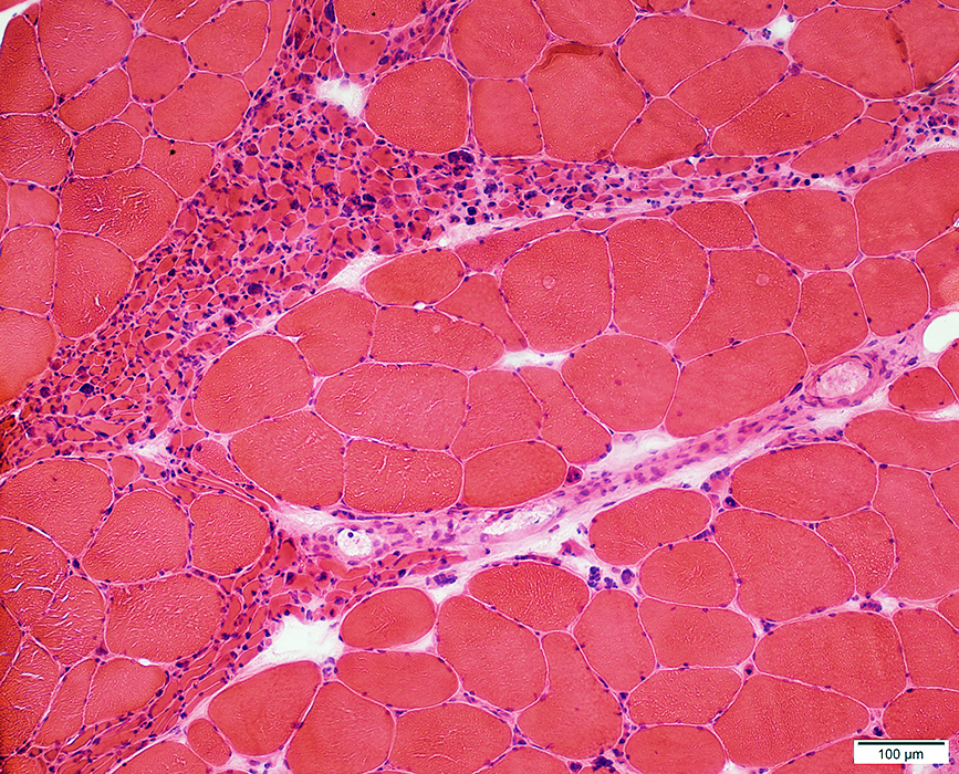
Muscle: Chronic Partial Denervation
|
Muscle Capillaries Fibers Atrophy Ultrastructure Grouped atrophy Nuclei Internal Pyknotic clumps Necrosis Regeneration, Clustered Split Types Grouped Patterns Chronic Features Moderate Severe: Pseudomyopathic Endstage Nerve Regenerating axon clusters |
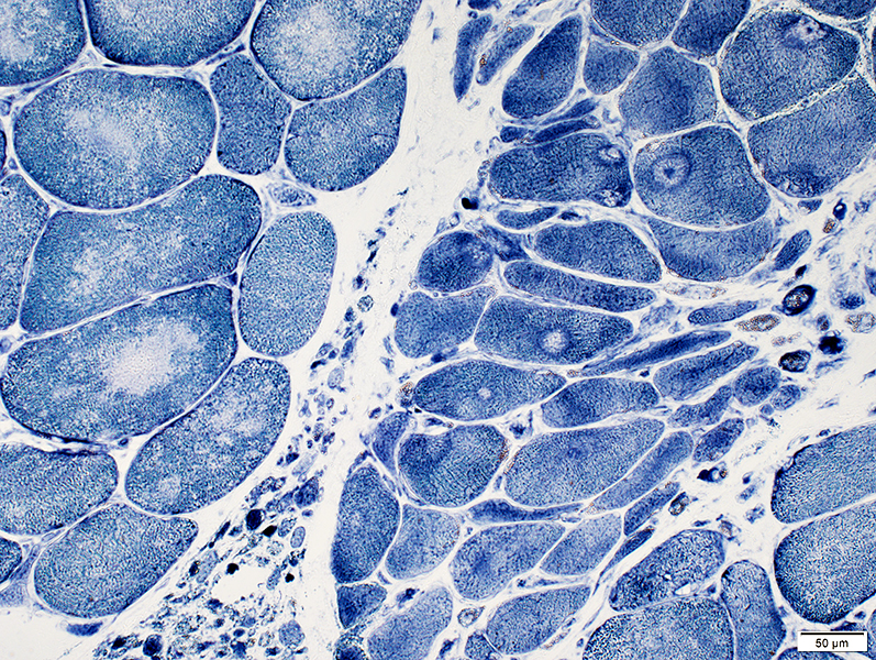
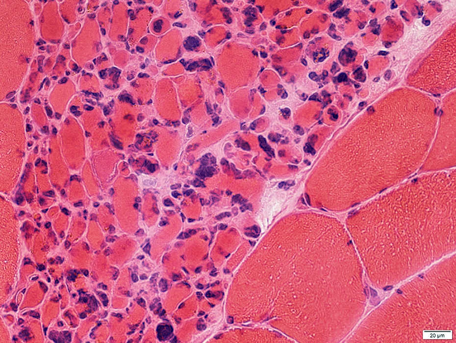
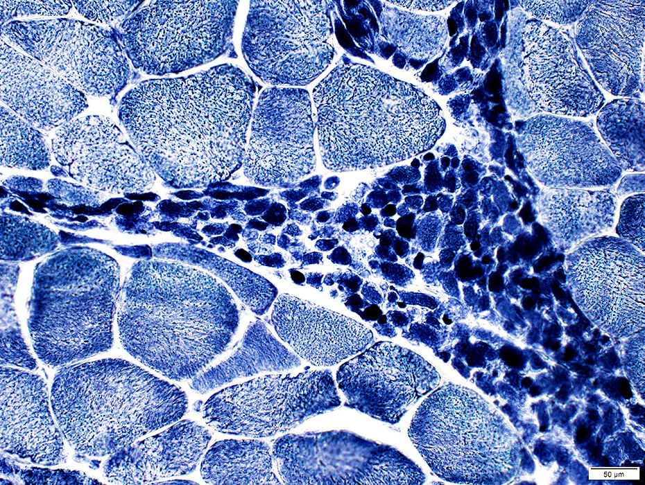
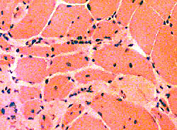
Groups (Size): Hypertrophic & Atrophic Muscle fibers
 H & E stain |
Distribution: Clustered
Size: Often hypertrophied
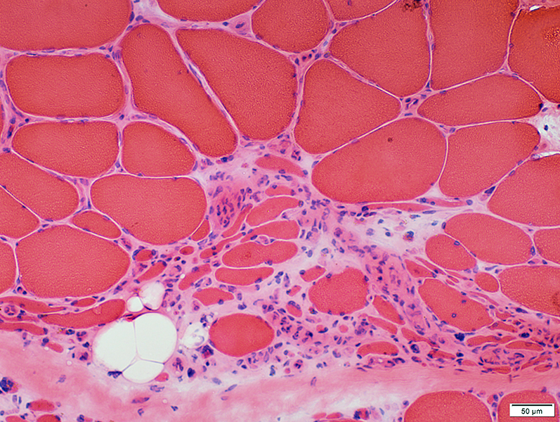
Small Muscle Fibers
Shape: Polygonal, Rounded or Nuclear Clumps
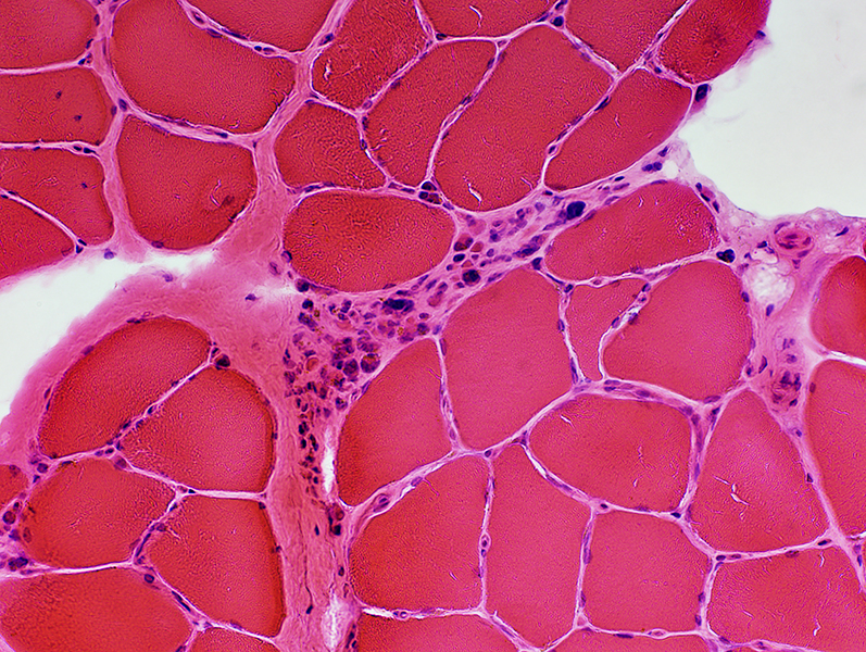
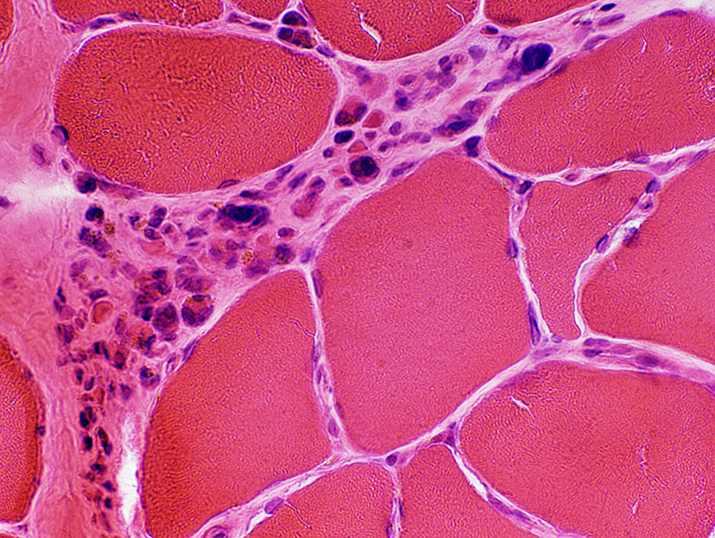
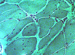
Endomysial connective tissue in regions of grouped muscle fiber atrophy
Distribution: Clustered or Grouped
Normal (Above) or Increased (Below)
 H & E stain |
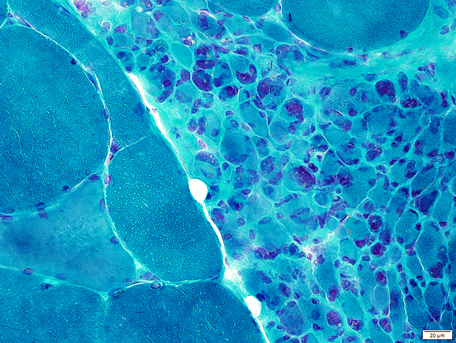
Clusters, or groups, of large, intermediate & small sized muscle fibers
 H & E stain |
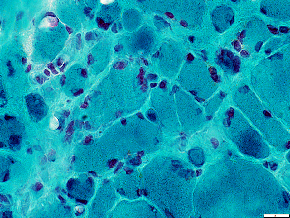
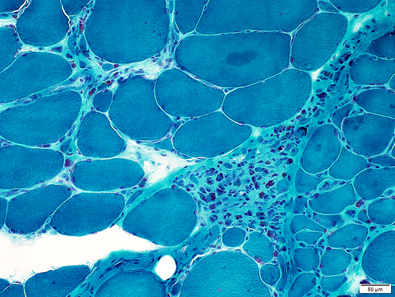 Gomori trichrome stain |
 VvG stain |
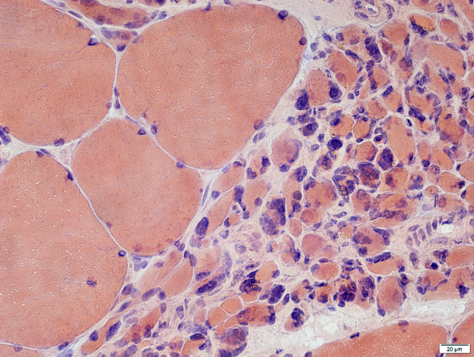
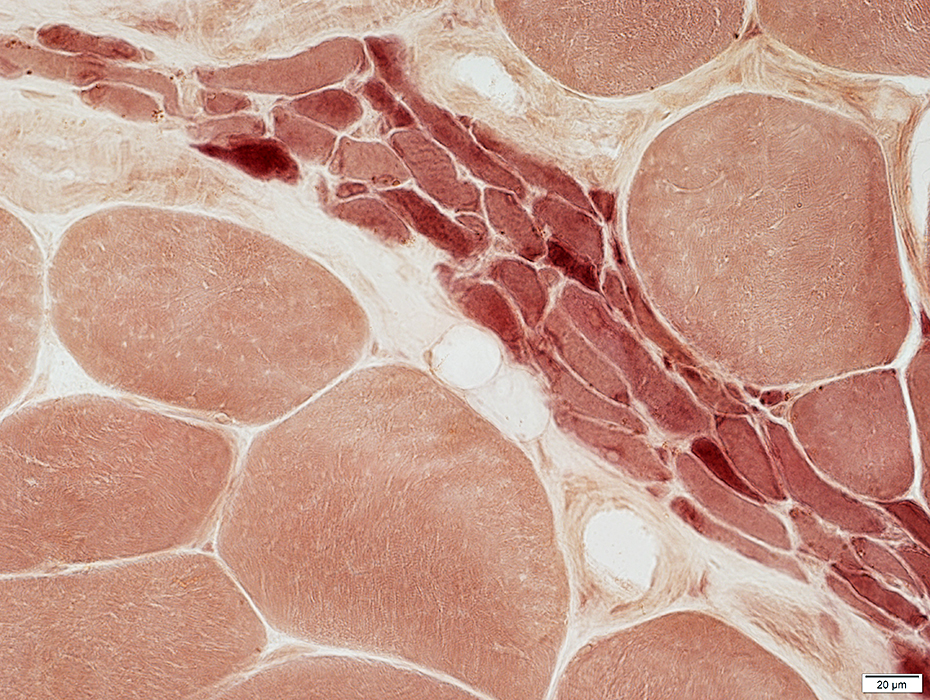
 NADH stain |
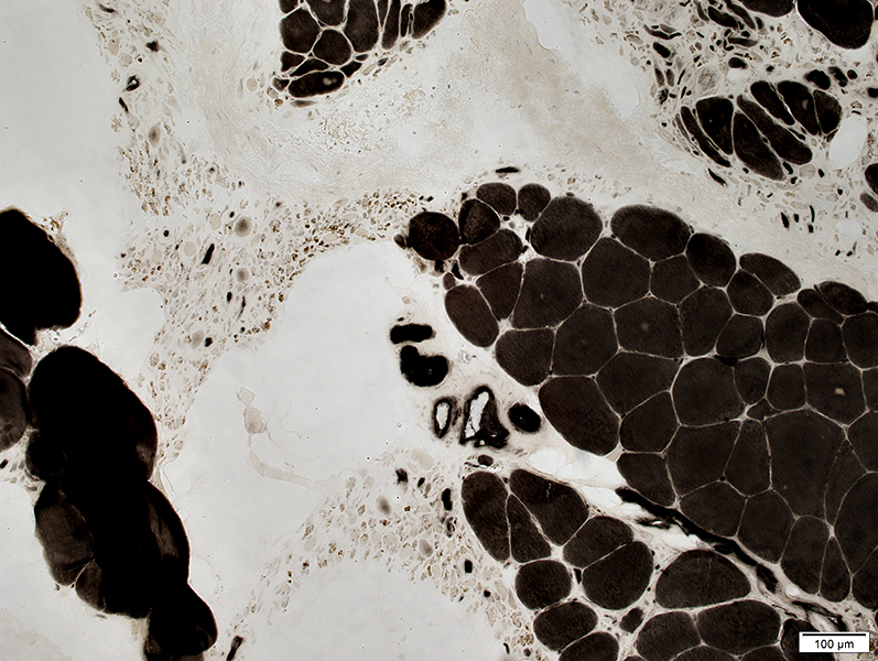
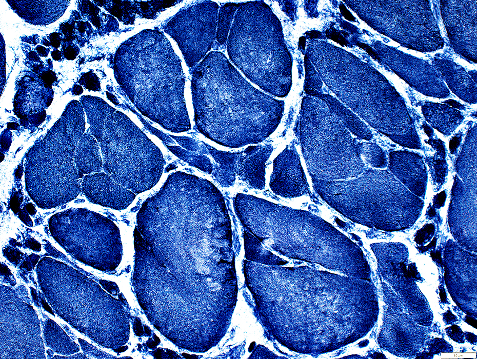
Chronic Partial Denervation: Fiber type abnormalities
Large fibers are commonly a single type: May be Type 1, 2, or Abnormal
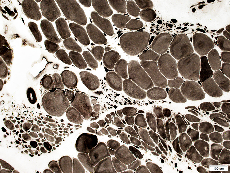 ATPase pH 9.4 stain |
 ATPase pH 4.3 stain |
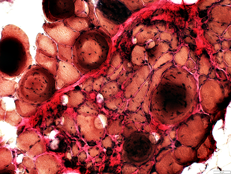
Large muscle fibers: All type 2
 ATPase pH 4.3 stain |
 ATPase pH 4.6 stain |
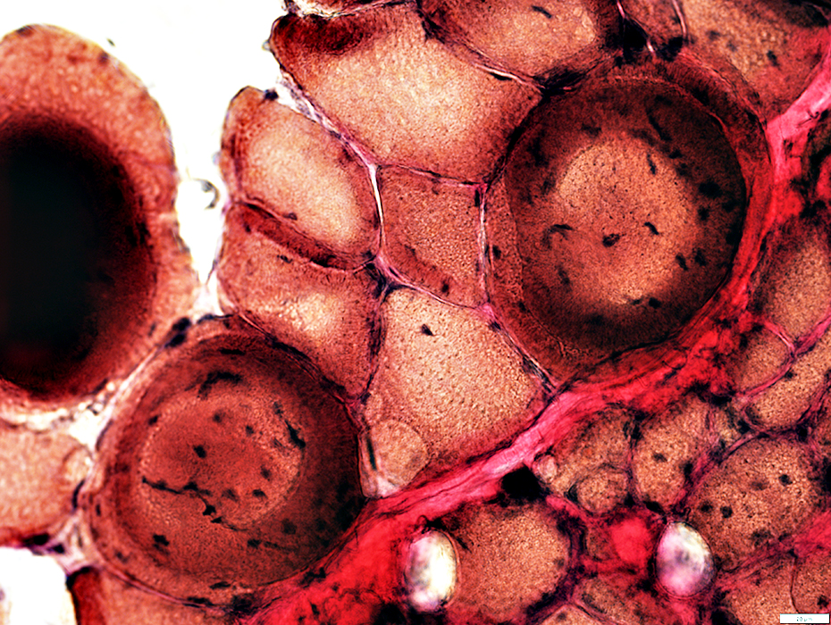
Type 2 with varied degrees of intermediate staining on ATPase pH 4.6 stain (Above)
Incomplete fiber type switch: Type 1 properties on COX stain (Below)
 COX stain |
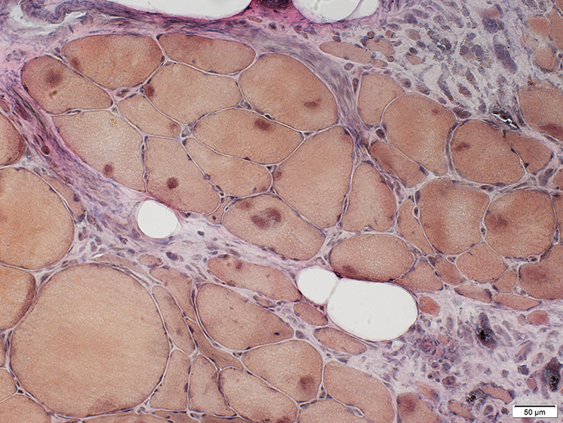
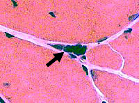
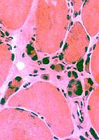
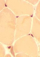
Atrophic Muscle fibers
 H&E stain |
 H&E stain |
 Gomori trichrome stain |
 Congo Red stain |
 Esterase stain |
 NADH stain |
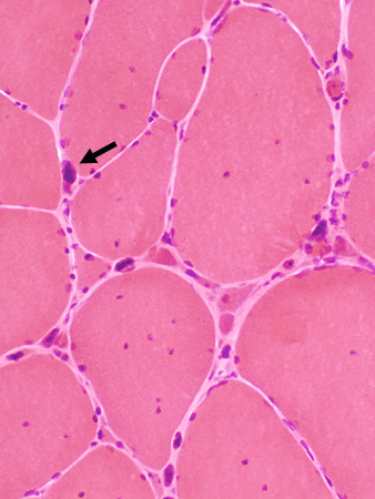
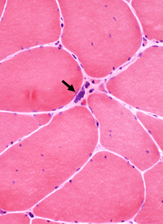
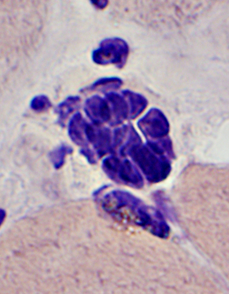
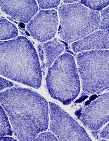
PYKNOTIC NUCLEAR CLUMPS
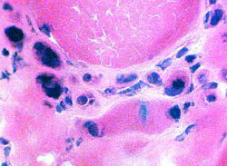
- Definition: End product of severe muscle fiber atrophy
- Nosology
- Nuclear clumps
- Pyknotic
- Term used to denote dark color
- Scientific definition (karyopyknosis with chromatin condensation): Not applicable
- Syncytial knots
- Clumps of myonuclei
- Larger (More nuclei) in muscles with previous fiber hypertrophy
- Lipofuscin: May also be present; In Older patients
- The remainder of the myofiber largely disappears
- Contractile apparatus
- Cytoplasm
- Pyknotic nuclear clump distributions
- Singular: May occur alone
- Clusters: Part of regions of grouped muscle fiber atrophy
- Disease associations
- Denervation without reinnervation
- Myasthenia gravis: Late stage, untreated
- Myopathies: Myotonic dystrophy 2; IBM3
- Histochemical staining
- Nuclei: Basophilic on H & E
- Muscle fiber: Dark on NADH & Esterase
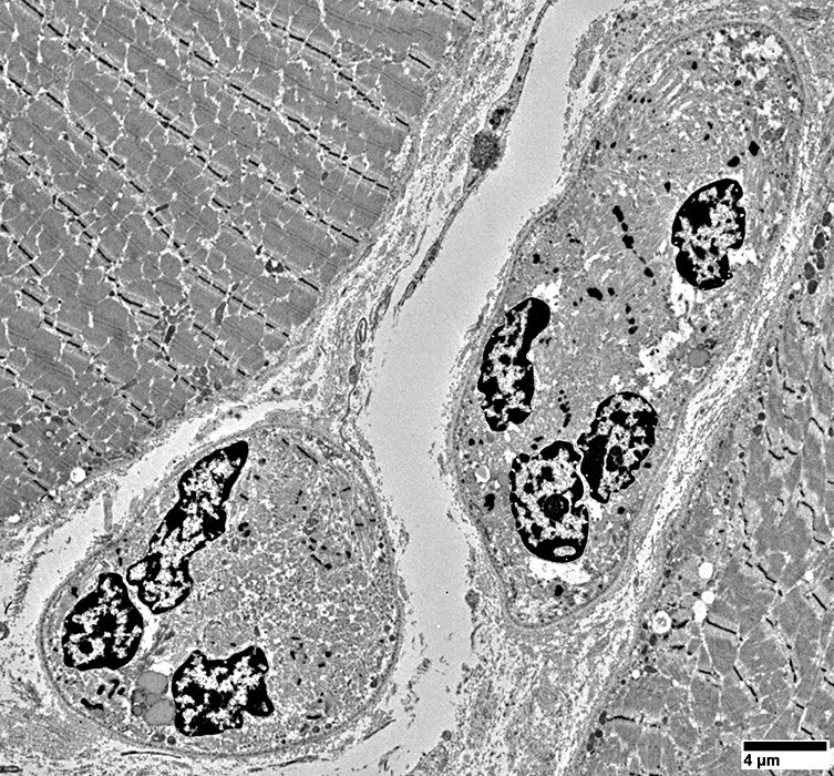
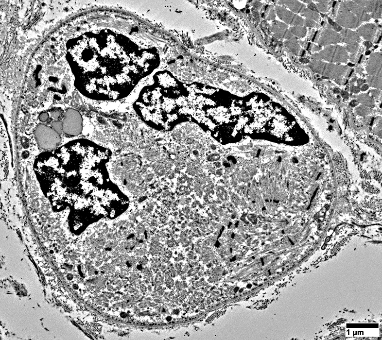
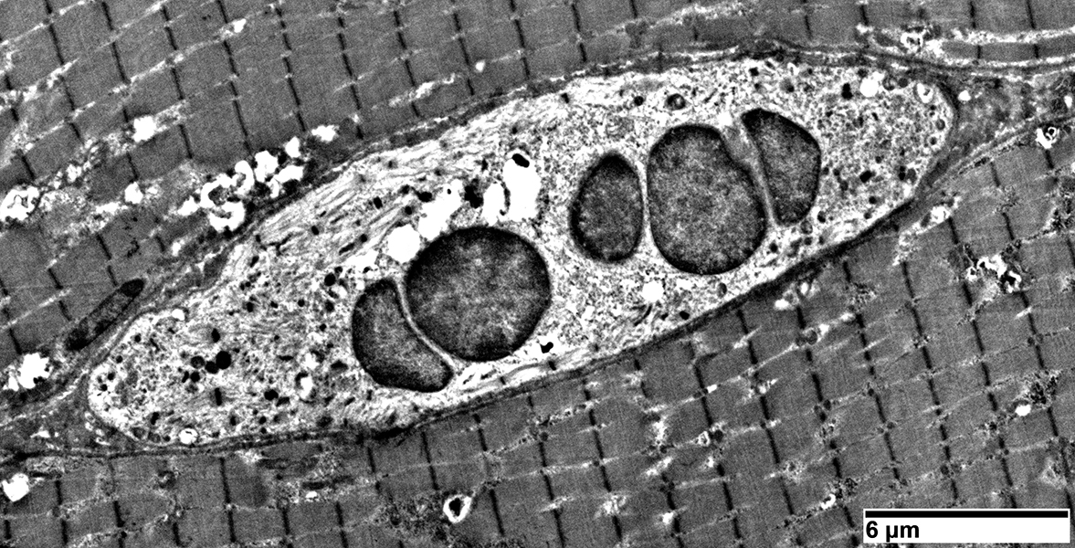
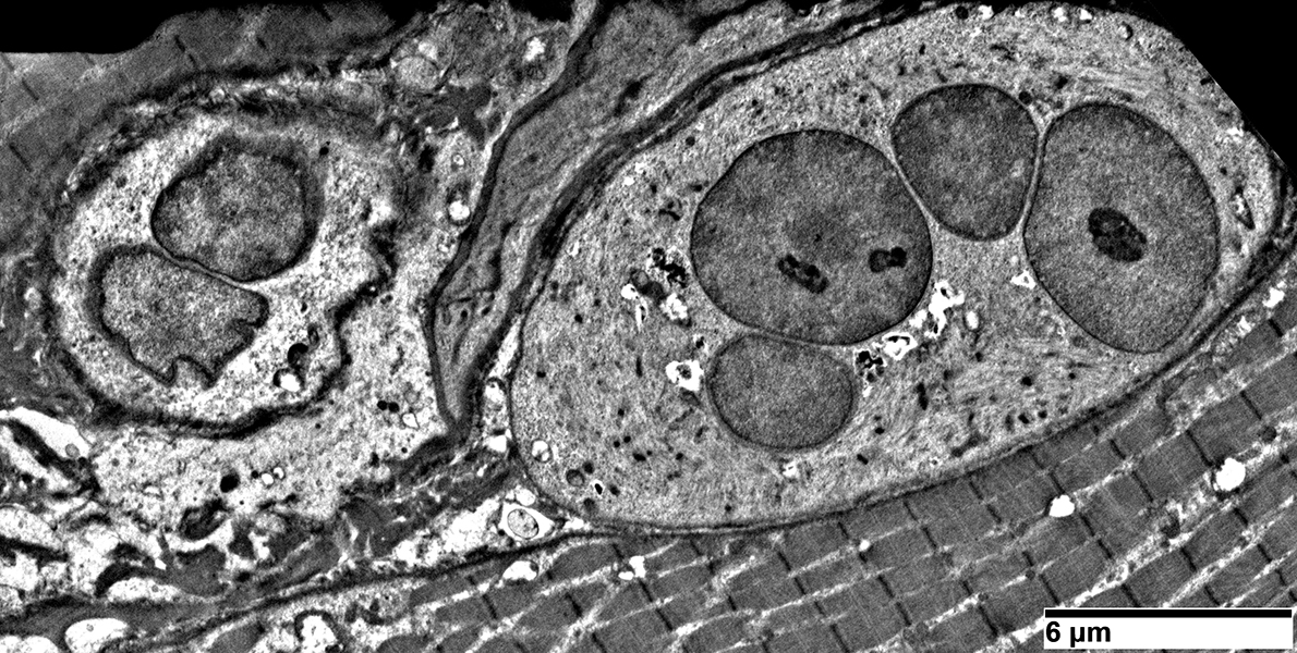
- Ultrastructure: 1; 2
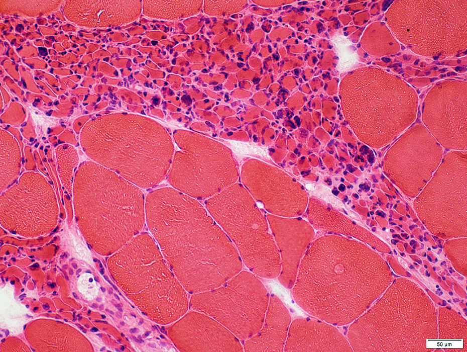
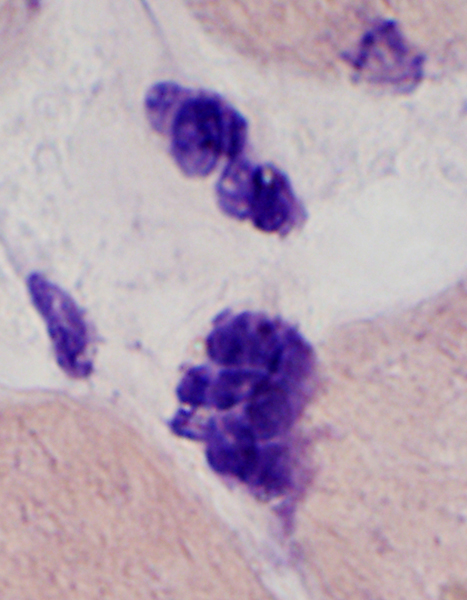
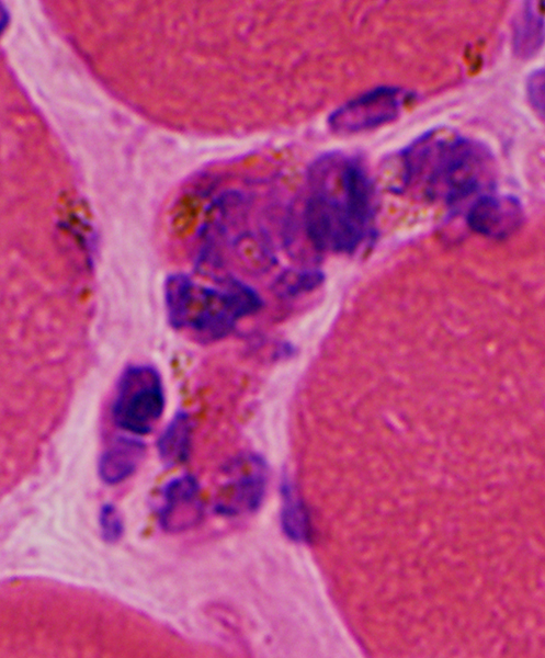
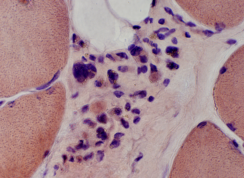
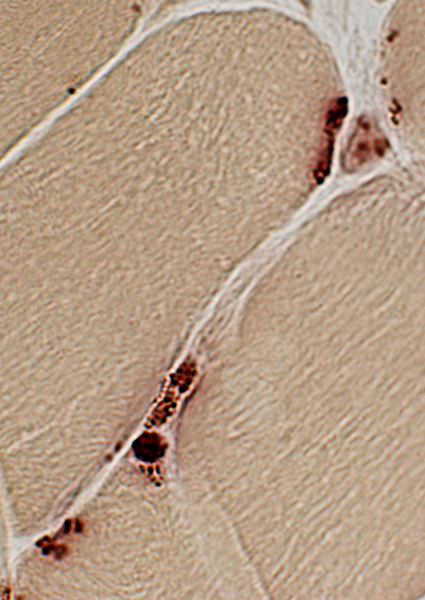
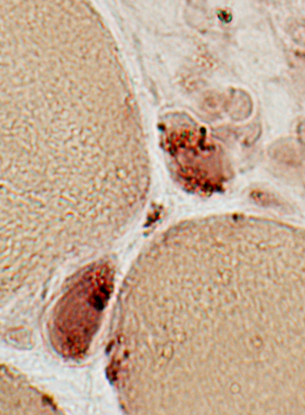
Pyknotic nuclear clumps: Morphology
 H & E stain |
 H & E stain |
  H&E stain |
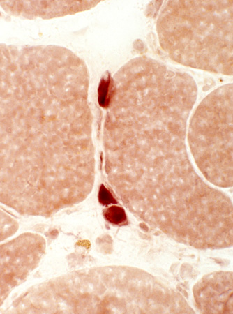
 Congo red stain |
 Congo red stain |
Clusters of myonuclei with no, Lipofuscin or little, visible cytoplasm
May also contain clustered lipofuscin
 H&E stain |
 Congo red stain |
 H&E stain |
Late outcome of grouped muscle fiber atrophy
 H&E stain |
 Congo red stain |
Nuclear clusters: Some are associated with lipofuscin
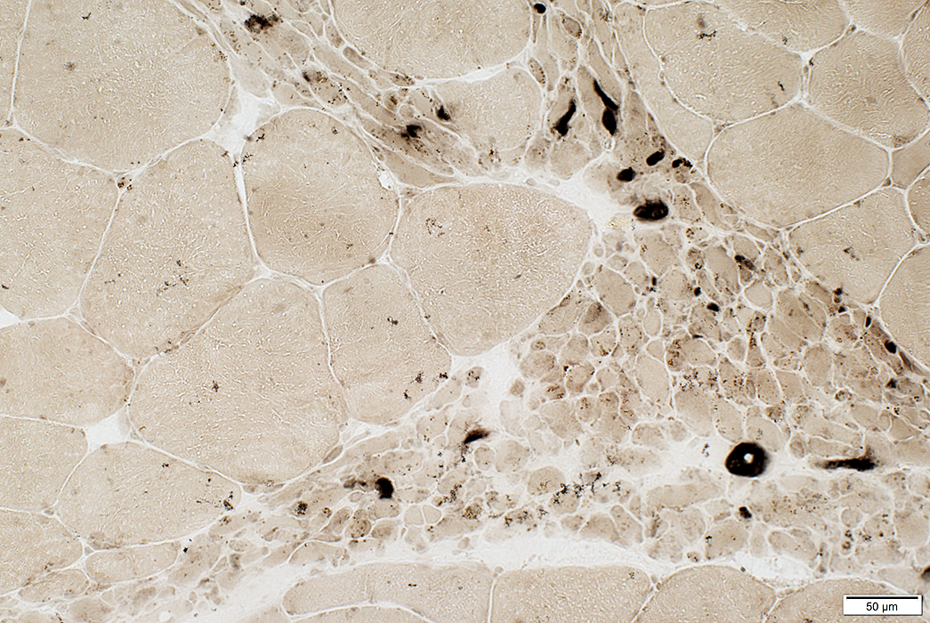
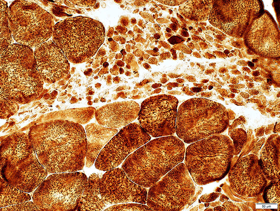
Pyknotic nuclear clumps: Stain dark with esterase & NADH
 NADH stain |
 Esterase stain |
 Esterase stain |
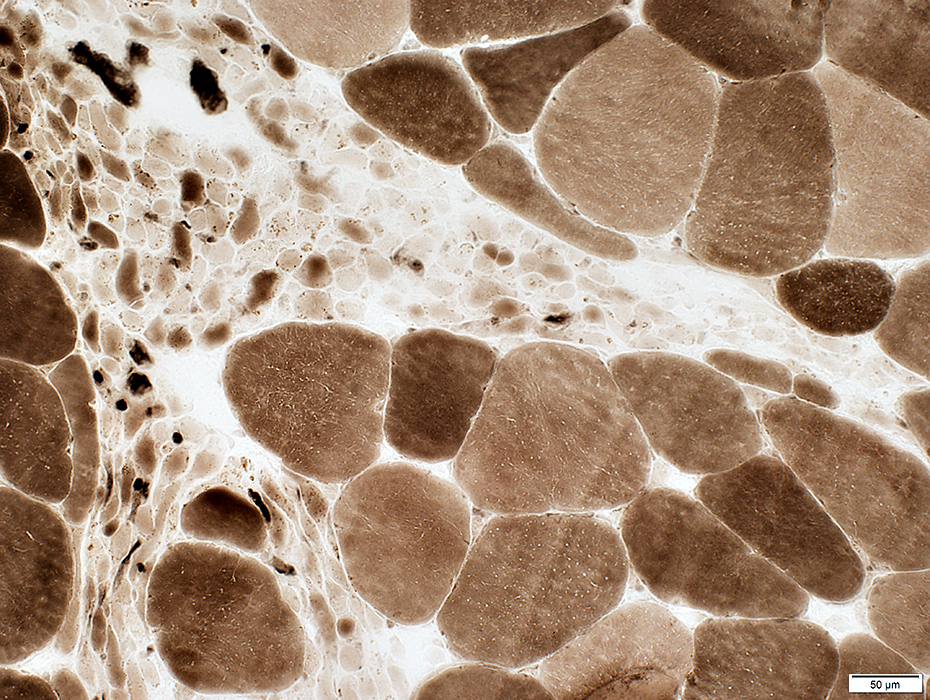
Pyknotic nuclear clumps: With lipofuscin
 Acid phosphatase stain |
 Acid phosphatase stain |
 From: R Schmidt |
 From: R Schmidt |
|
|
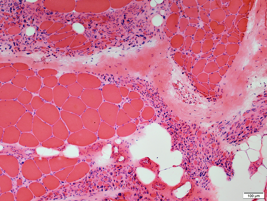
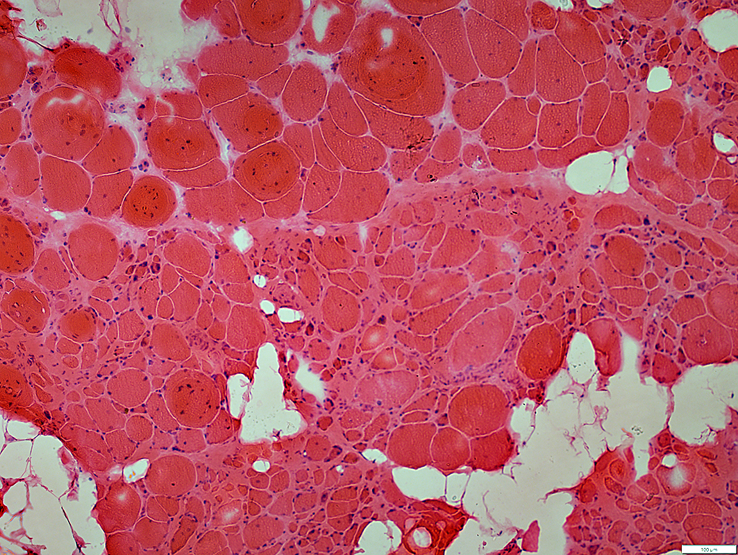
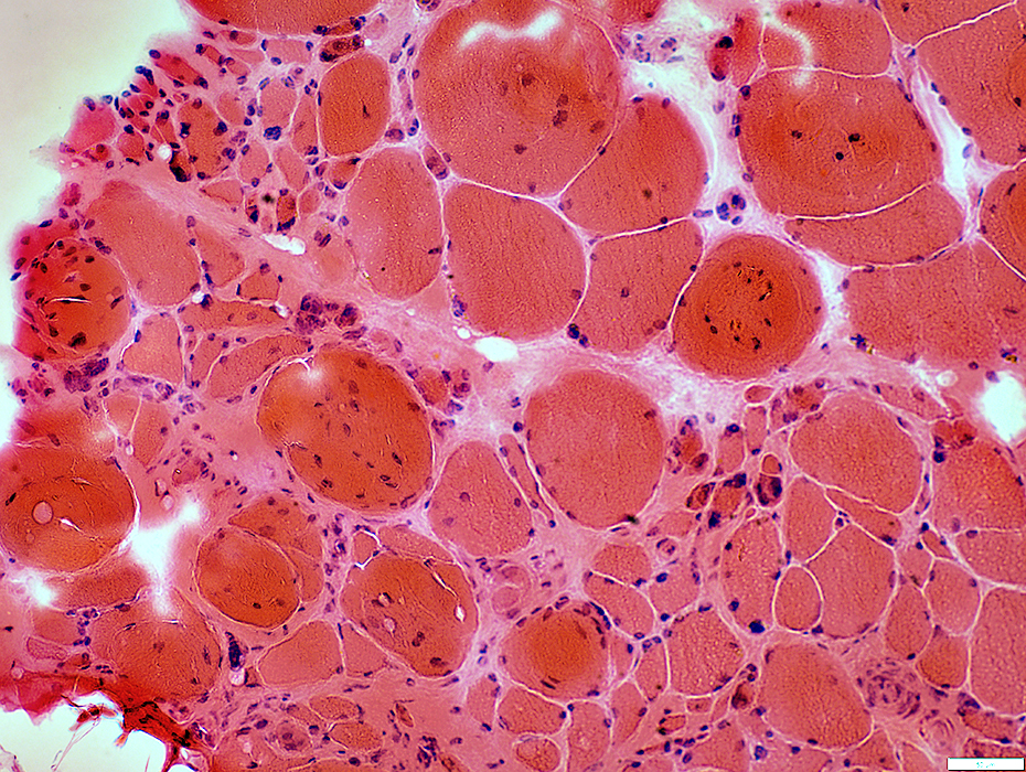
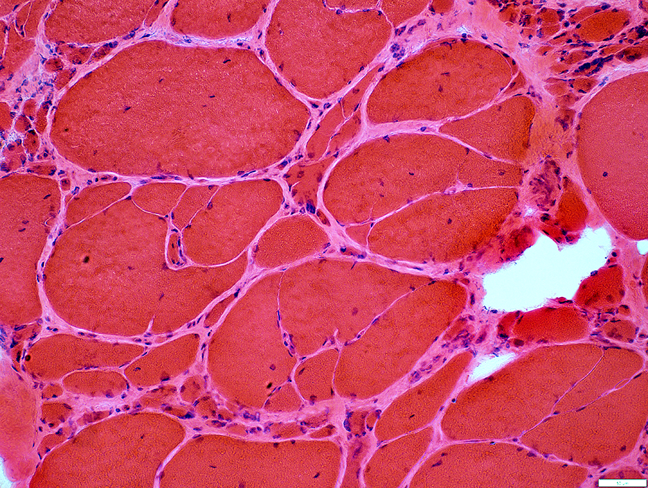
Chronic Partial Denervation: Pseudo-Myopathic Changes
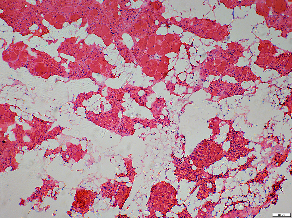 H& E stain |
- Endomysial connective tissue: Increased
- Internal nuclei: One or several in muscle fibers
- Muscle fiber sizes
- General: Widely varied
- Hypertrophic muscle fibers: Common
- Small fibers
- May be round or angular
- Pyknotic nuclear clumps: Common
- Grouped atrophy of muscle fibers
- Atrophic groups: Less well demarcated than in ongoing denervation
- Endomysial connective tissue: More prominent in these regions
- Perimysium: Replaced by fat
 H& E stain |
 H&E stain |
 Gomori Trichrome stain |
 Gomori Trichrome stain |
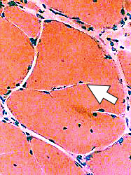
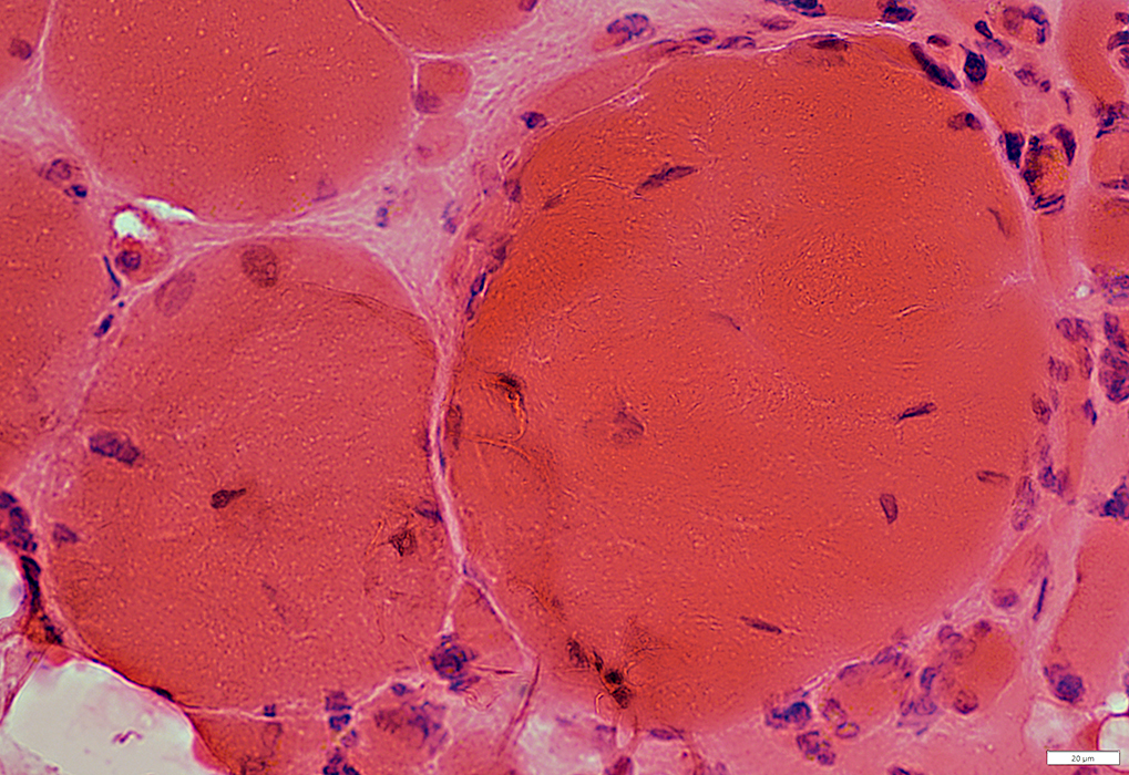
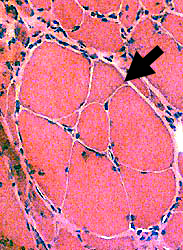
Large Muscle Fibers: Hypertrophy & Abnormal Internal Architecture
Split (Partially fused) Muscle Fibers (White arrows) 1
- Common features
- Very large (Hypertrophic)
- Have internal nuclei
- Probably result from: Partial fusion of regenerating fibers
- Associated with: Skeletal muscle fiber branching

|
 H&E stain 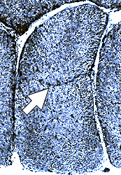 NADH stain |
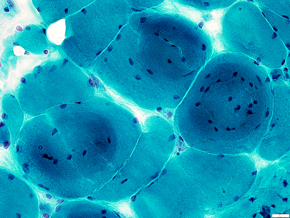
Internal Nuclei
 H&E stain |
Several myonuclei are scattered internally in cytoplasm of some muscle fibers
Normal muscle fibers have all myonuclei in subsarcolemmal regions
 H&E stain |
 H&E stain |
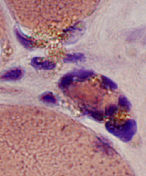
Necrotic large muscle fibers → Clustered regeneration
- Hypertrophied muscle fibers may become necrotic (left)
- Several different muscle fibers regenerate in its place & cluster within old basal lamina of large fiber
Muscle Fiber Necrosis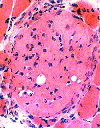 H&E stain Large necrotic muscle fiber Invaded by many histiocytes |
Clustered Regeneration (Arrows)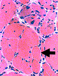
 H&E stain |
Clustered Regeneration
 H&E stain |
Areas of grouped atrophy are also present in this image
 NADH stain |
Hypertrophic Muscle Fibers: Abnormal Internal Architecture
 VvG stain |
 Gomori Trichrome stain |
 VvG stain |
 Gomori Trichrome stain |
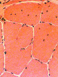
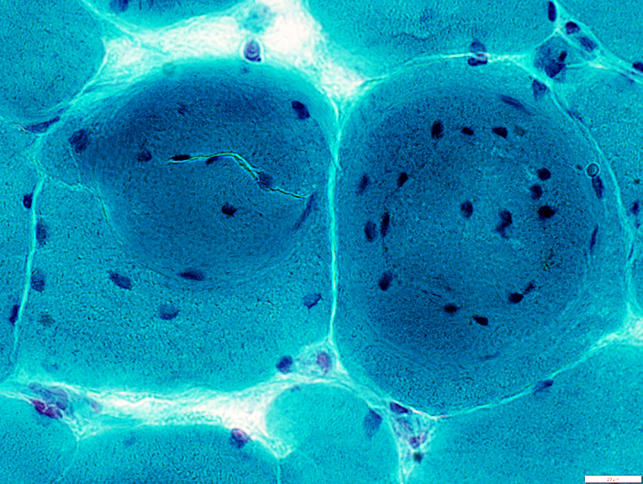
Chronic Denervation: Capillary Pathology
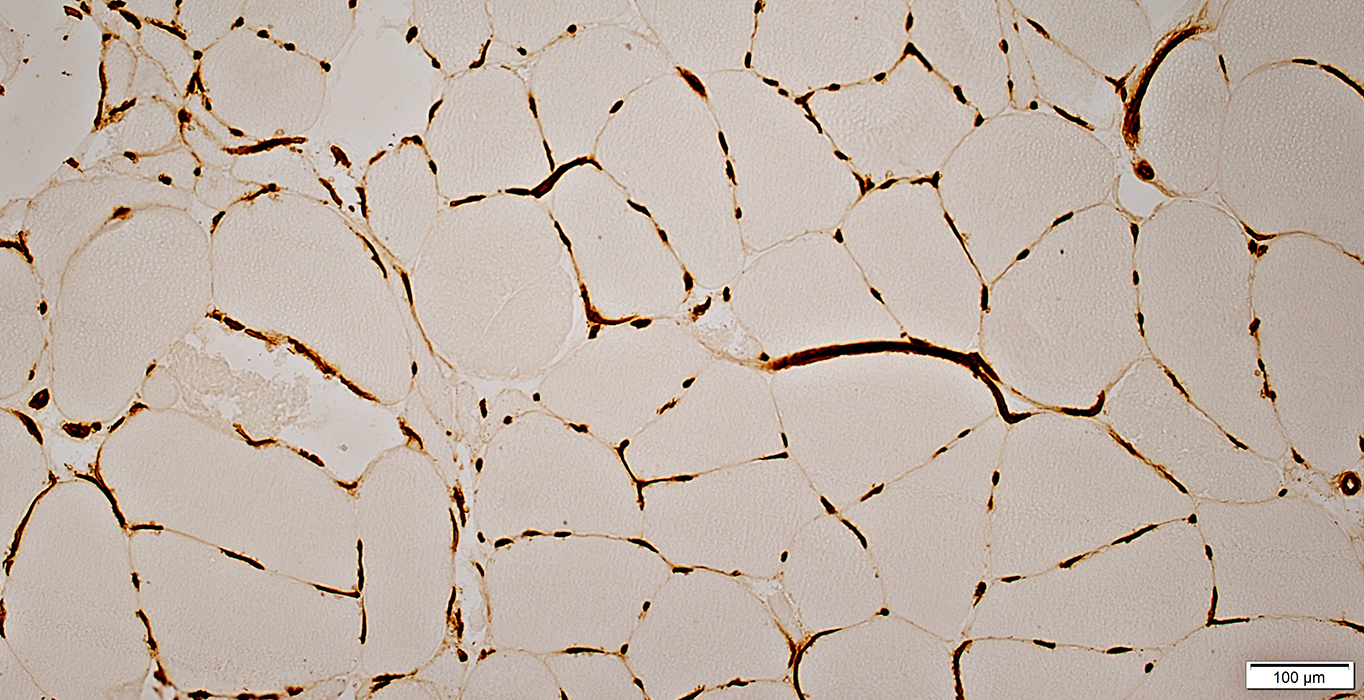 UEA I stain |
Abnormal orientation: Some become circumferential around muscle fibers
Increased numbers of capillaries adjacent to each muscle fiber
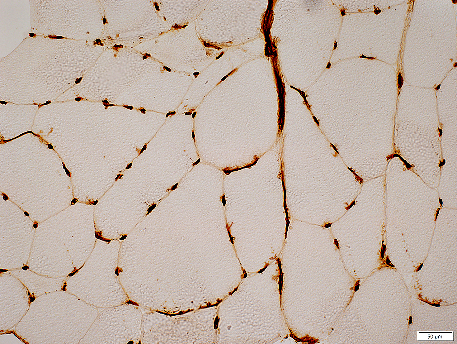 UEA I stain |
References
1. Skelet Muscle 2023;13:13
Return to Muscle biopsies
Return to Biopsy illustrations
Return to Neuromuscular home page
Return to Polyneuropathy Index
6/1/2025