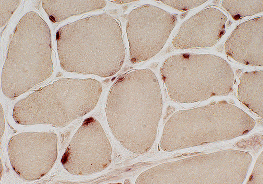
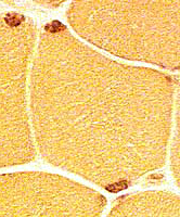
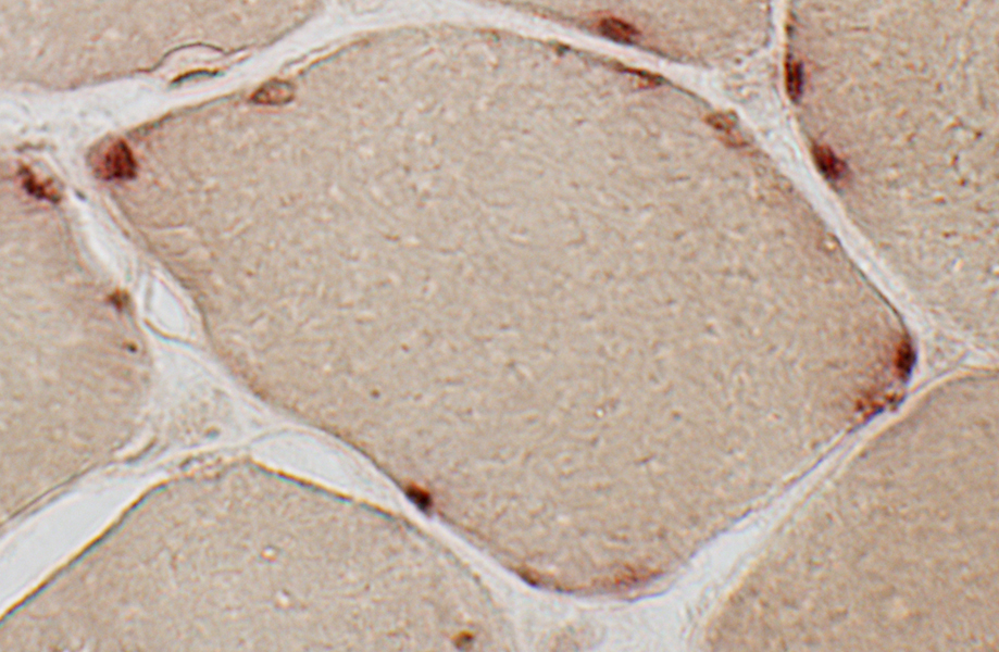
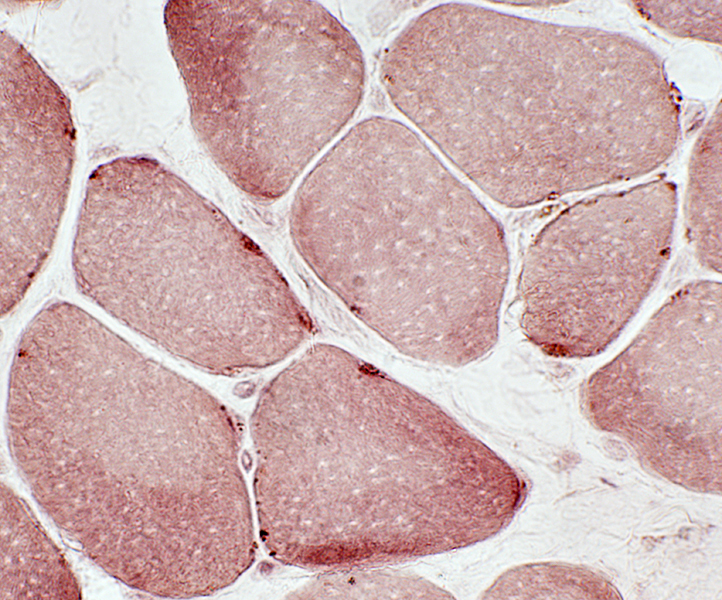
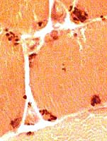
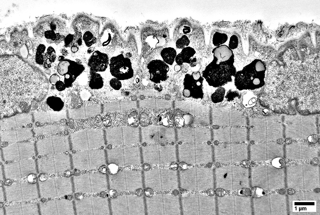
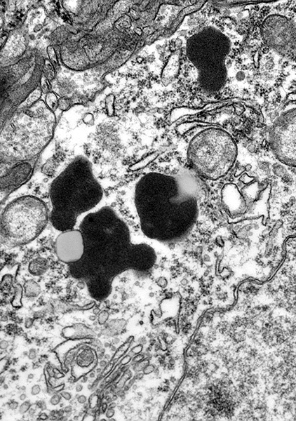
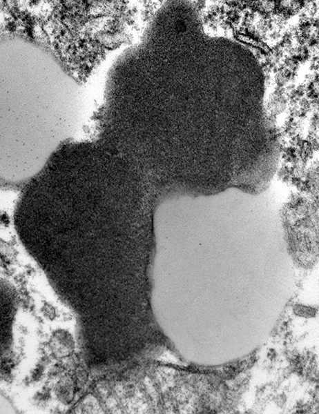
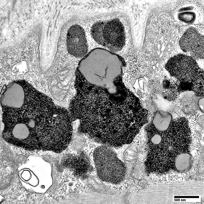
- Lipofuscin granules: General
- Yellowish brown
- Location: Subsarcolemmal region of muscle fibers; Near nuclei
- Highlighted by
- Lysosomal stains: Acid phosphatase (Strongly positive); Esterase
- Oil red O
- Ferric-ferricyanide reduction test
- Autofluorescence
- Ultrastructure: 12 nM granules; Vacuolar
- Lipofuscin granules become more abundant with
- Normal
- Non-dividing cells: Neurons; Muscle (Skeletal, Smooth, Cardiac)
- Fiber type: More in Types I & IIA than in Type IIB
- Increasing age
- Not degraded
- Accumulate over years
- Acromegaly
- Denervation atrophy: Increased concentration in small, denervated muscle fibers
- Lipid myopathies
- Chronic obstructive pulmonary disease
1
- Centronuclear myopathies, adult: Lipofuscin accumulates around nucleus
- Concentrated in: Pyknotic nuclear clumps
- Lipofuscin reduced
- Active myopathies: Duchenne muscular dystrophy
|