BULBOSPINAL MUSCULAR ATROPHY
Patterns of Pathology: Muscle
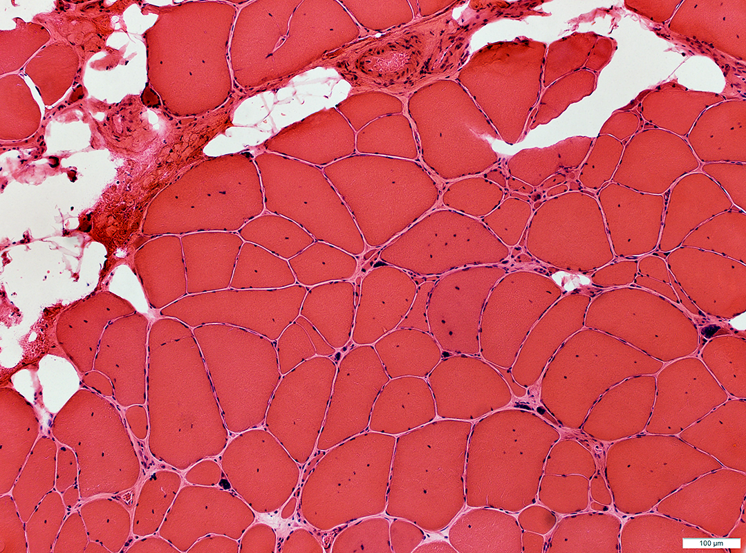
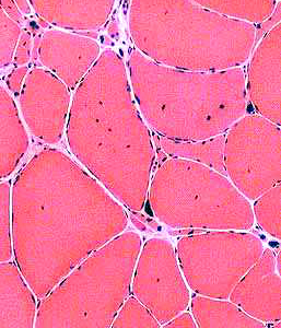
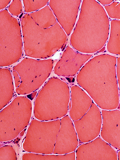
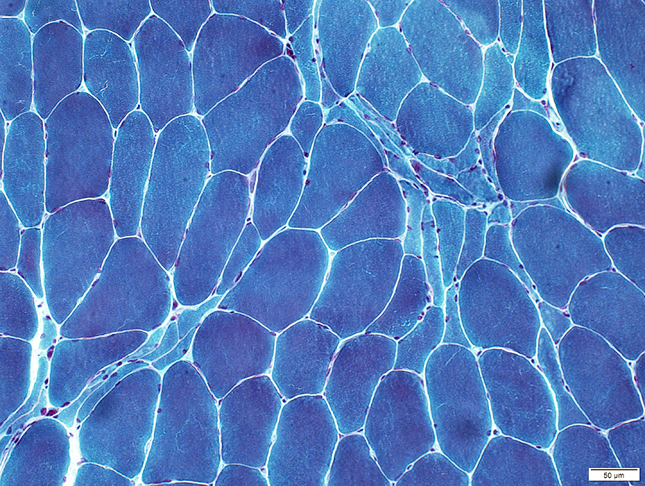
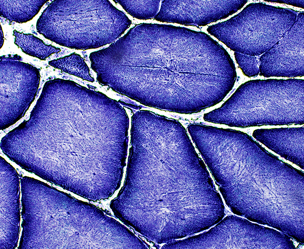
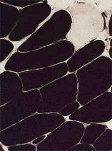
H & E stain |
|
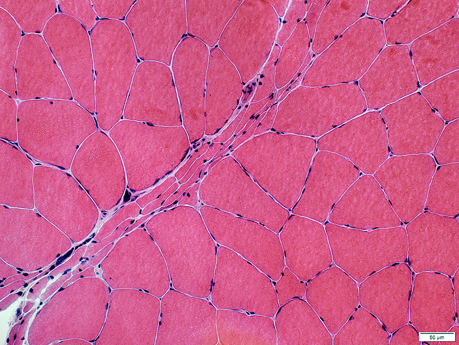
Muscle Fibers Sizes Hypertrophic Small: Angular or Rounded Grouped or Individual Pyknotic nuclear clumps (Large) Internal architecture Partially fused (Split) Internal nuclei Damage Necrosis: Scattered large fibers Regeneration: Clustered immature fibers |
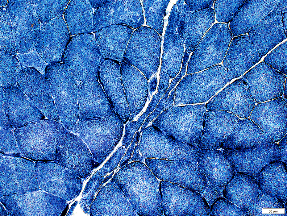
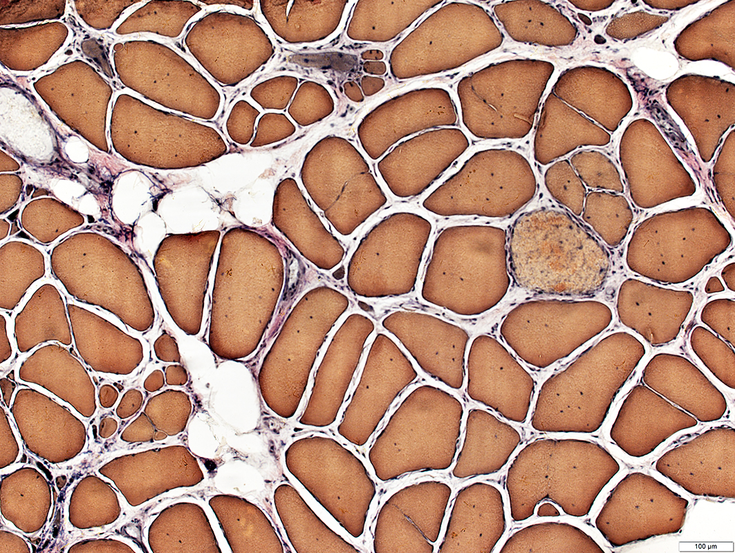 VvG stain |
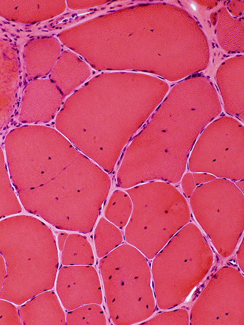
 H & E stain |
 VvG stain |
 H & E stain |
 |
 |
 |
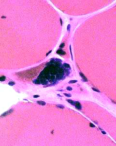
Internal nuclei
Partial, or incomplete, fusion
Hypertrophy
Pyknotic nuclear clumps
|
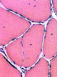
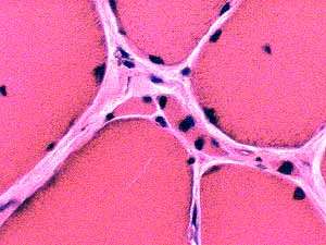
Muscle fibers: Small & Angular, or Pyknotic nuclear clumps |
|
 H & E stain |
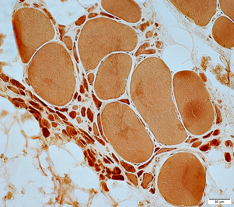
 Esterase stain |
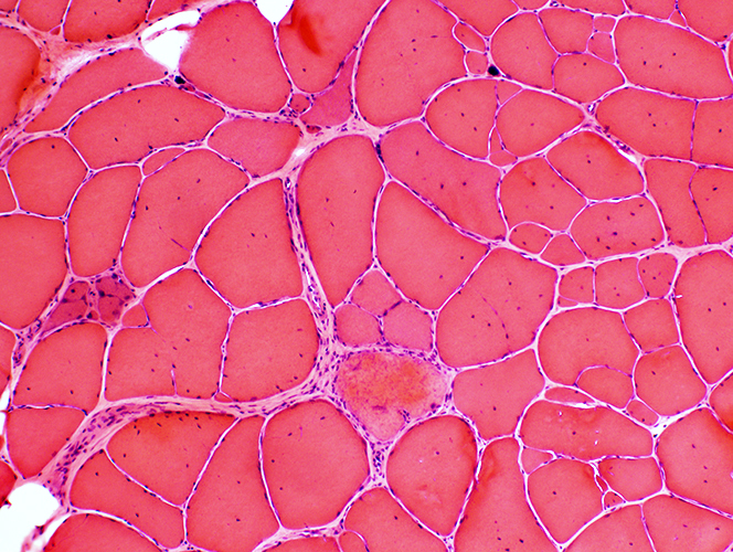
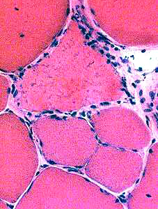
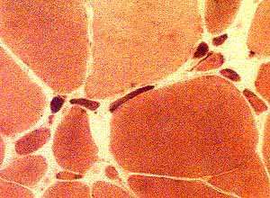
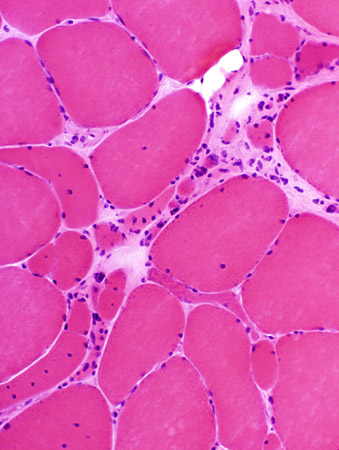
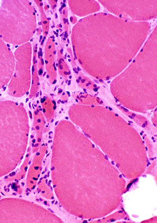
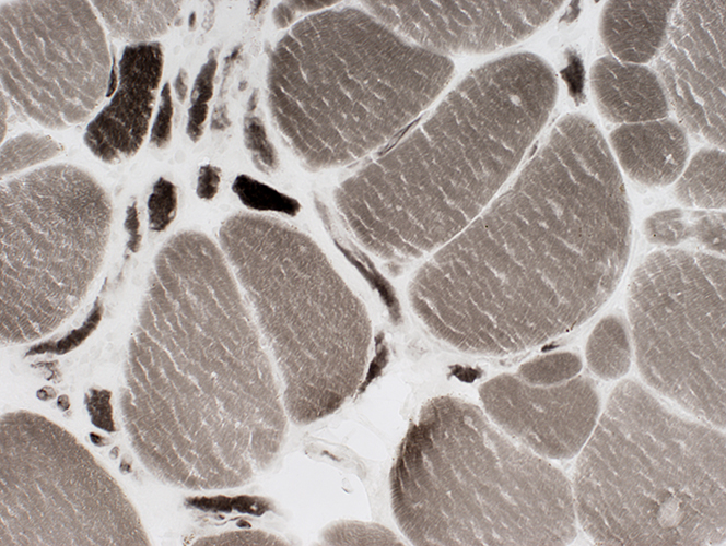
Muscle fibers: Grouped atrophy
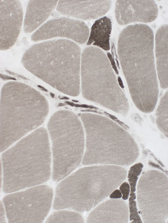
 H&E stain |
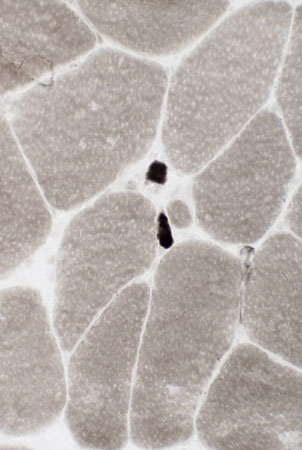
 H&E stain |
 H&E stain |
|
|
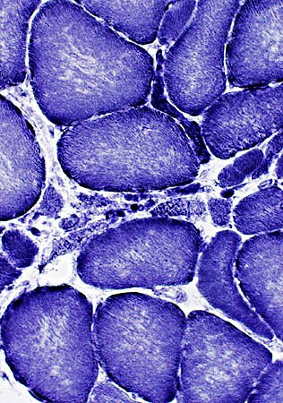
Grouped atrophy Smaller fibers may be rounded or angular Large muscle fibers are hypertrophied | ||
 VvG stain | ||
 Gomori trichrome stain |
 Esterase stain |
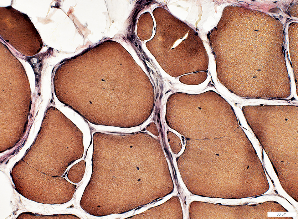
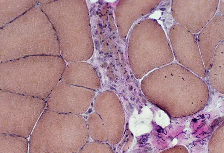
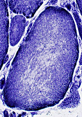
Muscle fibers: Internal architecture
 NADH stain |
 NADH stain |
|
Some small fibers are darkly stained Large muscle fibers Coarse internal architecture; Pale centers "Splitting" (Partial fusion)(Below) | |
 NADH stain | |
 NADH stain |
Muscle: Fiber types
 ATPase, pH 9.4 |
 ATPase, pH 9.4 |
 ATPase pH 4.3 stain |
|
Large muscle fibers: Type 1 predominance Small muscle fibers: Either type 1 or 2 Grouped atrophy: Small regions; Mixed fiber types |
 ATPase pH 9.4 stain |
Neuronal Intranuclear Aggregates
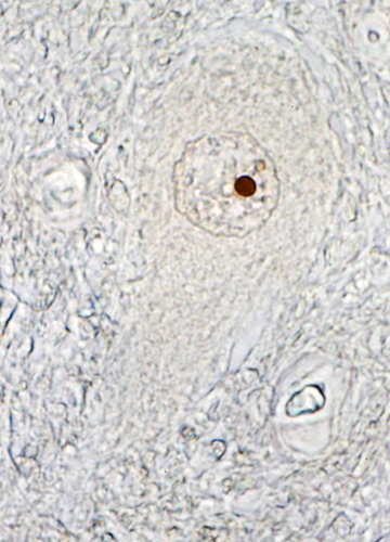 From Mei Li MD Androgen Receptor in Motor Neuron
|
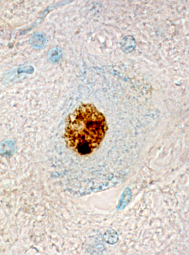 From Mei Li MD Polyglutamine in Motor Neuron
|
Return to Neuromuscular Home Page
Return to Pathology index
Return to BSMA (Kennedy's syndrome)
5/24/2019