IMPP (Also see IMPP with Jo-1 antibodies)
Clinical presentations
- General
- Onset age: Adult more common than Childhood
- Dermatomyositis: With weakness & skin rash
- Immune myopathy: With weakness, but no skin rash
- Myalgia syndrome: With high aldolase but normal strength & serum CK
- Interstitial lung disease
- Arthritis
General features
- Clinical & Pathologic classification: Immune myopathies with Perimysial Pathology
- Also see: Jo-1 antibody-related IMPP
- Onset age: More common in adults
- Pathology
- Perimysial damage: Common
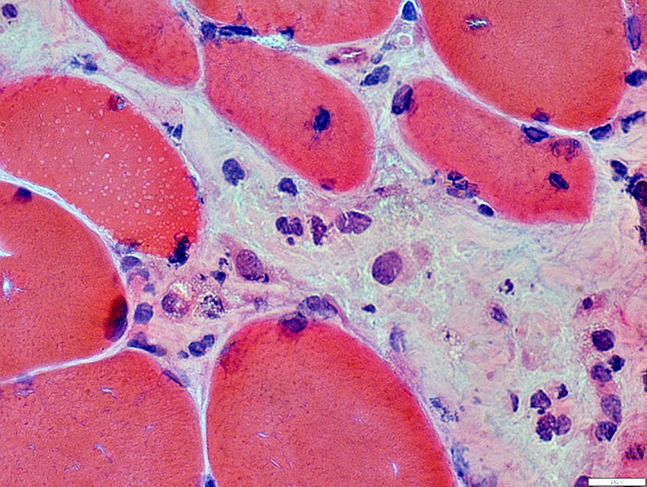
- Muscle fiber necrosis
- More in perifascicular muscle fibers
- More common than in DM-VP
- Cytochrome oxidase: No reduction in muscle fibers
Muscle Fiber Pathology
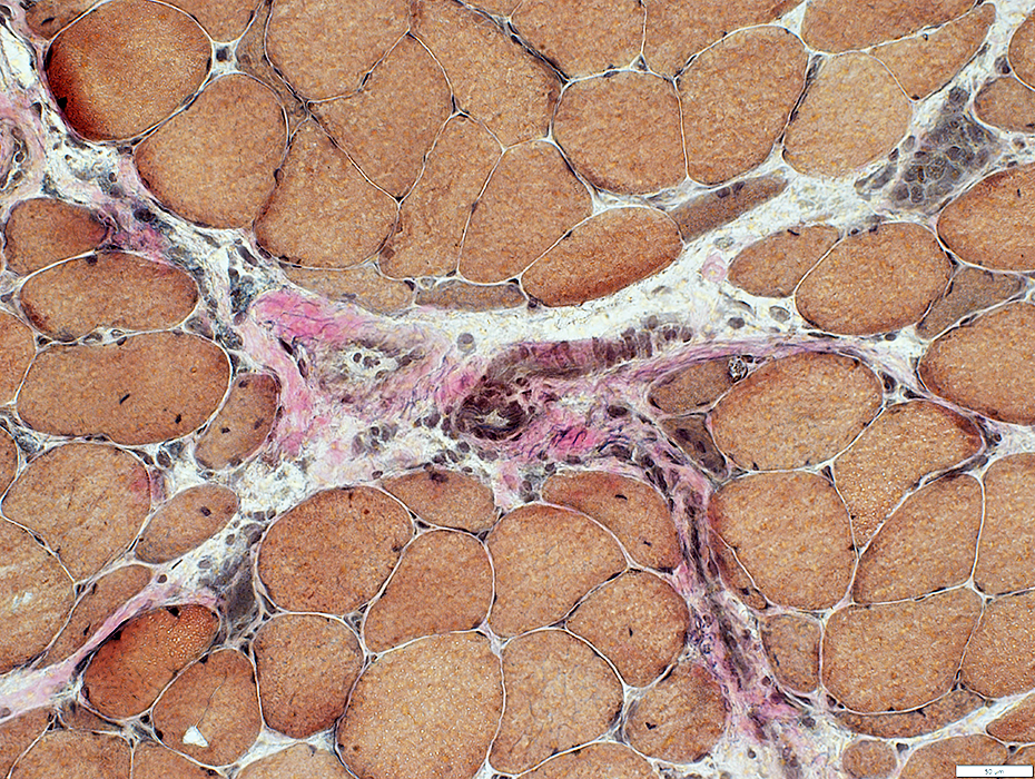 VvG stain |
Most in perifascicular regions
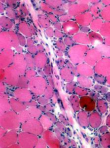
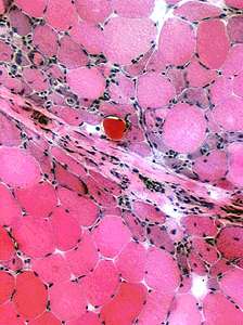
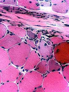 H&E stain |
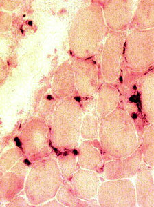 Acid phosphatase stain |
|
Acid phosphatase stained cells replace necrotic muscle fibers |
 Alcian blue stain |
 Alcian blue stain |
Muscle Fiber Internal Architcture
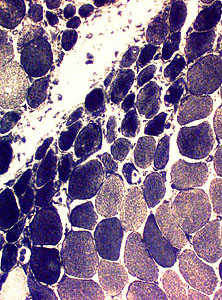
 NADH stain |
 Cytochrome oxidase stain |
| |
|
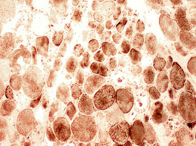
MHC Class I Often upregulated more on muscle fibers near the edge of fascicles in IMPP 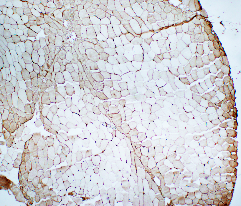 MHC Class I stain |
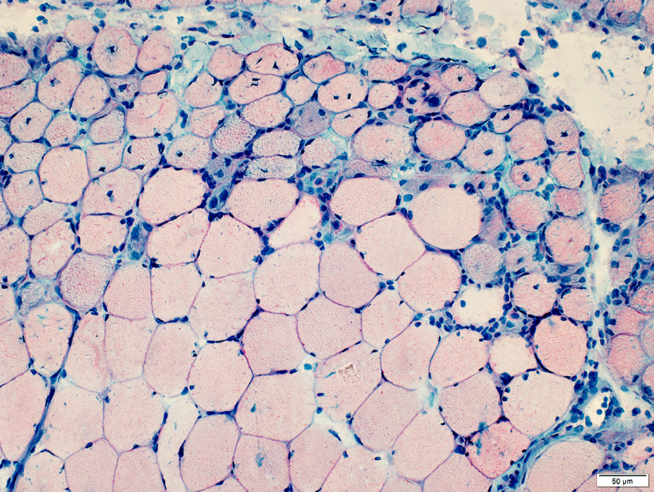
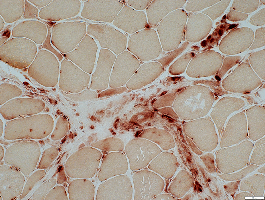
Perimysium: Immune cells (Histiocytes)
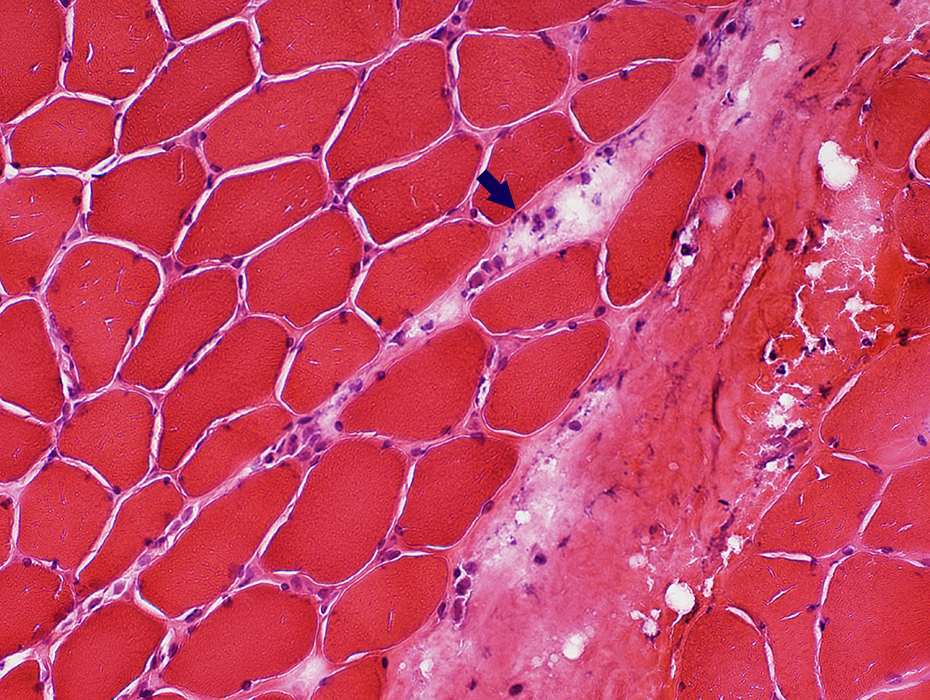 H&E stain |
 H&E stain |
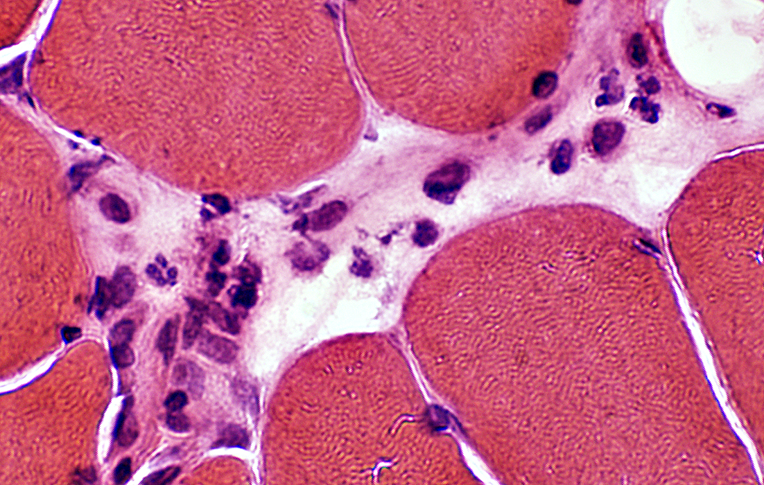 H&E stain |
 H&E stain |
|
 Acid phosphatase stain |
 H&E stain |
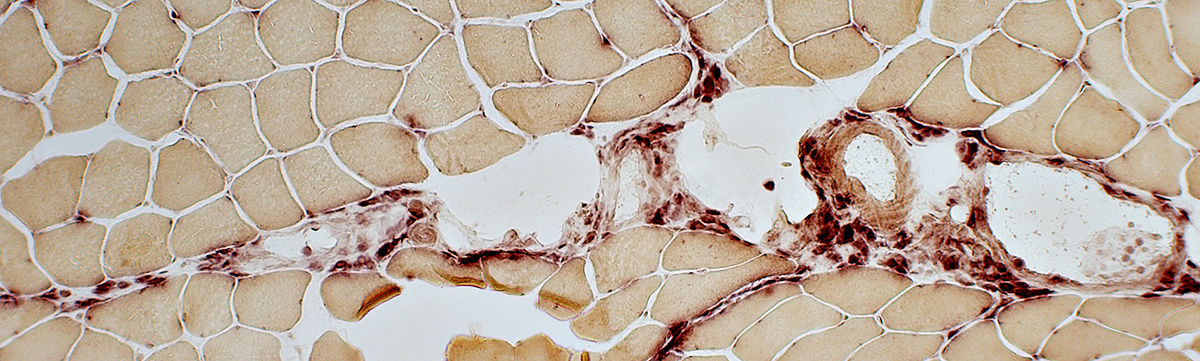 Acid phosphatase stain |
 Acid phosphatase stain |
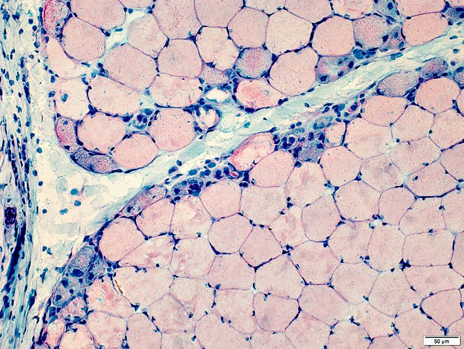
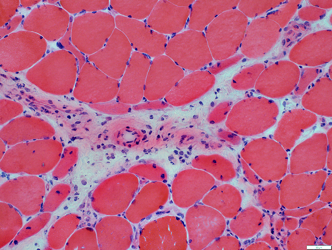
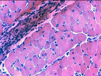
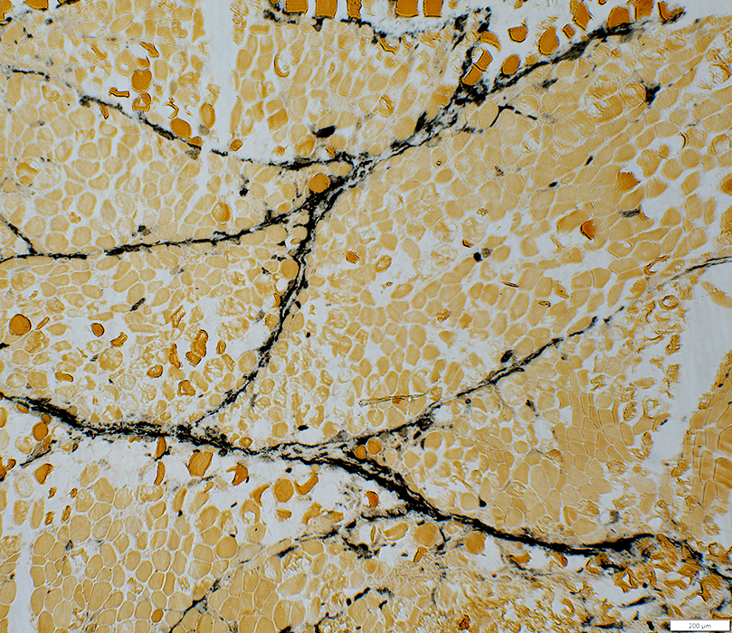
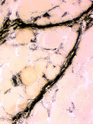
Perimysium: Damaged structure
Normal perimysium (Left) IMPP perimysium (Right)
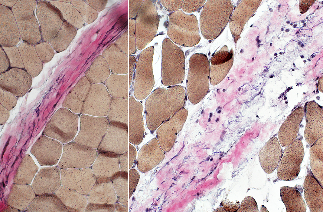 VvG stain |
Perimysium, Damaged structure: Fragmented & Widened
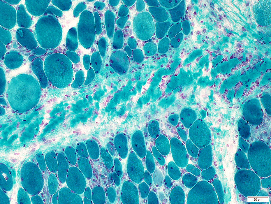 Gomori trichrome stain |
 VvG stain |
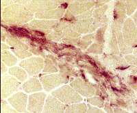
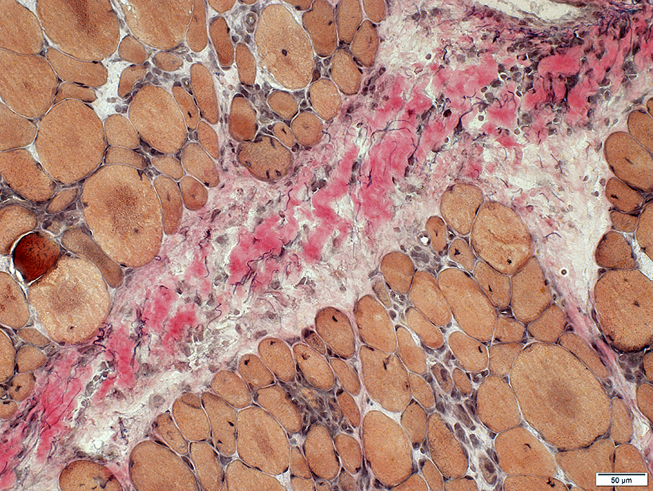
Perimysial Pathology: Alkaline Phosphatase staining
 Alkaline phosphatase stain Perimysial connective tissue: Alkaline phosphatase stained |
 Alkaline phosphatase stain |
|
C5b-9 Deposition on perimysial connective tissue extending into the endomysium Punctate deposition on the surface of some muscle fibers 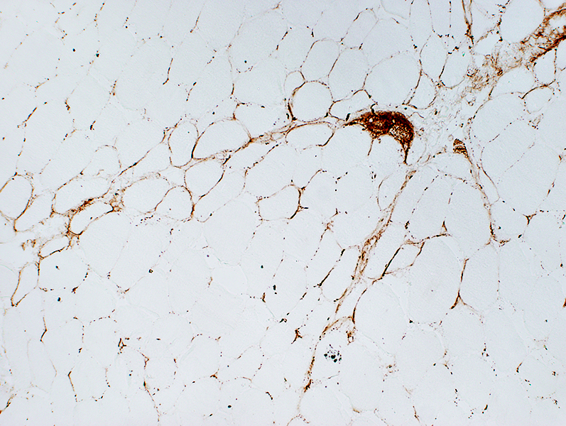 C5b-9 stain |
Return to Neuromuscular Home Page
Return to Dermatomyositis, Childhood pattern
Return to Inflammation
Return to Inflammatory myopathies
Return to Dermatomyositis
3/3/2021