Home
,
Search
,
Index
,
Links
,
Pathology
,
Molecules
,
Syndromes
,
Muscle
,
NMJ
,
Nerve
,
Spinal
,
Ataxia
,
Antibody & Biopsy
,
Patient Info
SYSTEMIC METABOLIC DISORDERS
Endocrine
Hepatic
Renal
RENAL FAILURE
Polyneuropathy
Epidemiology
Frequency: 60% with at least subclinical neuropathy
Male > Female
Associated medications: Colchicine; Nitrofurantoin; Alcohol
Clinical features
General
Distribution: Symmetric; Distal; Legs > Arms
More severe with long term dialysis
Sensory loss
Modalities: All (Large & Small fiber)
Distal > Proximal
Legs > Arms
Spontaneous sensations
Paresthesias & Dysesthesias: > 40%
Pain & Burning (Feet): 10%
Itch
Especially frequent with hemodialysis
Associated with presence of polyneuropathy
Motor: Weakness in patients with more severe neuropathy
Distal
Feet & Hands
Spontaneous activity
Cramps: 66%
Restless legs
Myokymia
Tendon reflexes: Absent distal or diffuse
Autonomic dysfunction: Mild
Loss of R-R interval variation
Sympathetic skin response: Abnormal in 45%
Course: Progression & resolution: Slow
Lab
CSF: Unremarkable
Electrophysiology
Mainly axonal loss: Low CMAP amplitudes
Mild slowing of NCVs
Not related to clinical signs
Correlates with creatinine clearance
Motor conduction velocity
May fall to 50% of mean control value
Peroneal correlates with creatinine clearance
Distal motor latencies: May be prolonged
SNAPs: Reduced amplitude
Nerve pathology
Distal axonal degeneration & loss
Axon size: May be reduced
Treatment
Renal transplantation
Improvement in most features
Little change in autonomic dysfunction
Dialysis: Stabilizes neuropathy
Rule out:
Additional drug toxicity
Amyloidosis
Mononeuropathy
Carpal tunnel syndrome
: ? related to vascular shunts or amyloid
Ischemic arm neuropathies: Related to fistulas
Neuromuscular junction
Aminoglycoside toxicity
Myopathy
Disorders
Cachexia
Nephrotic syndrome
Inanition
Electrolyte disturbances
Hypercalcemia
Hyperkalemia
Hypermagnesemia
Disordered vitamin D metabolism: With bone disease
Muscle infarction 2° arterial calcification:
Calciphylaxis
Skin necrosis
Myopathy;
Rhabdomyolysis
Nephrogenic fibrosis
Drug toxicity
Aluminum
Laboratory
Serum CK: Usually normal, except some drug toxicity
Pathology:
Type II muscle fiber atrophy
HEPATIC DISORDERS
Demyelinating neuropathy
Sensory neuropathy with 1° biliary cirrhosis
Vitamin E deficiency
Childhood cholestatic disease
Hepatitis C
Navajo neuropathy with corneal ulceration
Hepatic myelopathy
Pruritis
Hepatic Demyelinating Neuropathy
Frequency: 50% to 70%
Usually subclinical
Distal; sensory
Autonomic: Usually parasympathetic, mild
Hepatic myelopathy
1
Associated with
Chronic liver disease
Acquired hepatocerebral degeneration: White matter enhancement on T2 MRI
Myelopathy
Spasticity: Legs > Arms
Tendon Reflexes: Brisk
Sensory: Normal
Bladder: Normal
Other CNS: Ataxia; Encephalopathy
Pathology: Pyramidal tract loss of myelinated axons
Nephrogenic Fibrosis (Nephrogenic fibrosing dermopathy)
2
Epidemiology
Age: 4th to 6th decades
Sex: Males & Females affected
Cutaneous fibrosing disorder
Skin
Thickening & Hardening
Limbs: Usual
Legs: Most common; Distal > Proximal but sparing feet
Arms: Some patients; Distal or diffuse
Trunk: Occasional; Late
Face: Spared
Hyperpigmentation
Erythematous patches
Pain: Burning
Course: Progressive
Clinical: Other
Joints
Restriction of motion: Flexion contractures
Pain
Muscles
Hardening: Distal > Proximal
Skin changes over affected muscles
Strength: Normal
Neuropathy
Sensory: May be painful (Burning)
May be related to syndrome or renal disease
Eyes: Scleral injection
Associated disease
End-stage renal disease: Dialysis treatment; 2 to 7 years
Laboratory
Serum CK: Low (< 50)
Parathyroid hormone: Often high
Calcium-phosphate products: High
EMG: Myopathic changes
NCV: Reduced SNAP & CMAP amplitudes in most
Muscle MRI
High attenuation of fascial planes
Muscle signal: Patchy increase on T2 weighted image
Atrophy
Pathology: Skin
Dermal fibroblasts, spindle-shaped: Proliferation
Dendritic cells: Tissue accumulation
Collagen bundles: Thickened
Elastic fibers: Increased
Mucin deposition
Pathology: Muscle
Fibrosis: Perimysial (widening) > Endomysial
Myopathy: Varied muscle fiber size
Calcium deposition
Inflammation: Perimysium
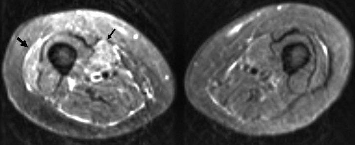
Nephrogenic fibrosis: MRI
Subcutaneous tissue: Thickened
Muscles
Patchy high T2 signal
MRI shows changes in: Vastus lateralis (Large arrow) & Gracilis (Small arrow) on right
Fascial planes: Increased signal
Hemochromatosis
3
General
Disorder of iron storage
Inappropriate increase in intestinal iron absorption
Deposition of iron in parenchymal cells
Several hereditary causes
Common adult form: HFE
with C282Y mutation; Recessive
Clinical
Onset
Age: Midlife
Symptoms: Fatigue, Abdominal Pain
Organ damage
Liver: Cirrhosis; Hepatomas
Pancreas (Diabetes)
Joints: Arthritis
Pituitary: Hypogonadotropic hypogonadism
Cardiac: Cardiomyopathy
Skin: Pigmentation
Polyneuropathies
Frequency (26%)
Sensory loss
Type
Large or Small fiber
Axon loss
May be associated with diabetes
Myopathy
Objective features: Rare
Associated with hemodialysis
Proximal weakness
Discomfort: Cramps, Fatigue & Pain
Hemosiderin deposits
Laboratory
Serum ferretin: High
Treatment: Removal of iron by phlebotomy or erythrocytapheresis
Return to
Neuromuscular Home Page
References
1. Neurology 2000;54:1011
2. Muscle Nerve 2004;30:569–577
3.
J Neurol 2010; Online April
4/29/2010