
Front,
Search,
Index,
Links,
Pathology,
Molecules,
Syndromes,
Muscle,
NMJ,
Nerve,
Spinal,
Ataxia,
Antibody & Biopsy,
Patient Info
|
RESPIRATORY FAILURE: Early or selective
Adult onset respiratory failure
- Muscle
- Neuromuscular junction
- Nerve
- Spinal
- CNS
Infantile respiratory failure
Respiratory failure: Evaluation & Management
- Testing
- Forced vital capacity (FVC): Reliable; May miss some respiratory problems
- Other testing
- FVC while supine
- Formal pulmonary function testis
- Nocturnal disorders of breathing
- Management
- Early: Non-invasive nocturnal ventilation, preferably NIPPV
- Assist cough when needed
- Avoid early tracheostomy
- Maintain ideal body weight.
- Control oral secretions
- Aggressively treat URI’s.
- Give Influenza vaccine & Pneumovax.
- Avoid supplemental O2 unless addressing hypoventilation fails
- Outcomes in patients with chronic neuromuscular disease,
- Prolongs life
- Reduces symptoms
- Improves quality of life
- Prevents acute respiratory failure & delays tracheostomy
- End of life
- Prevent dyspnea and discomfort with O2
- Use of narcotics & benzodiazepines
- Terminal weaning: Not sudden withdrawal of support
Respiratory failure: Exacerbating factors
- Reduced central drive
- Primary: Myotonic dystrophy
- Drugs: Sedatives hypnotics
- Supplemental oxygen
- Physical impediments
- Obesity: Increased work of chest wall movement
- Constipation
- Supine position: Inhibition of diaphragmatic movement
- Kyphoscoliosis: Reduced chest wall compliance
- Primary lung disease: Obstructive disease
- FEV1/FVC ratio reduced
- Increased airway resistance slows expiration
| PATTERNS OF SPIROMETRIC ABNORMALITIES |
|
Obstructive Diseases |
Restrictive Diseases |
| FVC |
Normal or ê |
ê to êêê |
| FEV1 |
ê to êêê |
ê to êêê |
| FEV1 /FVC |
ê to êêê |
Normal or é |
| FEF25%-75% |
ê to êêê |
ê to êêê |
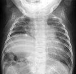
Restrictive ventilatory defects are characterized by proportional
decreases in FVC and FEV1, leaving the FEV1/FVC
normal or even slightly elevated. Any lesion affecting the lung, chest
wall, or respiratory muscles that reduces the ability to take in a normal amount of
air but does not affect the conducting airways is classified
as a restrictive lung disease.
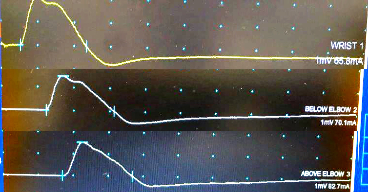
Obstructive defects are characterized
by their involvement of the airways and the resultant reduction in
expiratory flow. Spirometric studies of obstructive
lung disease generally show a reduced FEV1,
FEV1/FVC and flow rates with a relatively normal FVC. In
severe obstructive lung disease, the FVC may also be reduced.
Home ventilation: Types
Pressure-limited: BiPAP
- Mechanisms
- Patient initiates breath
- Machine senses negative pressure
- Air delivered until set pressure is reached
- End inspiration pressure drops to set (positive) level
- Typical settings
- IPAP: 12 to 20 cm H2O
- EPAP: 3 to 4 cm H2O
- Advantages
- 1st step: When hypoventilation in only nocturnal
- Can deliver breaths without patient triggering: For sleep apnea
- Simple to operate
- Inexpensive
- Semi-portable
- Disadvantages
- Can't deliver large volumes: No cough assist
- Most unit have no battery
Volume-limited
- Mechanisms
- Patient initiates breath
- Machine senses negative pressure
- Air delivered until set volume is reached
- No compenstaion for air leakage: Set for large volumes (10 to 15 cc/kg)
- Advantages
- Battery back-up
- Back-up alarms
- Better for full time respiratory support
- Adjustable flow rates
- Can increase voice volume & cough
- Disadvantages
- Expensive
- Units may be heavy: But can be place on power wheelchair
MYOSIN-LOSS (Acute Quadriplegic; Critical Illness) MYOPATHIES2
- General Syndrome
- Rapidly Evolving Myopathy with Myosin-Deficient Fibers
- Clinical features
- History
- Progressive proximal > distal weakness over days to weeks
- Unexplained, persistent weakness after respiratory support or neuromuscular paralysis
- Epidemiology
- Age: Mean 6th decade
- Sex: Males & Females involved
- Weakness
- Diffuse: Proximal > Distal; Neck flexors
- Symmetric
- Respiratory failure (80%)
- Severe cases: Face & occasional EOM
- Mild cases: Monophasic; May have Proximal arm weakness
- Often associated with some rhabdomyolysis
- Sensory: Normal, or Mild distal loss
- Tendon reflexes: Reduced
- Prognosis
- Slow improvement in weakness (months) when steroids tapered
- High mortality (30% to 50%) from associated disorders
- Associated factors
- Corticosteroids (80%): Especially higher doses
- Transplant: Lung; Liver
- Systemic disease
- Post-Paralysis Paralysis
- History
- Paralytic treatment for Status asthmaticus or other disorders
- Treatment with NMJ blocking agents
- Non-depolarizing agents; e.g. Vecuronium; ?Corticosteroids
- Treatment > 1 week
- Inability to wean from respirator
- Persistent weakness
- Laboratory
- Serum CK: Elevated or Normal
- High in 1st 2 weeks in status asthmaticus patients
- Normal in all after 2 weeks
- Electrodiagnostic
- NCV
- CMAP: Small
- Normal: Nerve conduction velocity; Distal Latencies; Sensory potentials
- EMG
- Often normal, or mild non-specific changes
- Irritative changes
- Occur early in course: Some patients
- Fibrillations
- Positive sharp waves
- Myopathic changes
- Occur in some patients: Later in course
- Action potentials: Low amplitude, Short duration
- Repetitive nerve stimulation
- Occasional decrement early: 1st week after discontinuation of paralytic agent
- Normal late
- Muscle fibers may be electrically inexcitable
- Muscle Pathology
- Myosin loss in muscle fibers on ATPase stain (ATPase, pH 9.4 & 4.3)
- ATPase
- pH 9.4: Fibers with staining intensity less than type 1
- pH 4.3: Fibers with reduced staining intensity at pH 9.4 also show reduced staining at pH 4.3
- Some biopsies show loss diffuse loss within muscle fibers
- Others show focal regions of
myosin loss within fibers
- Myosin loss present in scattered muscle fibers throughout musle biopsy: Not focal or diffuse regions
- Quantitation3
- Normal myosin/actin ratio in muscle: 1.31 to 1.57
- Critical illness myopathy myosin/actin ratio is low: 0.37
- Differential diagnosis
- Dermatomyositis in focal regions of muscle fiber damage
- HIV with nemaline rods
- Muscle fiber necrosis
- Atrophic muscle fibers
- Basophilic, atrophic type II muscle fibers (60%)
- All fibers atrophic (30%)
- Enlarged nuclei in atrophic muscle fibers
- Degeneration & Regeneration of muscle fibers: Occasional; Early
- Weakness: Pathophysiology
- Muscle membrane inexcitability
- Animal (rat) model
- Denervate hindlimb + High dose dexamethasone
- Loss of myosin with preservation of actin
- Increased glucocorticoid receptors in muscle membrane
- Muscle membrane changes
- Depolarization
- Reduced Membrane impedance 2° to Increased Cl- conductance
- Reduced Na+ current amplitudes
- Reduced number of Na+ channels
- Reduced Na+ channel conductance or voltage-dependent gating
- Myosin loss
- ? via Calpain pathway
- ? Related to Increased general muscle catabolism
- Protein degradation pathways possibly involved4
- TGF-β/MAPK
- Atrogin-1/proteasome
- Also see: Critical illness polyneuropathy
CONGENITAL DEFECTS OF THE DIAPHRAGM
Familial Congenital Agenesis of the Diaphragm

l
? Chromosome 1
Congenital Anterior Diaphragmatic Hernia

l
? X-linked vs. multifactorial with high male:female sex ratio
Diaphragmatic defects with skull ossification & limb deficiencies

l
? Autosomal recessive
Cough & Neuromuscular Disorders
Hoarse voice & Neuromuscular Disorders
Return to
Neuropathy Index
Return to
Myopathy & NMJ Index
References
1. J Neurol Neurosurg Psychiatry 1999;67:223-226
2. Muscle Nerve 2000;23:1785-1788
3. Intensive Care Medicine 2003;Online August
4. Ann Neurol 2004;55:195-206
12/2/2005