MYOPATHIES WITH MYOSIN LOSS
|
Clinical Histochemistry patterns: Myosin loss in Small muscle fibers Myosin loss in fibers Abnormal myonuclei Intermediate- & Larger-sized muscle fibers Severe muscle fiber atrophy Cushing disease Ultrastructure |
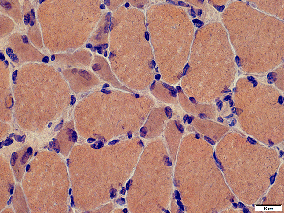
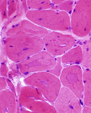
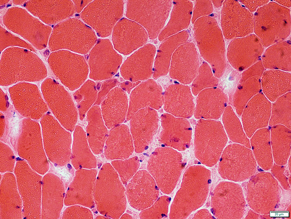
Myosin loss: Pathology predominantly in Small muscle fibers
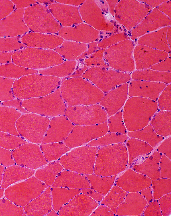
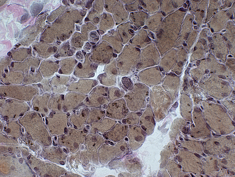
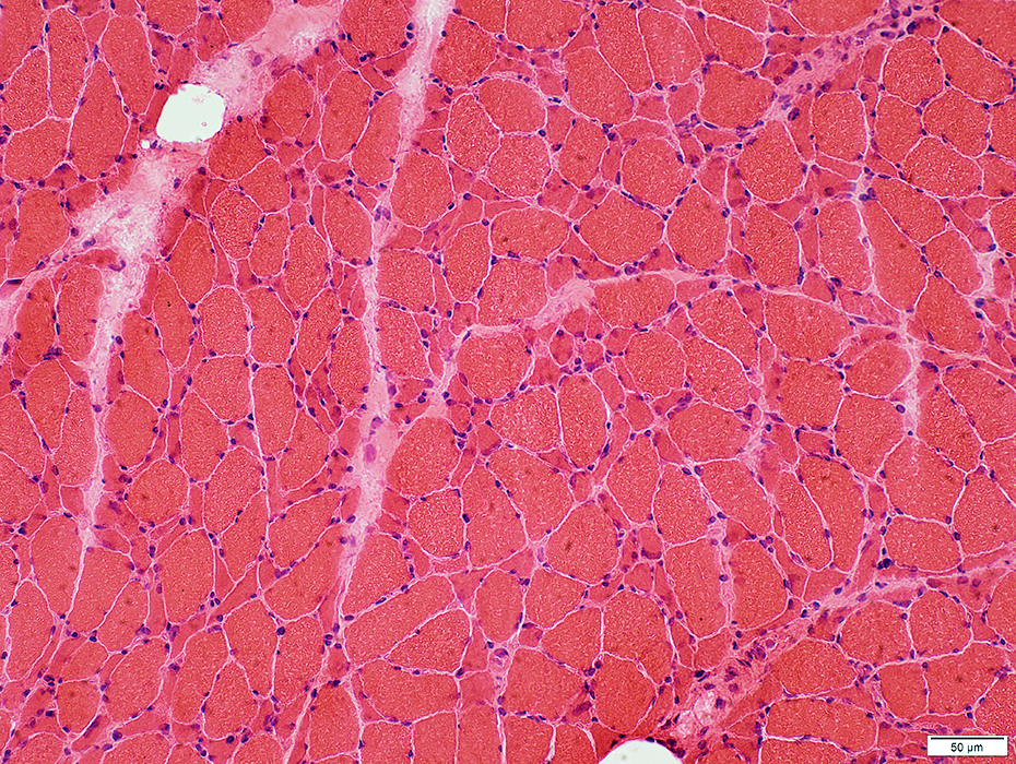 H&E stain |
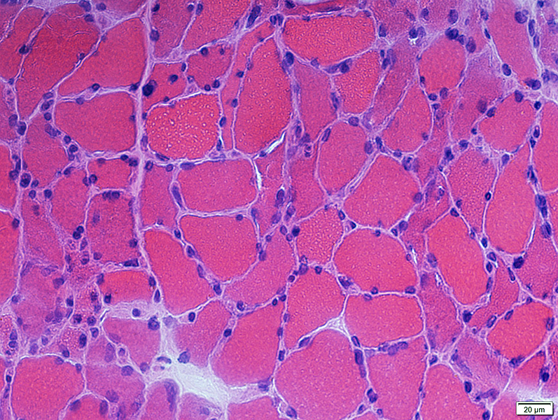
Shape: Angular
Cytoplasm: Basophiic
Nuclei: Large & Irregular shaped
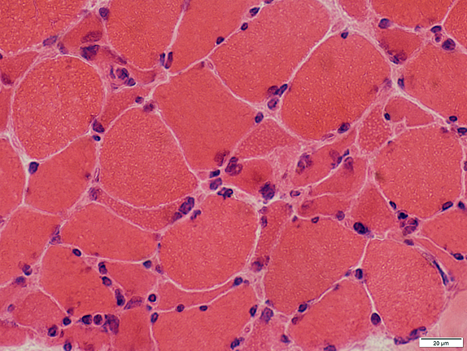 H&E stain |
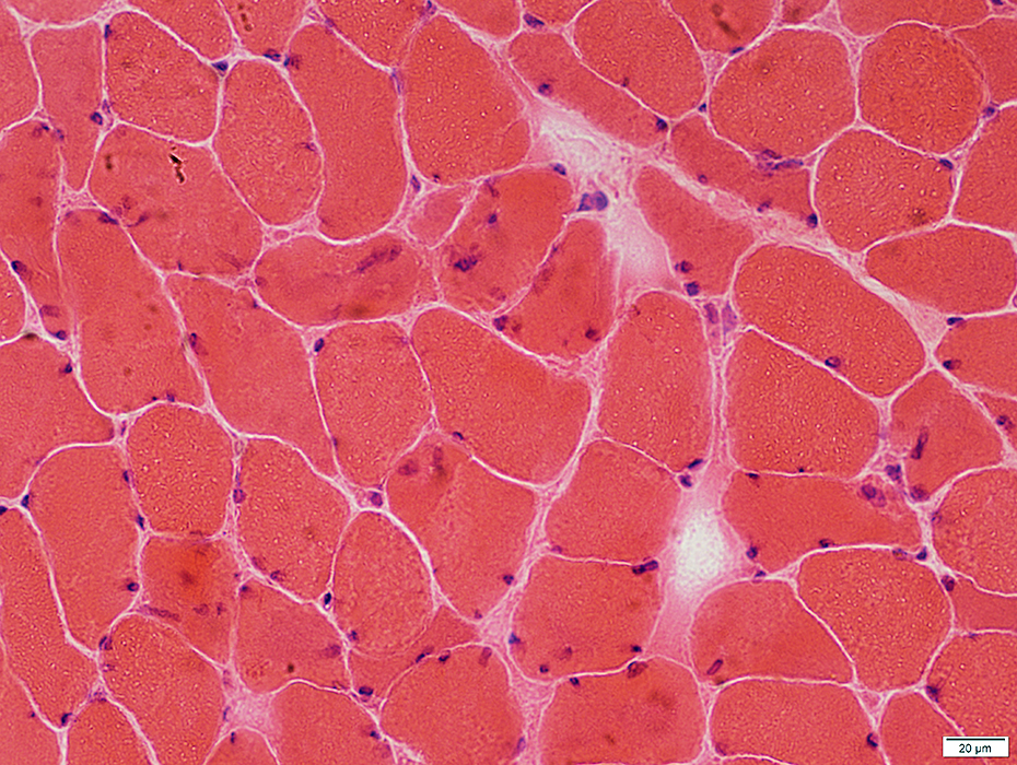
 H&E stain |
Shape: Angular
Cytoplasm: Basophiic
Nuclei: Large & Irregular shaped
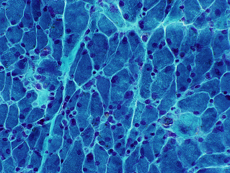
 Congo red stain |
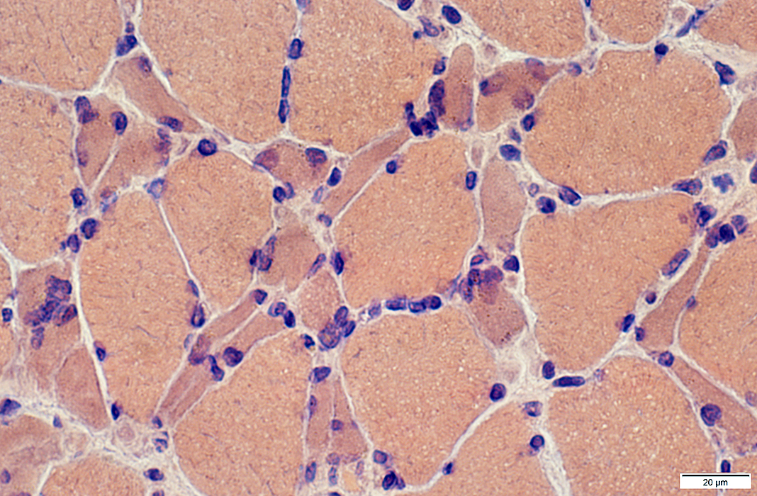
Myosin loss ("Critical illness") myopathy
Intramuscular nerves
Normal numbers of myelinated axons
Muscle fibers
Small
Angular
Cytoplasm: Loss of internal architecture
 VvG stain |
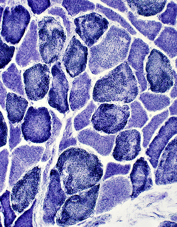
 Gomori trichrome stain |
 Gomori trichrome stain |
 NADH stain |
 NADH stain |
 NADH stain |
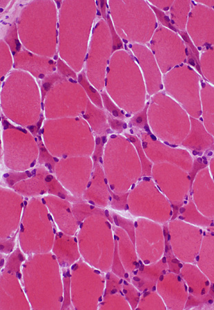
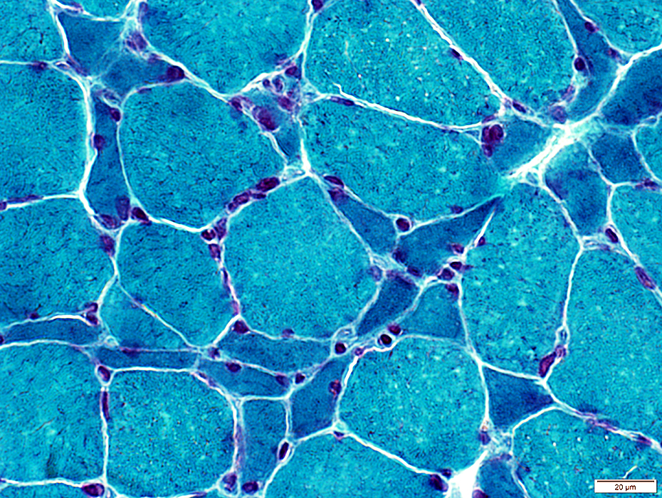
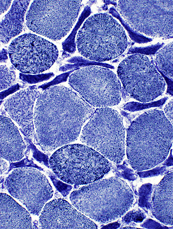
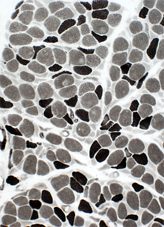
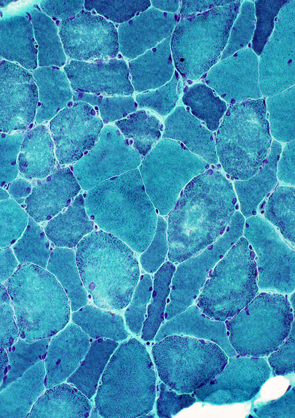
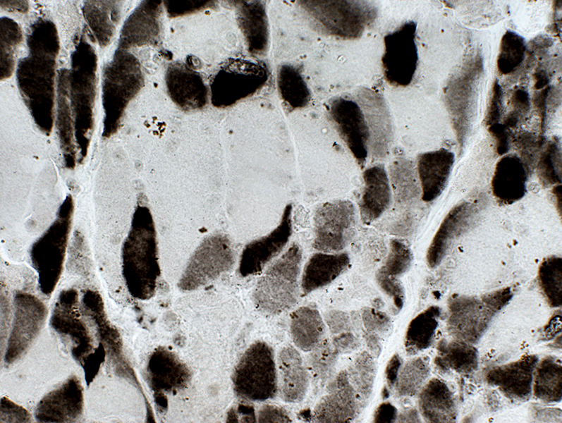
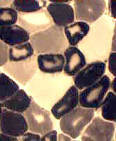
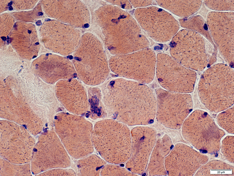
Myosin loss, Moderately Severe: Occurs in many small muscle fibers
Many small muscle fibers have staining that is less than in Type I fibers
 ATPase pH 9.4 stain |
|
Myosin loss, Moderately Severe: Occurs in many small muscle fibers Many small muscle fibers have staining that is less than in Type I fibers | |
 ATPase pH 9.4 stain |
 |
|
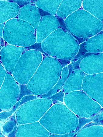
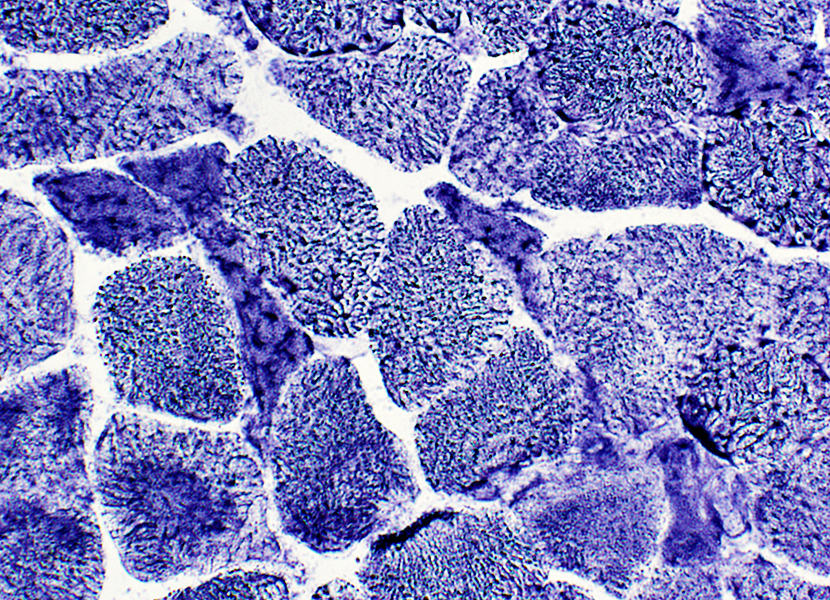
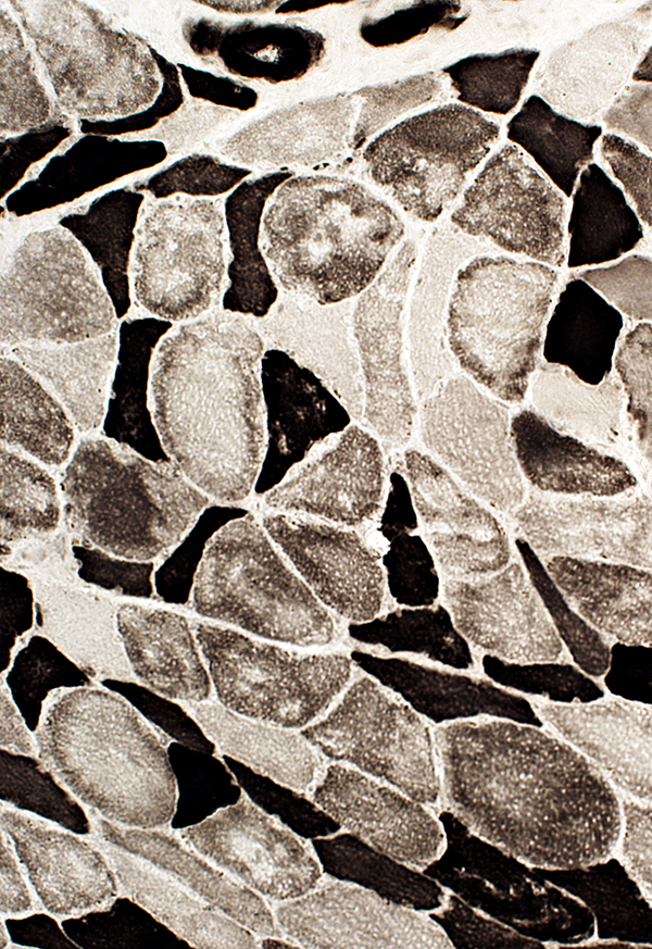
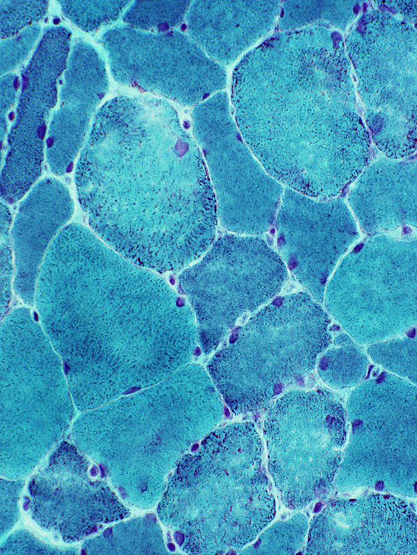
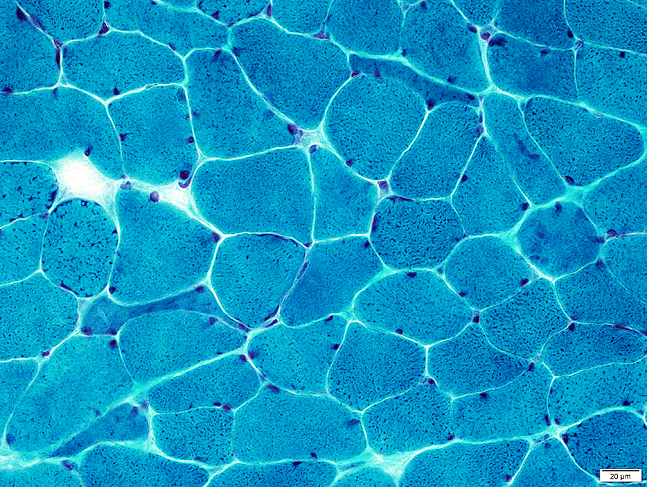
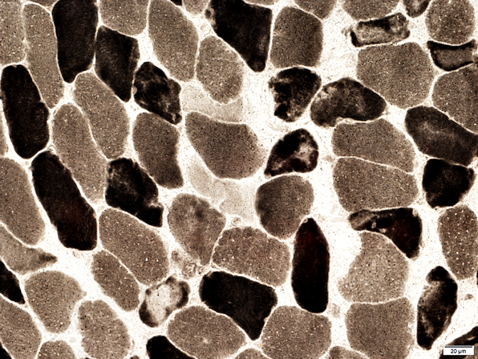
Muscle fibers with myosin loss: Less staining than type I muscle fibers on ATPase pH 9.4 stain. Varying degrees of severity of loss among fibers |
 ATPase pH 9.4 stain |
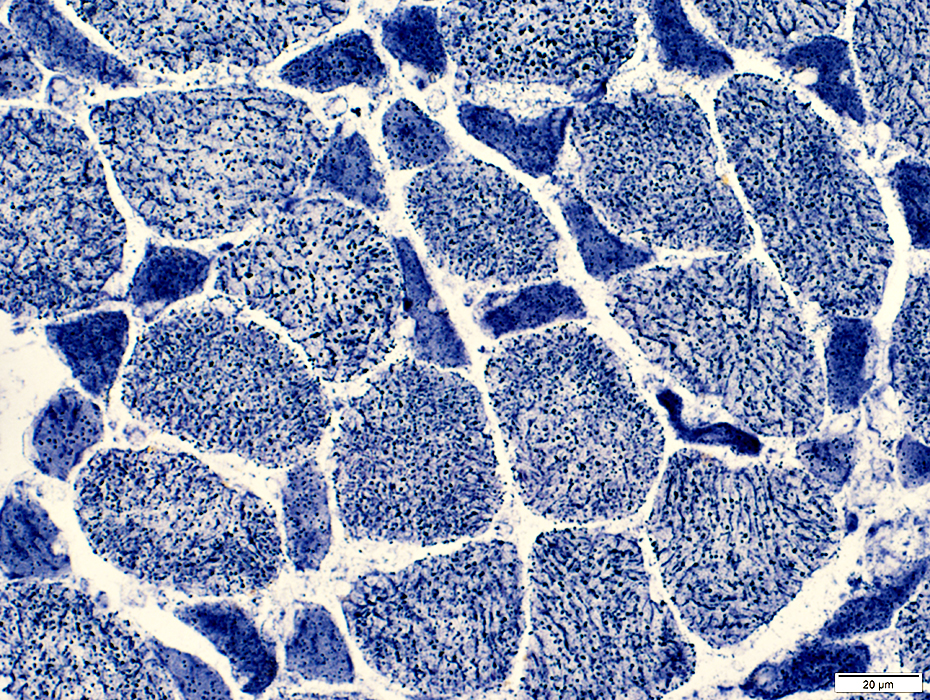
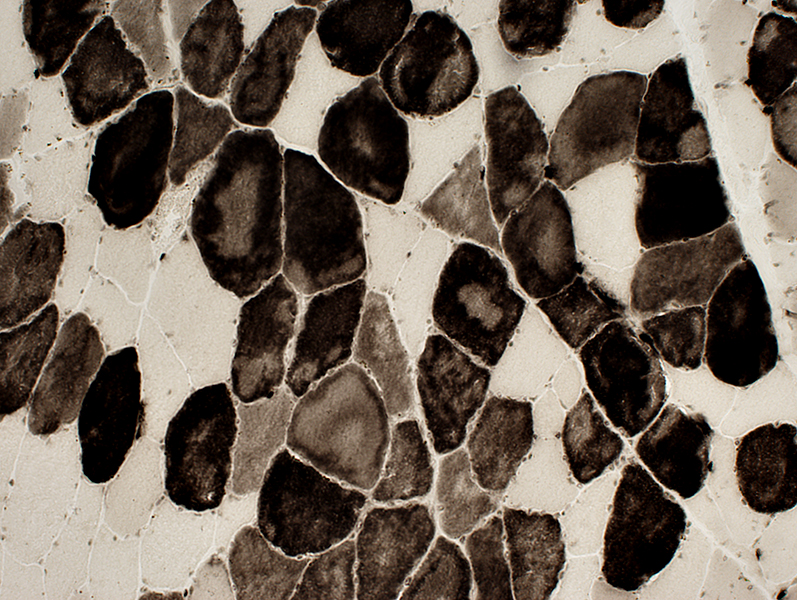
| Myosin Loss: Varied degrees of severity in different muscles | |
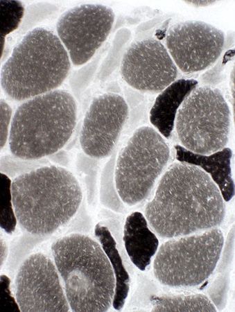
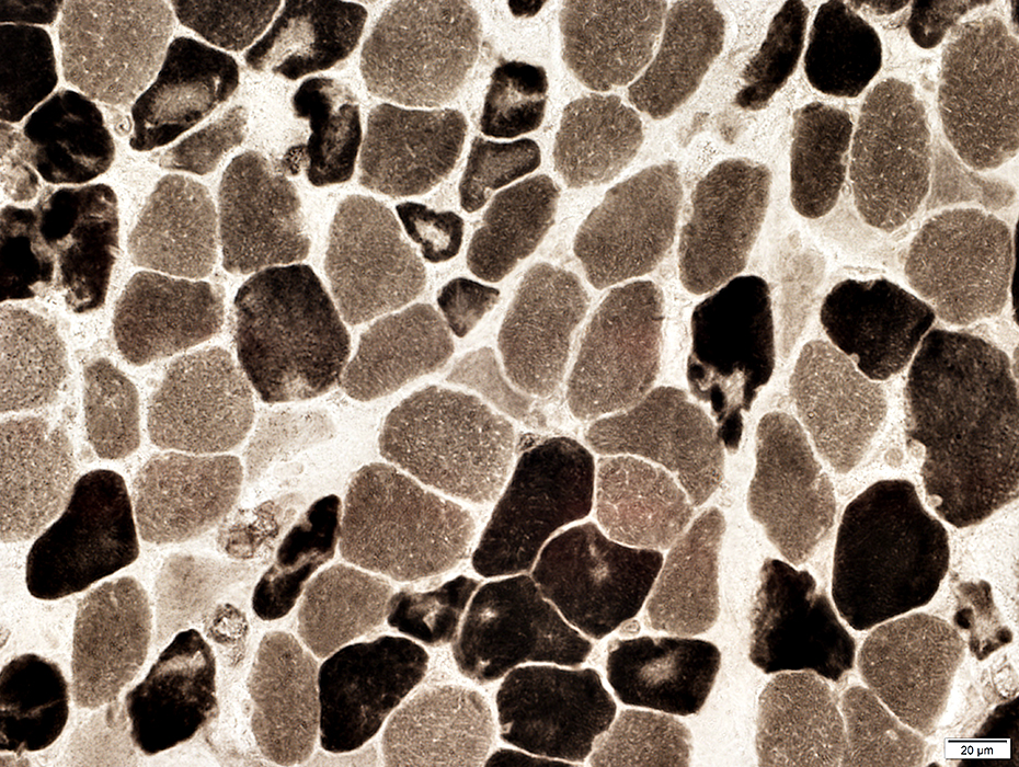
 ATPase pH 9.4 stain Many small muscle fibers with myosin loss |
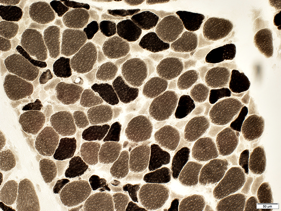
 ATPase pH 9.4 stain Few intermediate-sized fibers with myosin loss |
|
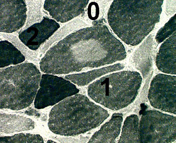
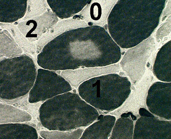
Comparison of ATPase pH 9.4 (Left) & 4.3 (Right) in serial sections |
|
|
|
|
Type I fibers (1): Darker staining at pH 4.3 than 9.4 Type II fibers (2): Darker staining at pH 9.4 than 4.3 Myosin loss fibers (0 or arrows): No staining at both pH 9.4 and pH 4.3 |
|
 ATPase pH 9.4 stain |
 ATPase pH 4.3 stain |
Autophagy marker (LC3): Increased in cytoplasm of many small muscle fibers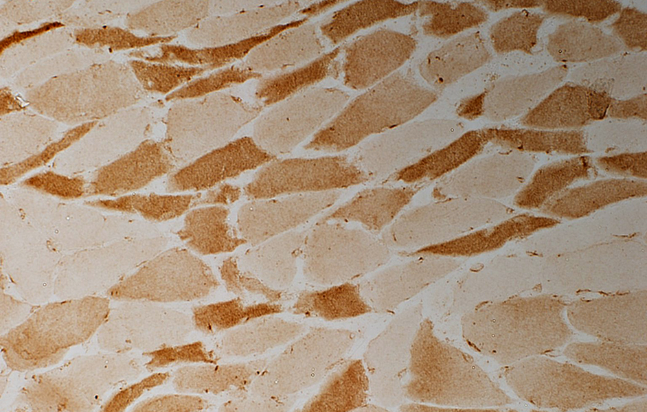 LC3 stain |
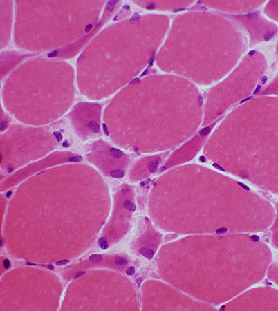
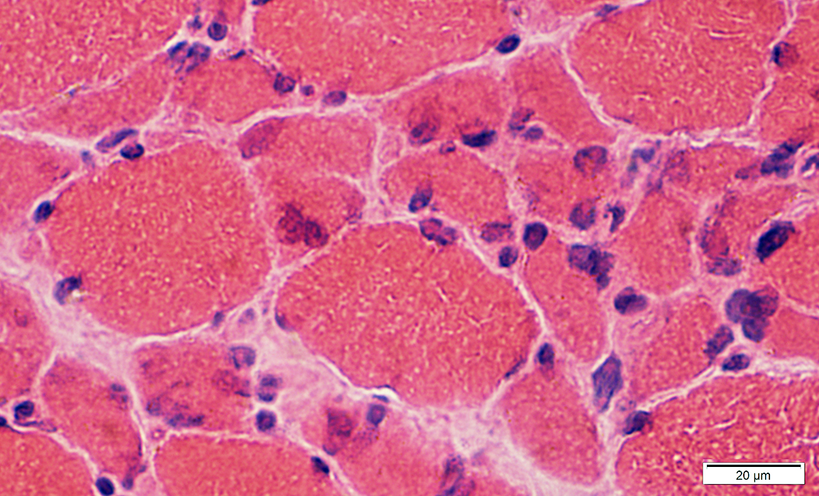
Myosin Loss: Myonuclei
 H&E stain |
Large
Irregular shapes
 H&E stain |
 H&E stain |
Large
Irregular shapes
 Congo red stain |
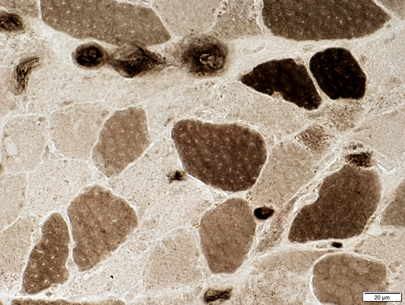
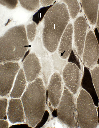
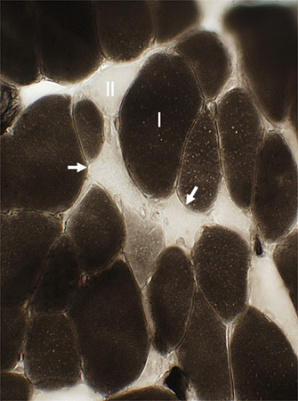
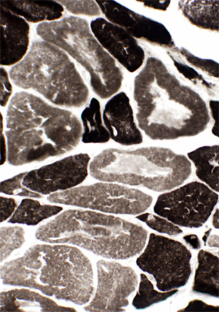
Myosin loss: Variant pathologyIn intermediate- and large-sized muscle fibersVaried degrees of loss in individual muscle fibers 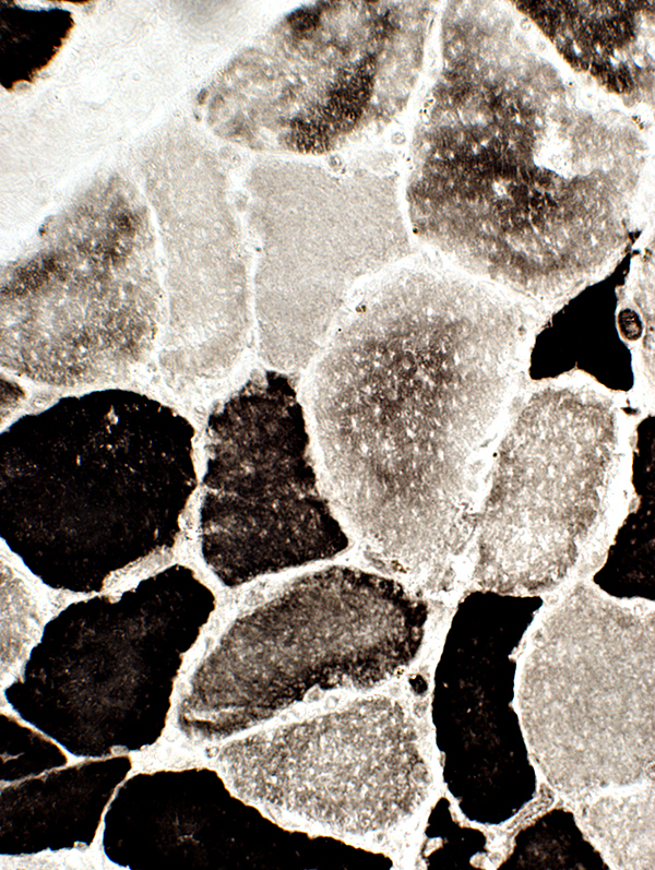 ATPase pH 9.4 stain |
 ATPase pH 9.4 stain |
 ATPase pH 4.3 stain |
|
 Gomori trichrome stain |
 Gomori trichrome stain |
 NADH stain |
 H&E stain |
Myosin loss: LC3 expression in scattered muscle fibers
 LC3 stain |
 LC3 stain |
 LC3 stain |
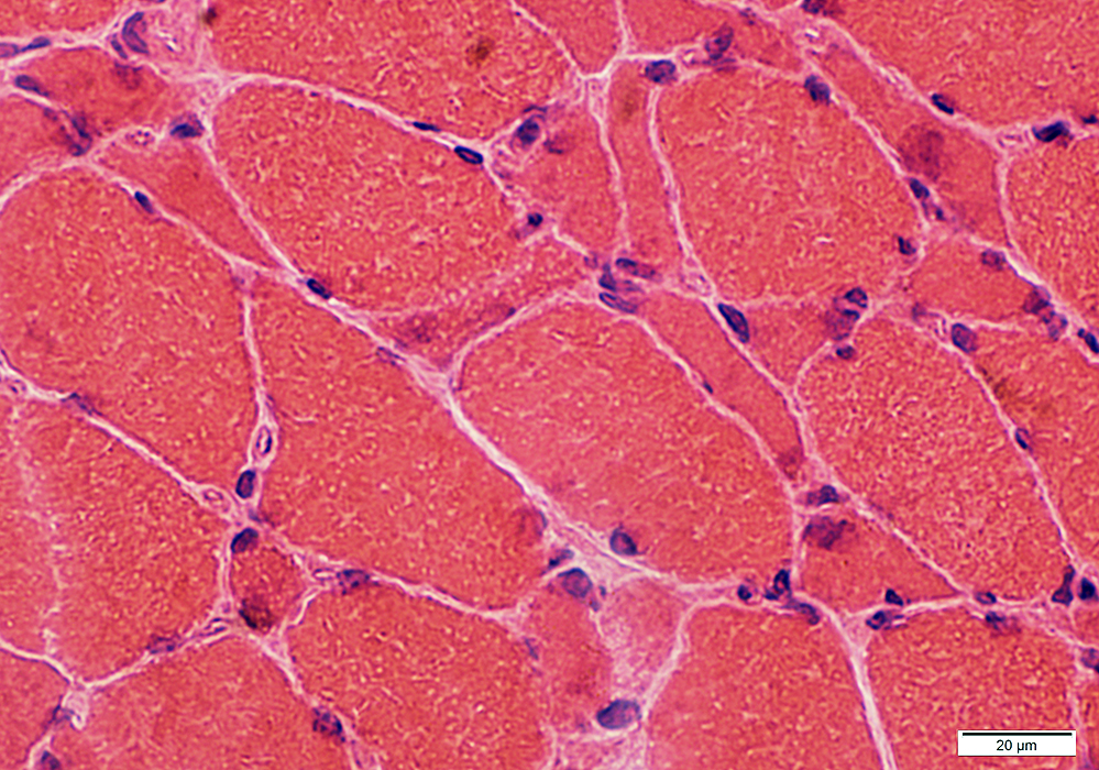
Myosin loss: Severe with diffuse muscle fiber atrophy
 H&E stain Muscle fibers: Severe atrophy Large nuclei in small fibers |
 VvG stain |
 Gomori trichrome stain Muscle fibers: Severe atrophy Large nuclei in small fibers |
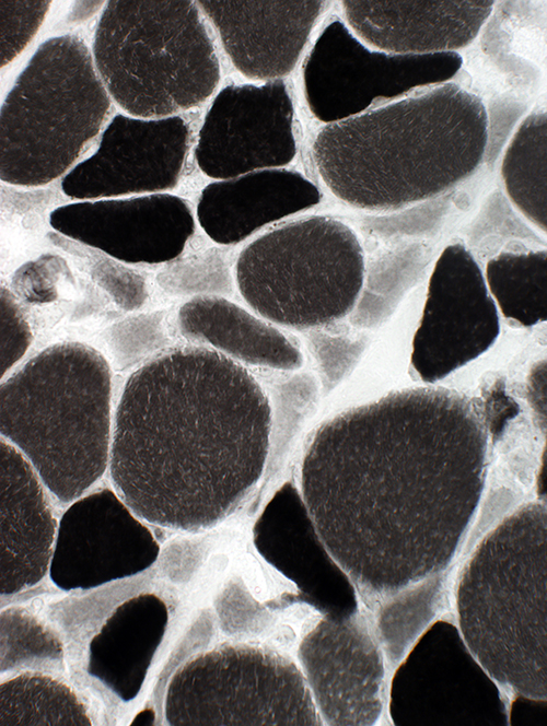
 ATPase pH 9.4 stain Myosin loss: May occur in large or small muscle fibers |
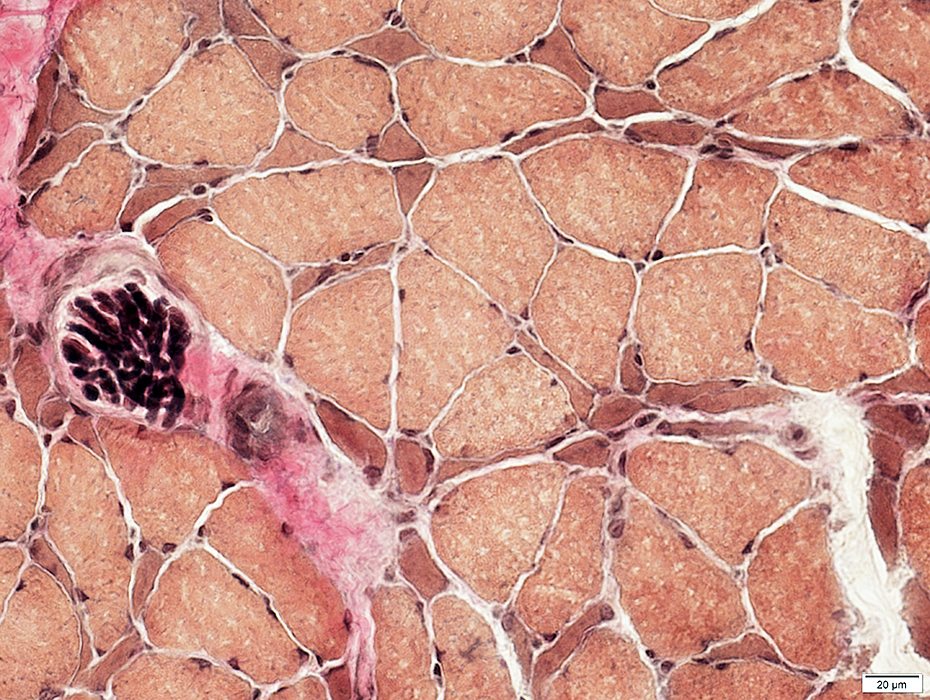
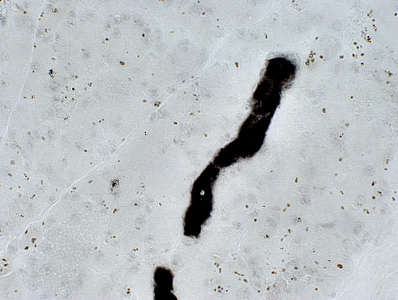 ATPase pH 4.3 stain Myosin loss: No staining in muscle fibers; Retained staining in an intermediate-sized vessel |
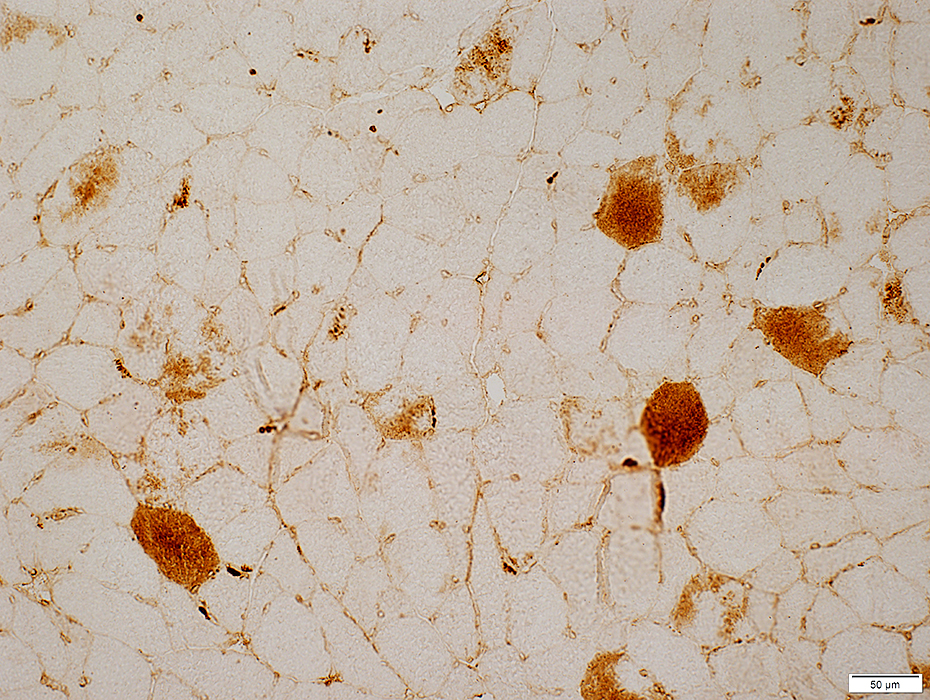
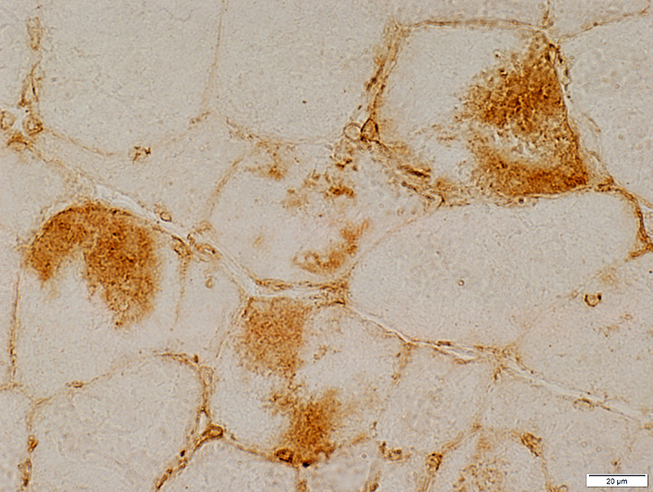
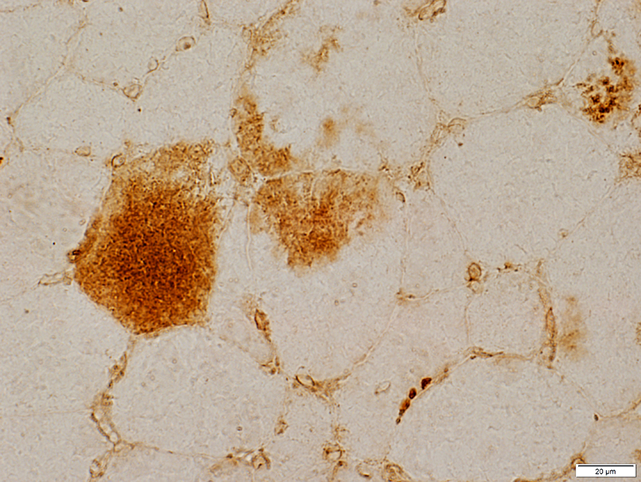
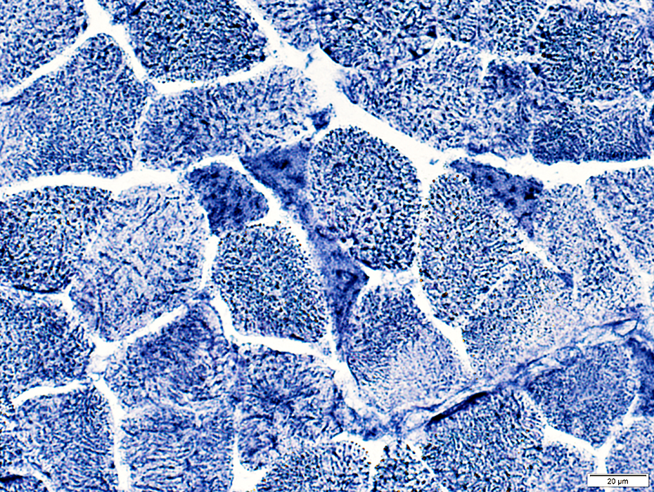
Myosin Loss: Anti-myosin antibody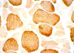 Reduced myosin In many small muscle fibers |
 H & E stain |
  |
 Myosin ATPase, pH 9.4 stain |
|
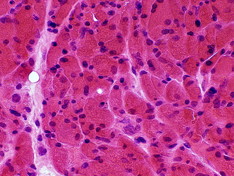
A focal pattern of myosin loss may also occur in muscle fibers. Focal regions of myosin loss in type I and II muscle fibers. These regions appear basophilic on H & E stain. Myosin loss may occur in large as well as small fibers |
Myosin loss: Ultrastructure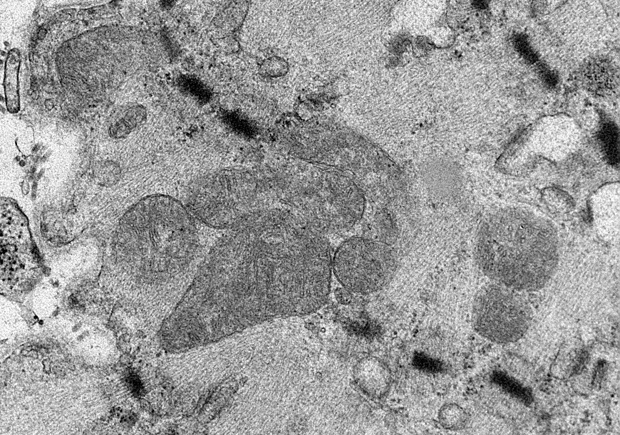
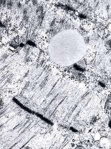 From: R Schmidt |
|
Thick filament loss: Variable; Can involve some regions within muscle fibers but not others
 |
Myosin Loss: Cushings disease
 |
Nuclei: Large; Irregular shapes
 |
 |
Muscle fibers
Shape: Varied shape; Some are small and angular
Internal architecture: Irregular
 |
Small, angular muscle fibers: Clustered internal architecture
 |
 |
Small angular fibers: Lost in whole fiber crossection
Larger fibers: Patchy loss
< br> br>
|
Return to Respiratory failure
Return to Myosin-loss myopathies
8/11/2025