Subperineurial Edema
|
Associations Differential diagnosis Hemorrhage Histochemistry Plastic sections Vessels, Endoneurial Ultrastructure |
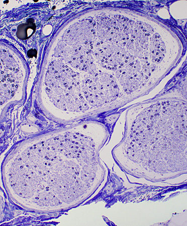
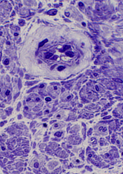
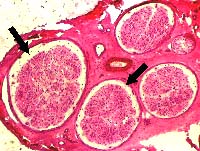 Subperineurial edema (Arrows) Variable severity in different fascicles |
Subperineurial edema: Contents
- Better visualized with plastic or frozen sections than fixed sections
- Granular material: May stain with Alcian blue (Mucopolysaccharides)
- Cells: Few & Scattered; Fibroblasts; Mast cells; Macrophages
- Endoneurial microvessels: May have thick wall & Large endothelium
Subperineurial edema: Diferential Diagnosis
- Systemic
- Thiamine deficiency
- Leprosy
- Ischemic (Atherosclerotic) disease
- Immune
- Toxic
- Hereditary
Subperineurial Edema: General Clinical Associations 1
- May occur with: Either axonal or demyelinating neuropathies
- More area with: Shorter disease duration in inflammatory disorders
- May persist despite previous immunmodulation treatment
- Possible cause: Leakage through blood-nerve barrier
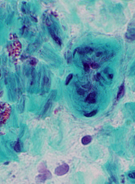
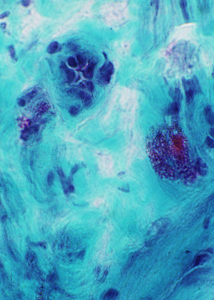
SUBPERINEURIAL EDEMA ± INFLAMMATION
 H&E stain |
 Gomori trichrome stain |
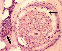 Subperineurial edema (Narrow arrow) In neighboring fascicle Inflammation Near a small epineurial vessel (Wide arrow) |
|
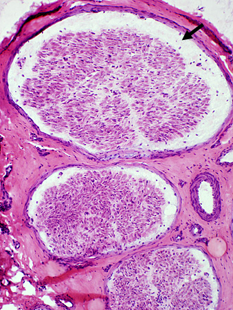
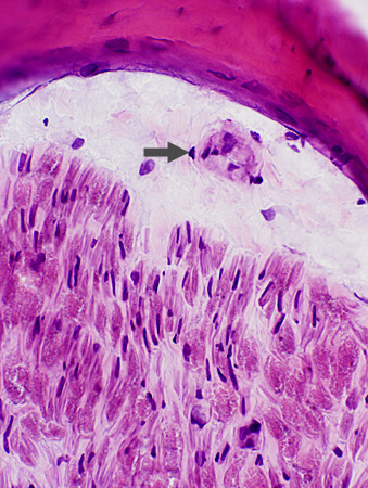
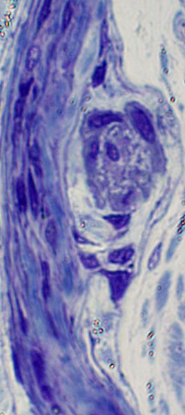
Clear space between the perineurium and endoneurial contents (arrows). |
||
 H&E stain |
 H&E stain |
|
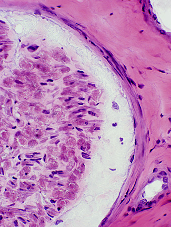
Clear space contains hyaline material and occasional vessels and cells. |
|
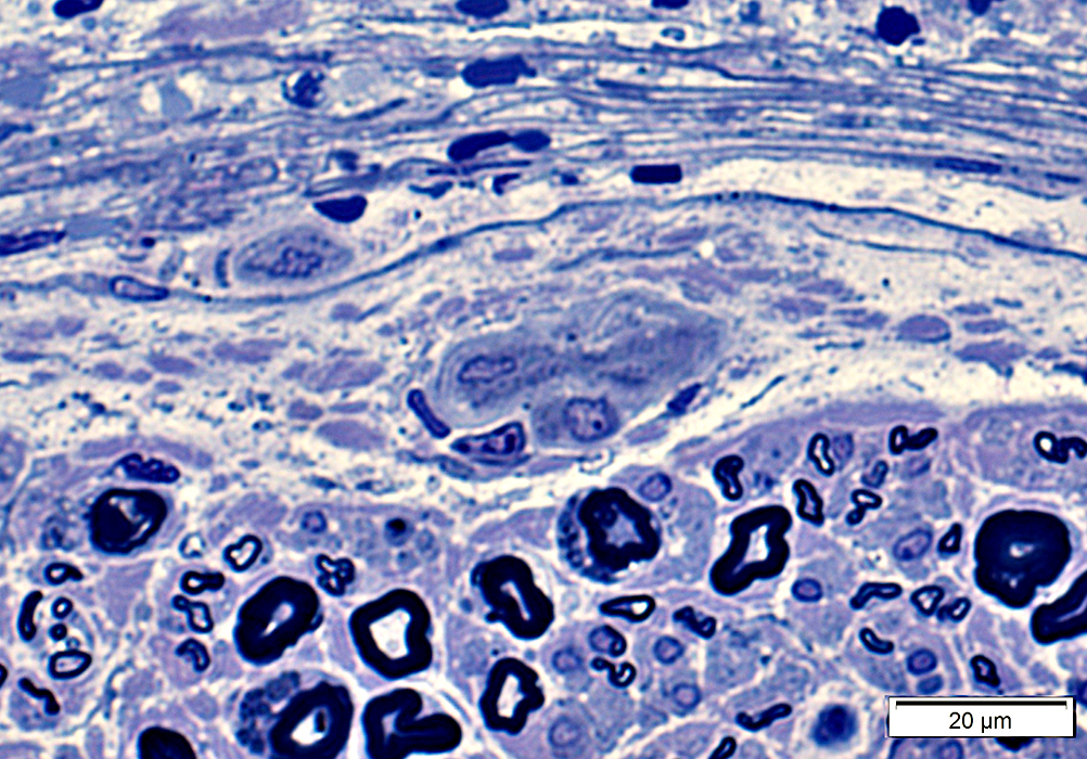
 Toluidine blue stain |
 Toluidine blue stain |
|
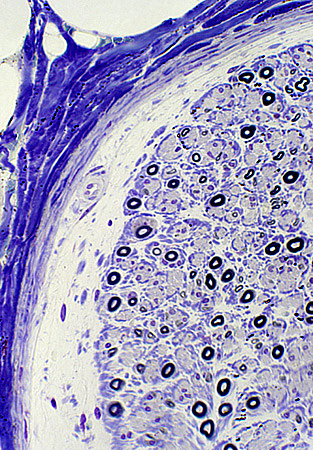
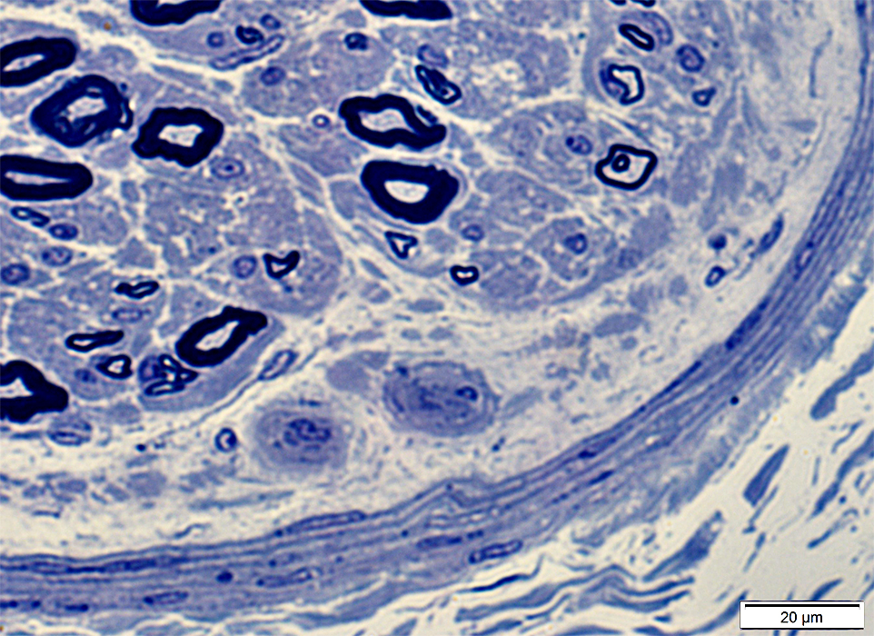
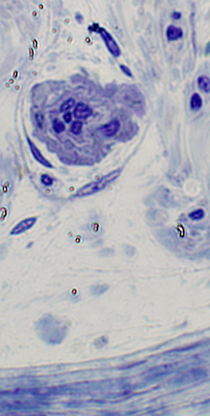
Subperineurial space contains wispy material, clear space, vessels and cells. There may be a variable degree of axon loss within fascicles. |
|
 Toluidine blue stain |
 Toluidine blue stain |
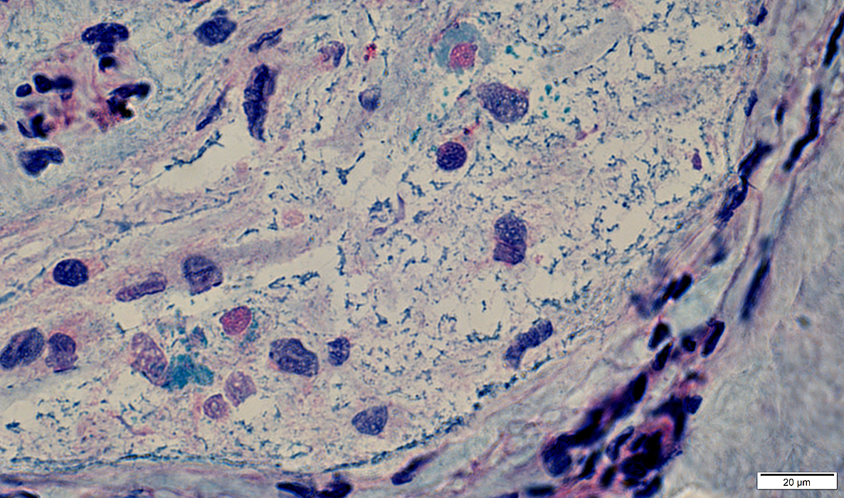
Subperineurial Edema (Arrow): Contents
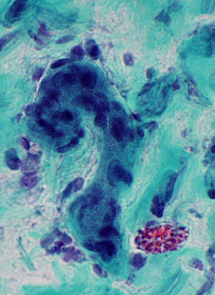
 Alcian blue stain |
 Alcian blue stain |
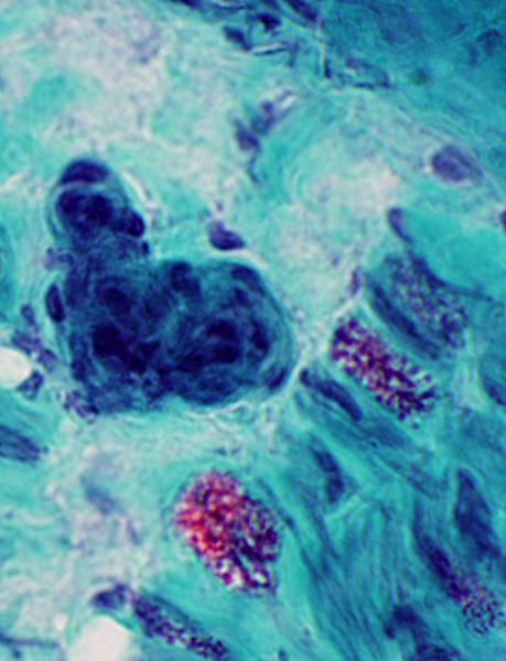
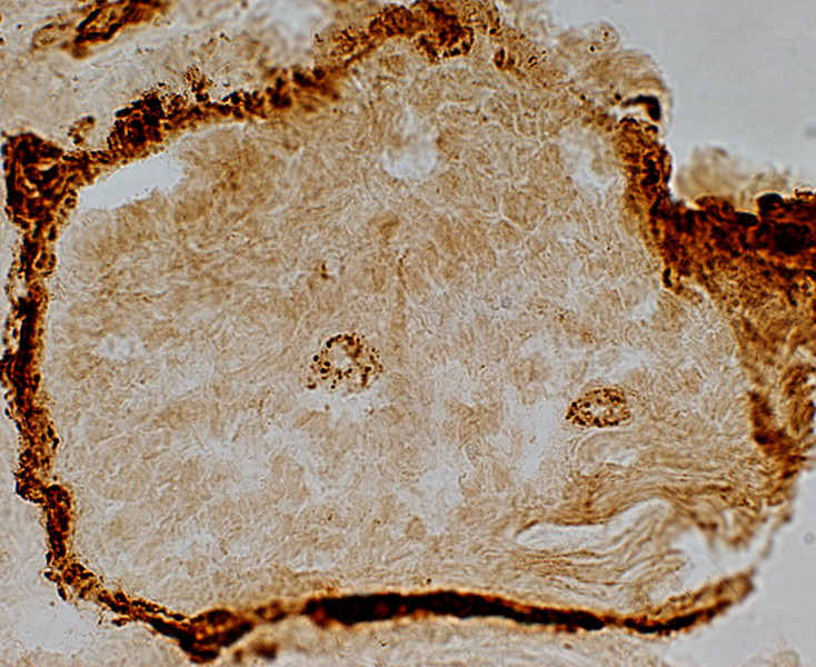
Background: Irregular & punctate clusters of mucopolysacharides (Alcian blue positive)
Cells: Large nuclei; Some have Alcian blue positive cytoplasm
 Alcian blue stain |
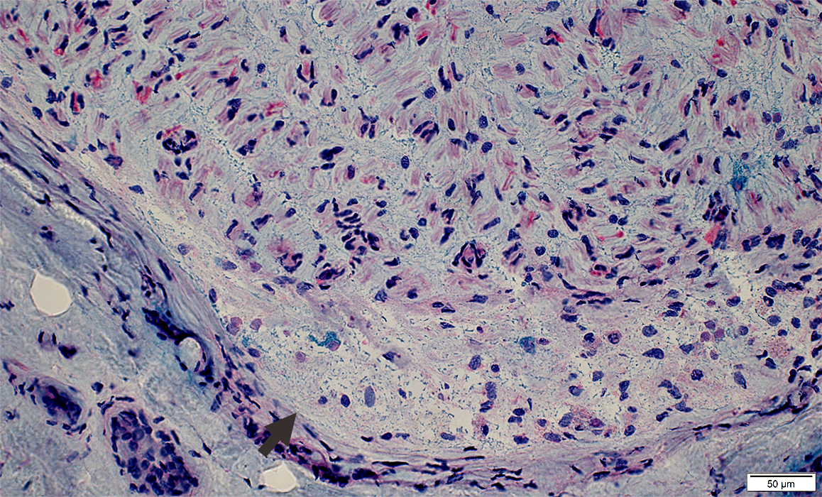
Subperineurial Edema: Subperineurial vessels
 Toluidine blue stain |
 Toluidine blue stain |
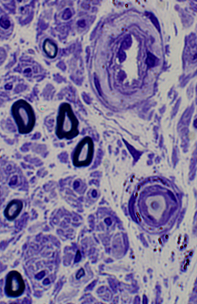
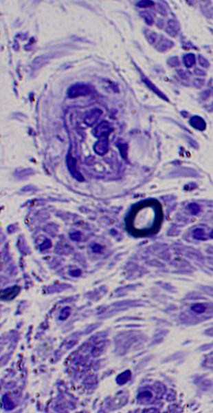
Subperineurial Edema: Endoneurial vessels
 Gomori trichrome stain |
 Gomori trichrome stain |
| Endoneurial vessels: Large with large endothelial nuclei | |
 Gomori trichrome stain |
 Gomori trichrome stain |
 Toluidine blue stain |
 Toluidine blue stain |
 Toluidine blue stain |
 Toluidine blue stain |
 Toluidine blue stain |
 Toluidine blue stain |
 C5b-9 stain C5b-9 complement: Deposited on endoneurial microvessels (capillaries) |
Subperineurial Edema: Ultrastructure
Contains
Granular, Proteinaceous fluid
Cells: Fibroblasts & Macrophages, Few
Bordered by
External: Perineurium
Internal: Endoneurial collagen, axons & Schwann cells
|
Granular material
Collagen: Small clusters
Fibroblasts (Above) & Macrophages, Rare
Endoneurial microvessels: May have thick wall & Large endothelium
|
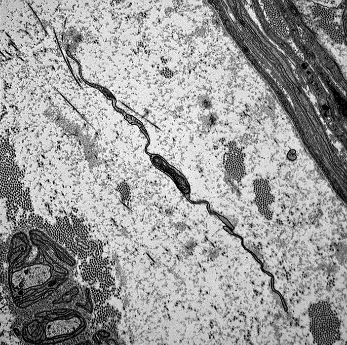
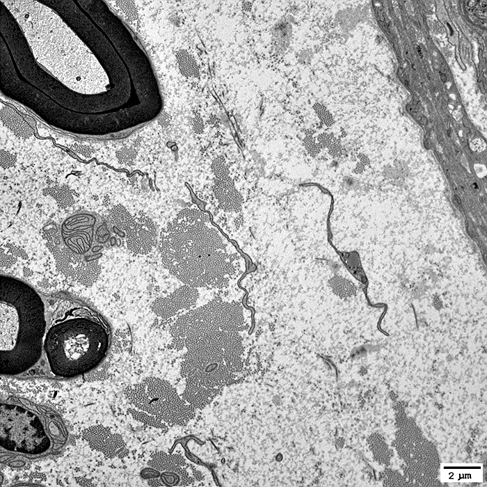
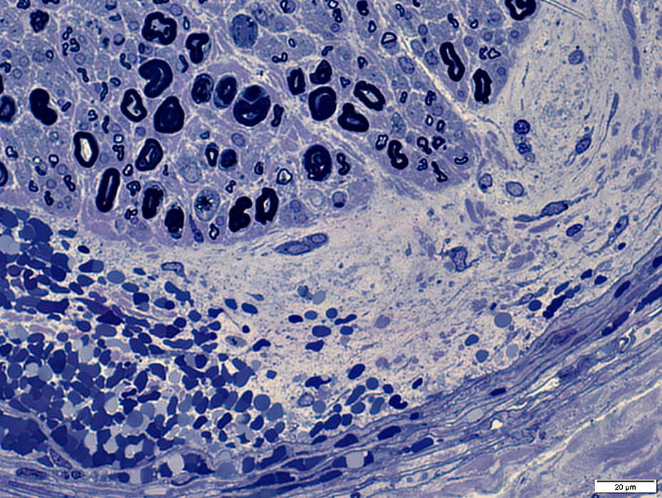
Subperineurial Edema: Region contains
Fibroblast processes
Granular material
Collagen: Clusters; Fibrils (Dark)
Internal edge of region (Bottom)
Contains Büngner band & Small, unmyelinated axons surrounded by Schwann cell processes
|
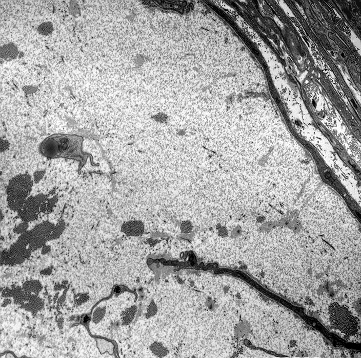
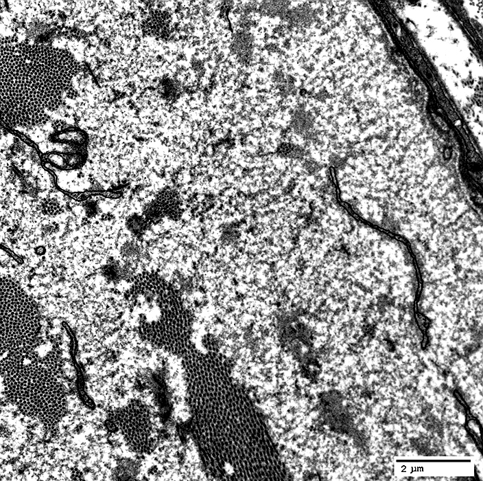
Subperineurial Edema: Region contains
Macrophage (Elongated)
Granular material
|
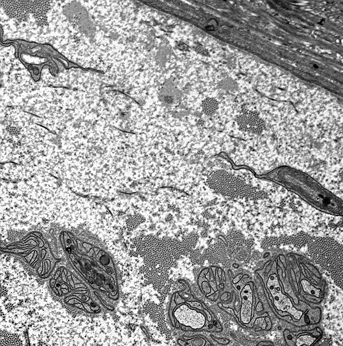
Subperineurial Edema: Region contains
Fibroblast processes
Collagen: Clusters
Internal edge of region (Left)
Contains: Myelinated axons & Büngner band
|
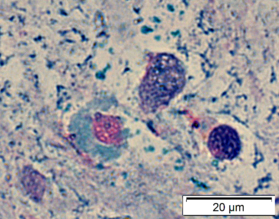
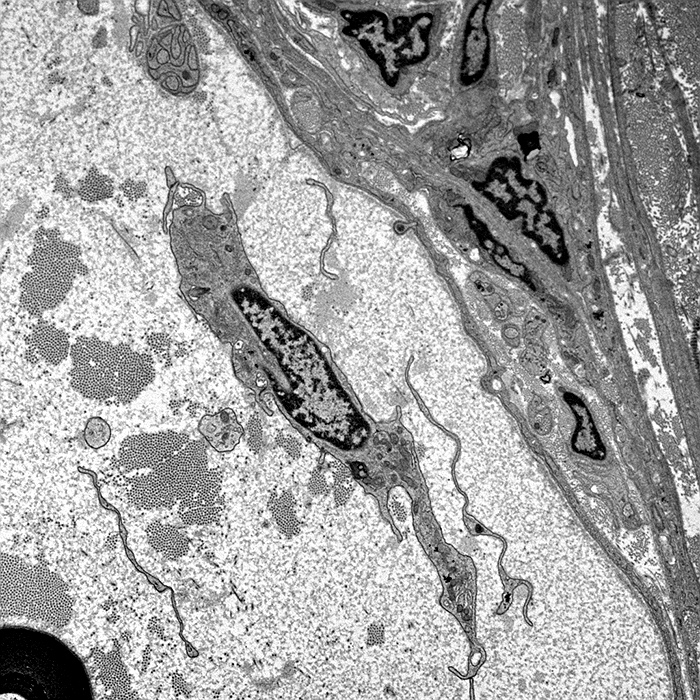
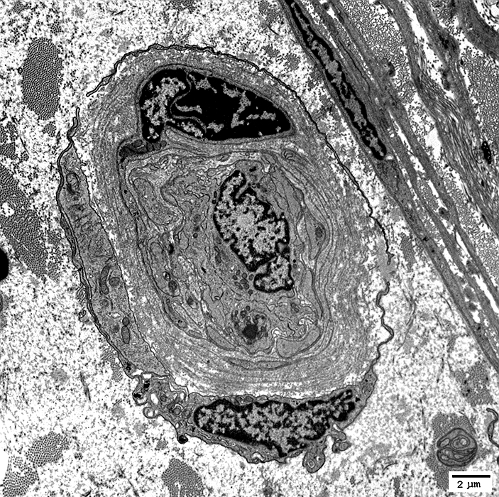
Endoneurial microvessel (capillary) within region of subperineurial edema
Contains
Endothelial cells (Large): In center of vessel One with nucleus
Pericyte with nucleus: In vessel wall (Top)
Fibroblasts & Processes (Dark): Surround vessel
|
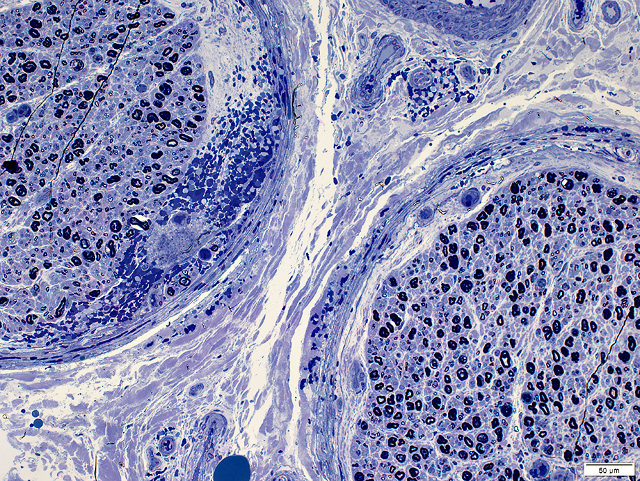
Subperineurial edema
Space under perineurium: Contains granular, mostly acellular material
|
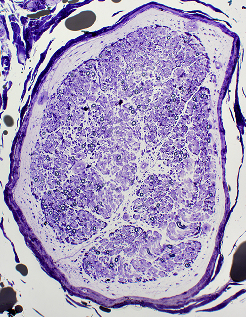
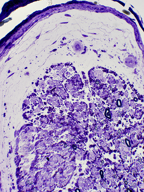
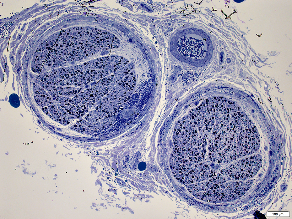
Subperineurial edema
Axon loss pattern: Differential fascicular
Varies within and among fascicles
Some regions (Left fascicle) have complete loss of large and small axons
Other regions (Right) have relatively preserved populations of large and small axons.
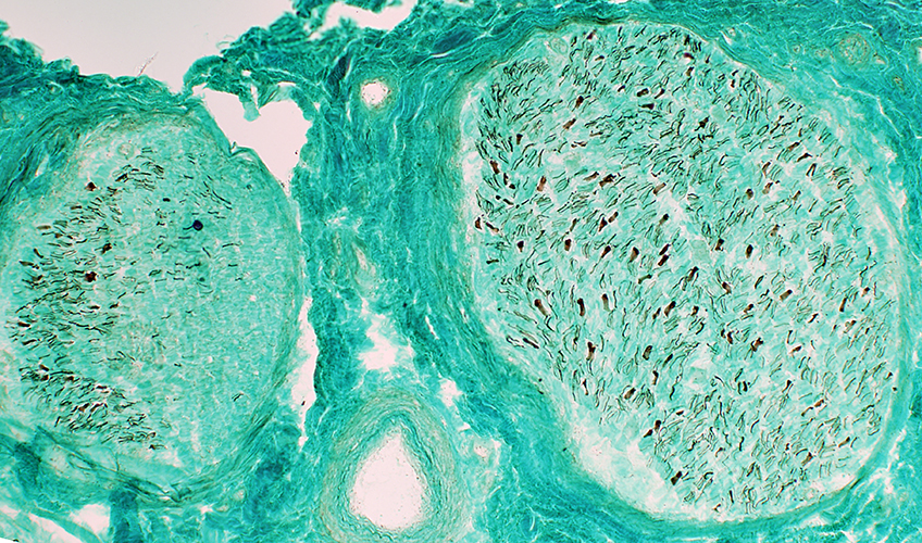 Neurofilament stain |
Subperineurial Edema & Hemorrhage
 Toluidine blue stain |
 Toluidine blue stain |
 Toluidine blue stain |
Return to Subperineurial edema DDx
Return to Pathology index
Return to Neuromuscular Home Page
References
1. Muscle Nerve 2016;53:705-710
9/8/2025