CIDP: Subacute Onset
Note: Patient features- Clinical
- 55 yo female
- Onset: Progressive over 8 weeks
- Weakness: Severe; Proximal & Distal
- Corticosteroid treatment: Improvement to normal strength & ADL
- NCV
- Demyelination
- Conduction velocities, motor: Variably slowed (12 to 31 M/s)
- Distal latencies: Long
- Temporal dispersion
- Axon loss
- SNAPs: Reduced amplitude or Absent
- Demyelination
- Pathology
- Demyelination
- Axon loss: Variable
- Subperineurial edema
- Vessels
- Endoneurial: Diameter enlarged; Endothelial cells large
- Epineurial: Normal structure; No immune cells
- Muscle
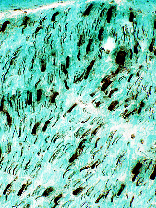
Nerve: Axon loss, Variable & Myelin loss, Diffuse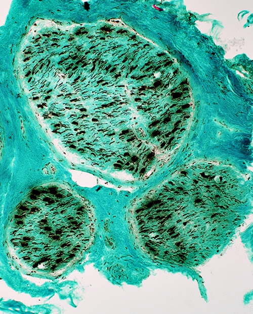 Neurofilament stain Axons (Same nerve as on right ) Variable loss of large & small axons within & among fascicles Large axons: Demyelinated Preservation of some large axons without surrounding clear myelin rim |
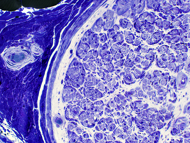
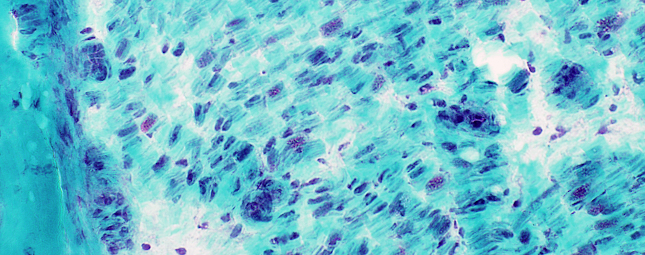
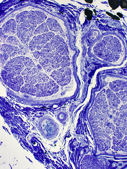 Toluidine blue stain Myelinated axons Apparent nearly complete myelinated axon loss in some fascicles Subperineurial edema |
Differential fascicular involvement: Variable loss of myelinated axons among fascicles
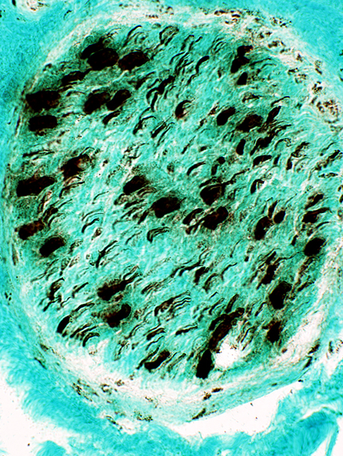
 VvG stain |
Nerve: Variable loss of large axons
 Neurofilament stain |
 Neurofilament stain |
|
Axon Loss Some fascicles have more loss of large (thick) axons (Right) than others (Left) Small axons are moderately reduced in both fascicles | |
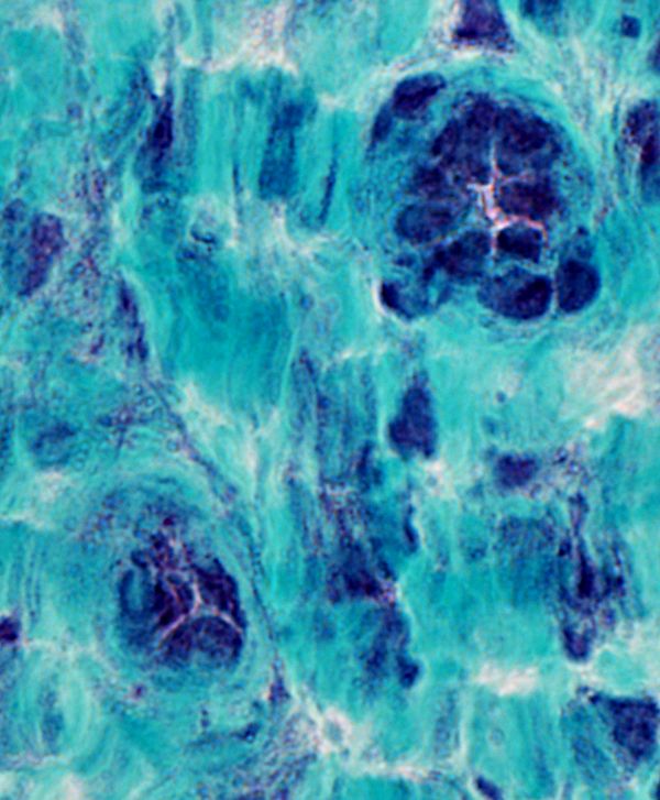
Large axons: Demyelinated & Poorly formed onion bulbs
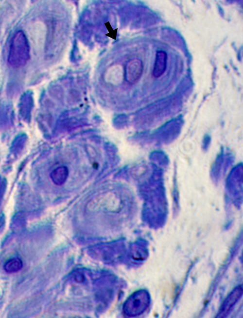 Toluidine blue stain |
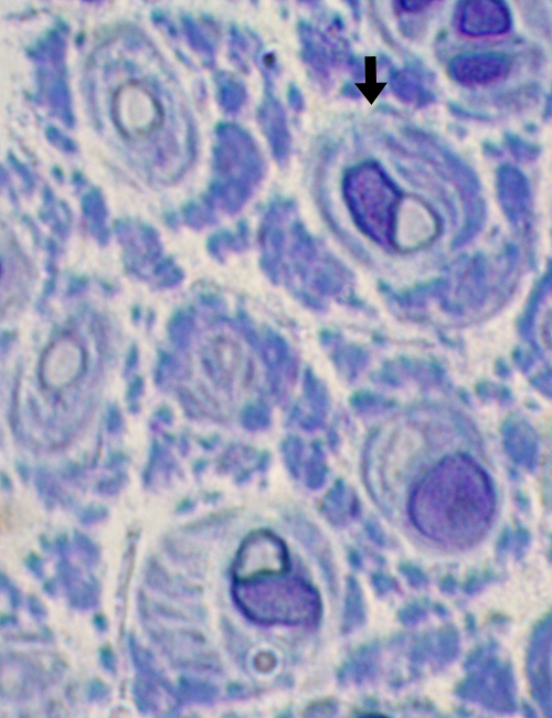 Toluidine blue stain |
|
Demyelinated axons Demyelinated, or thinly myelinated axons, often surrounded by poorly formed oniuon bulbs and Schwann cell nuclei | |
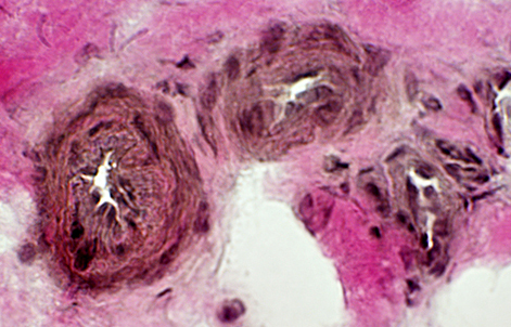
Subperineurial Edema
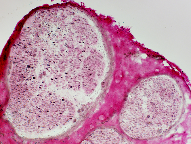
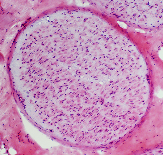 H & E stain Subperineurial Edema: Clear region beneath long area of perineurium |
 Toluidine blue stain Subperineurial Edema & Few normally myelinated axons |
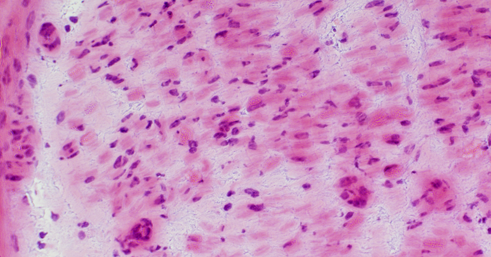
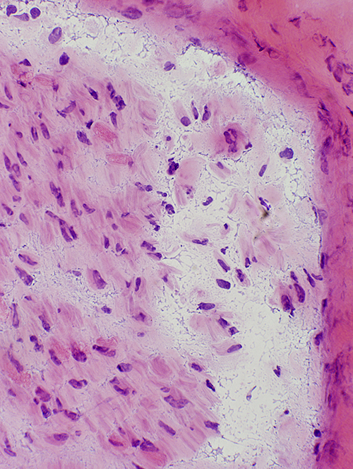 H & E stain Region of subperineurial edema: Contains amorphous material and small vessels |
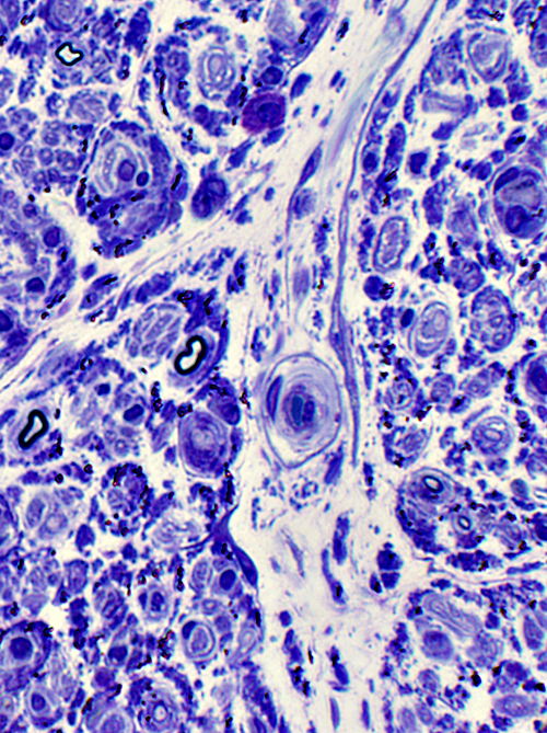 Toluidine blue stain Intrafascicular septa: Widened & Cellular Contains enlarged endoneurial vessel Myelinated Axons: Few remaining; Small |
Endoneurial vessels: Large size; Abnormal endothelial cells with large nuclei
 H&E stain |
Endoneurial capillaries: Enlarged with large endothelial cells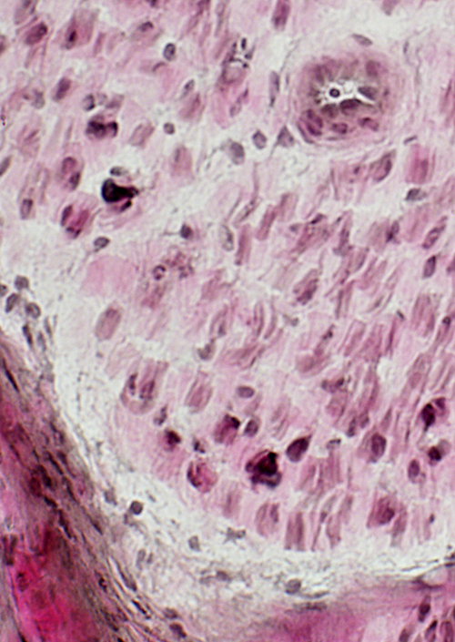 VvG stain |
 Gomori trichrome stain |
 Gomori trichrome stain |
 H&E stain |
|
Endoneurial capillaries: Alkaline phosphatase stained 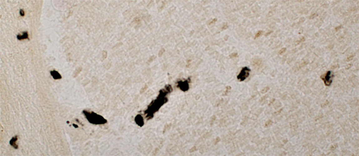 Alkaline phosphatase stain |
Endoneurial cells: Scattered small & punctate acid phosphatase staining
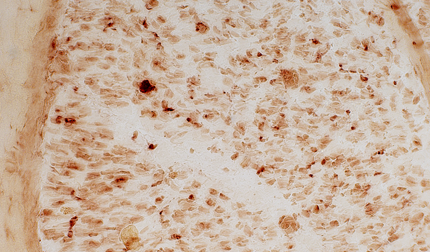 Acid phosphatase stain |
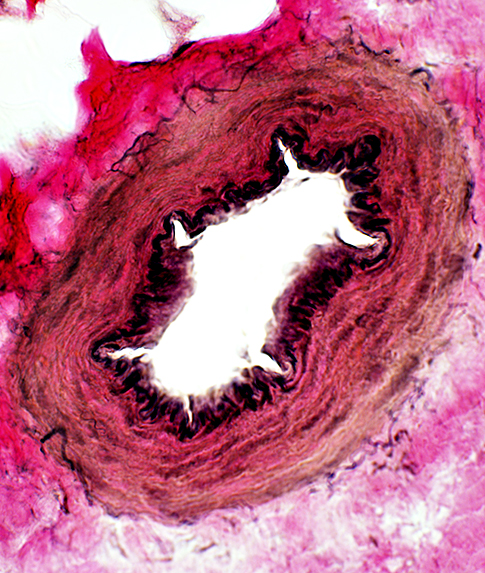 VvG stain Artery |
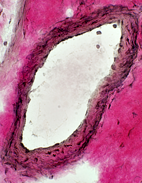 VvG stain Vein |
Epineurial vessels: Normal |
 VvG stain Smaller epineurial vessels |
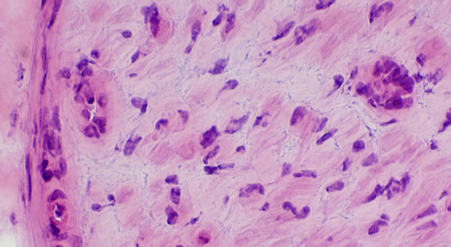
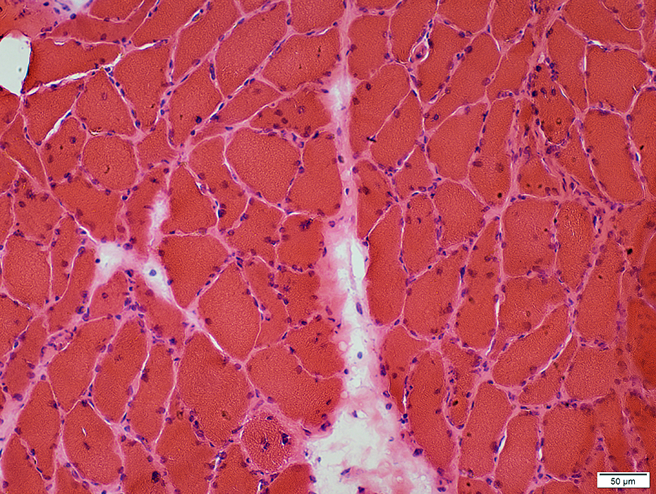
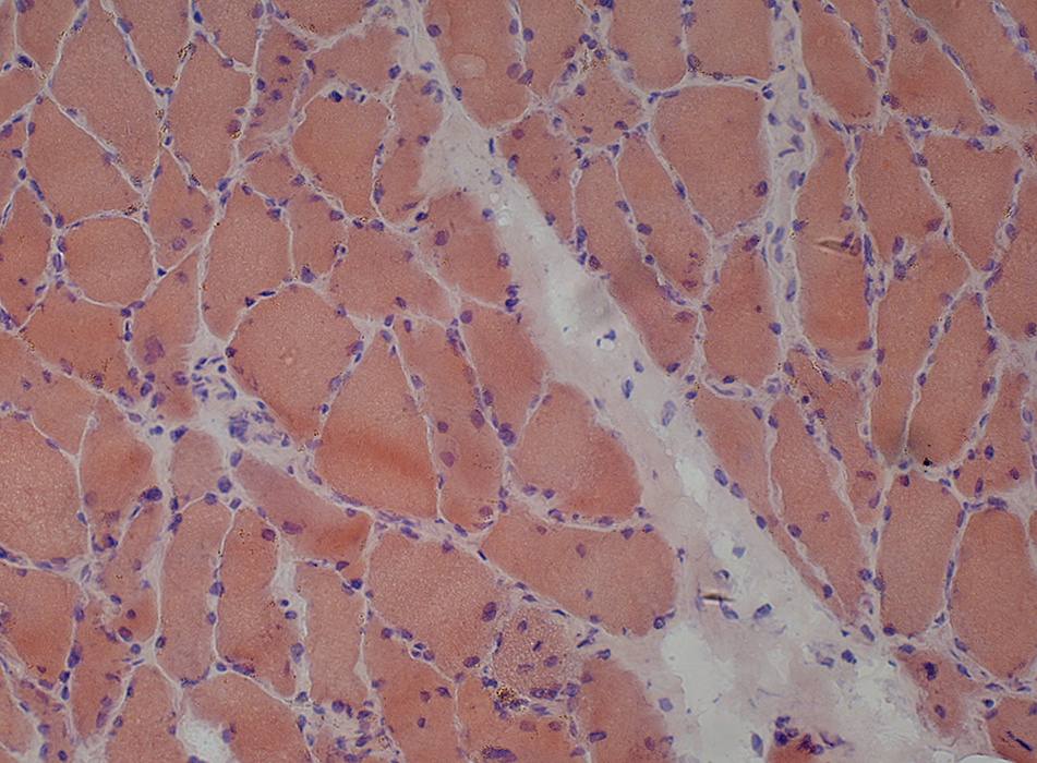
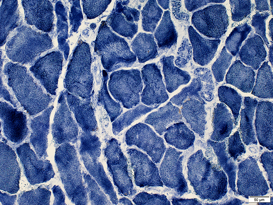
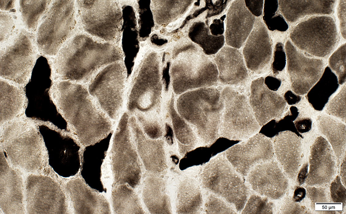
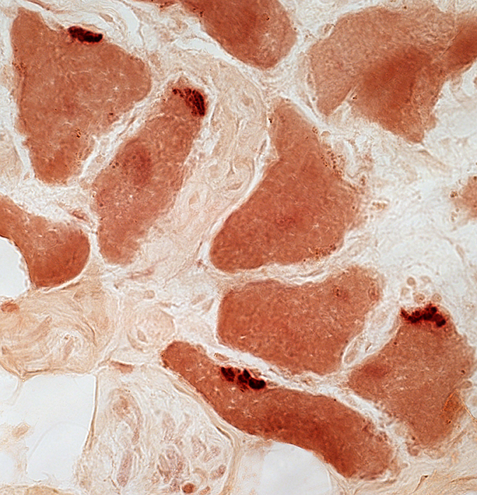
CIDP Sub-Acute Onset: Muscle
|
Nuclei: Large
Sarcolemma: Irregular
|
|
Fiber sizes: Type 2 often smaller than type 1
|
Neuromuscular junctions: Patchy; mildly elongated
|
Return to Neuromuscular Home Page
Return to CIDP, Subacute onset
9/22/2015