Eosinophilic Vasculitis
|
Axon loss Granuloma Muscle & Myopathy Subperineurial edema Vasculitic pathology Wallerian degeneration |
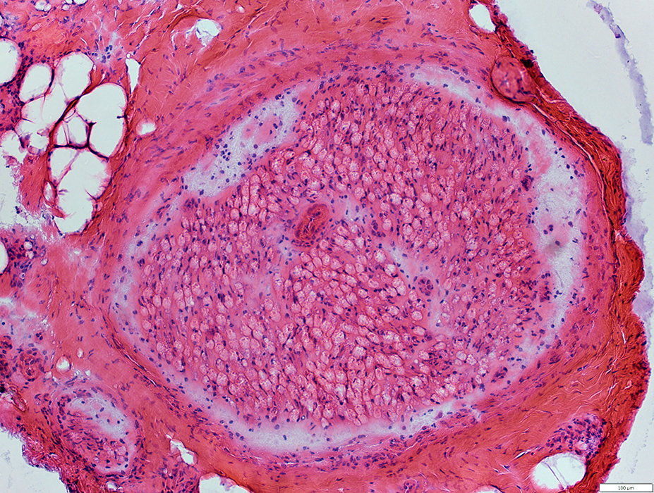
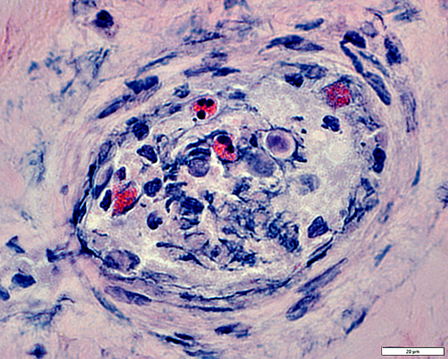 H&E stain Eosinophils in Vessel wall
|
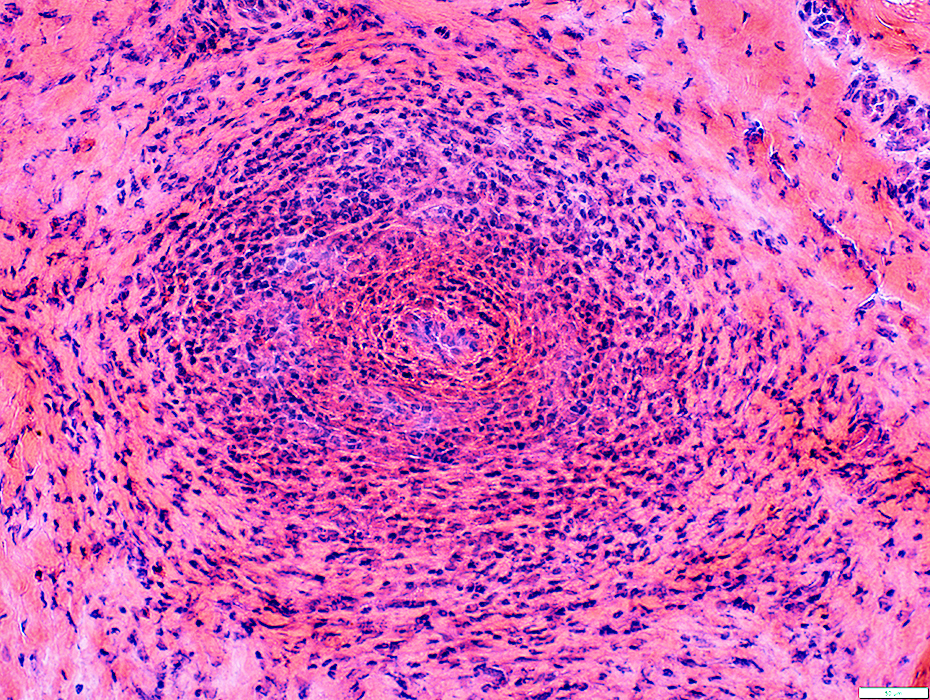
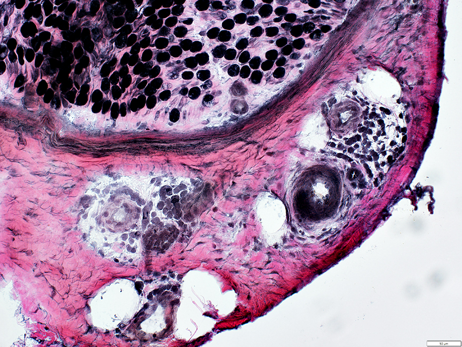
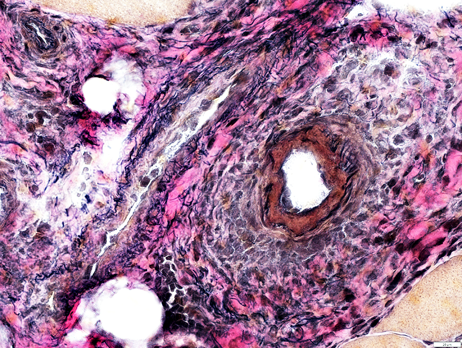
Vasculitic Pathology
Serum testing: ANCA+ with pANCA- & cANCA-; RNP mildly+
 H&E stain |
Wall: Damaged; Cellular
Lumen: Lost
Epineurium
Eosinophils (Scattered) & other Cells
Nerve fascicle
Subpoerineurial edema
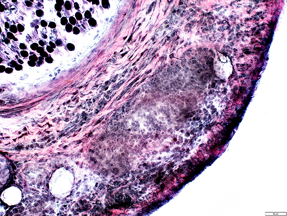
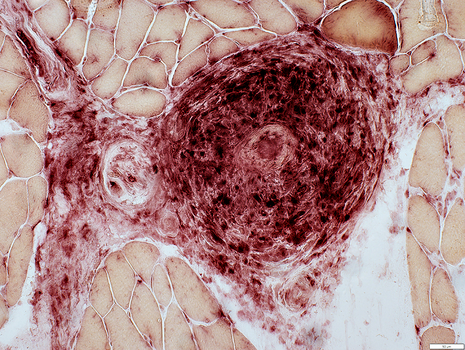
 Congo red stain |
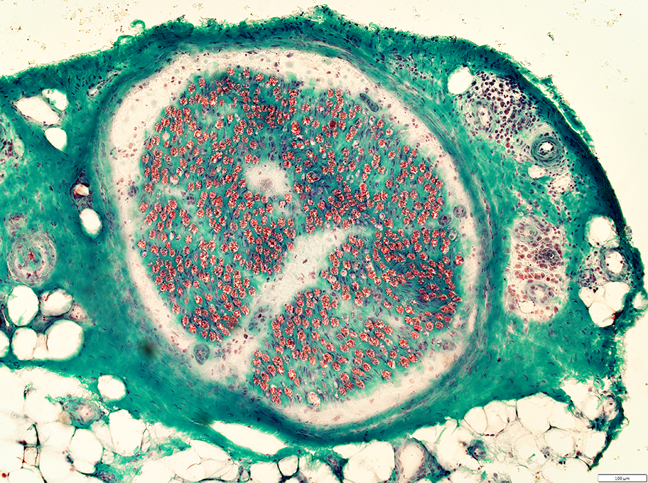
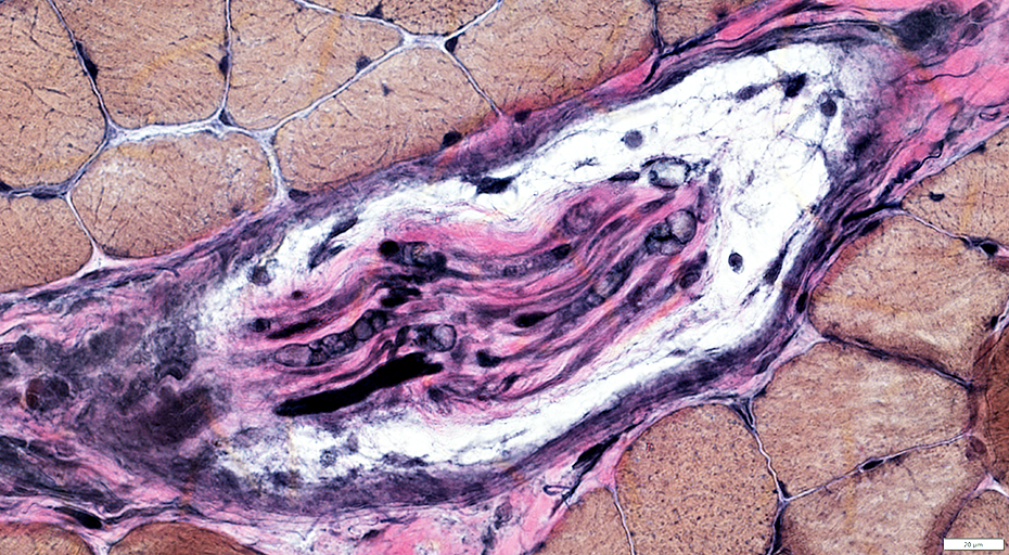
 VvG stain |
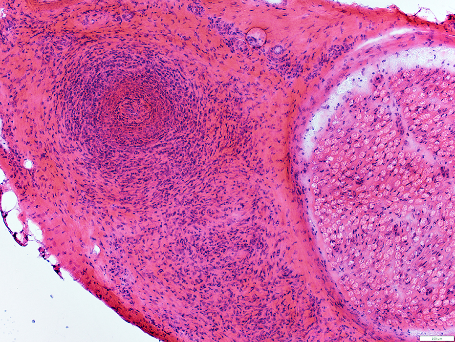
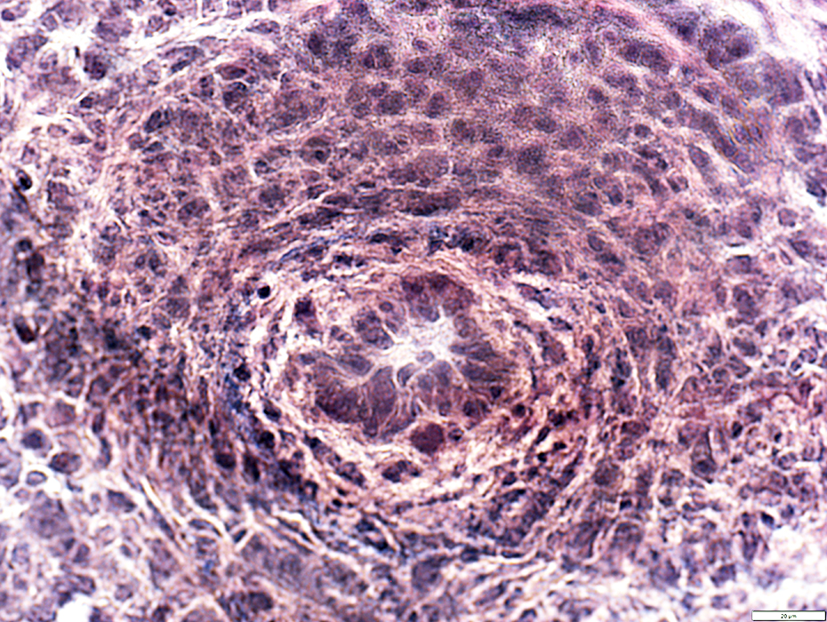
Wall: Damaged; Cellular; No fibrils
Endothelial cells: Large
Lumen: Lost
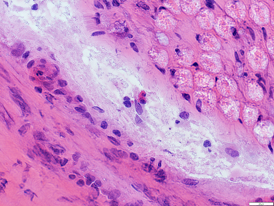
 H&E stain |
Wall: Damaged; Cellular; No fibrils
Endothelial cells: Large
Lumen: Lost
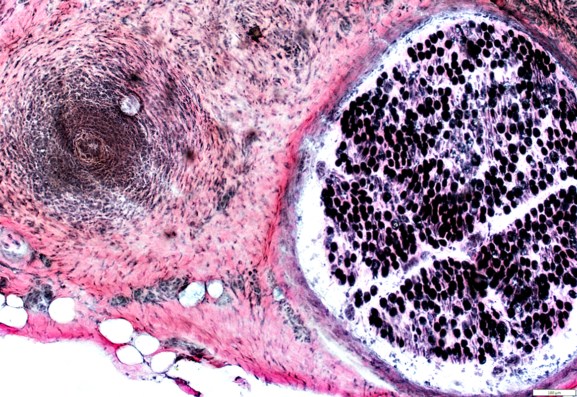
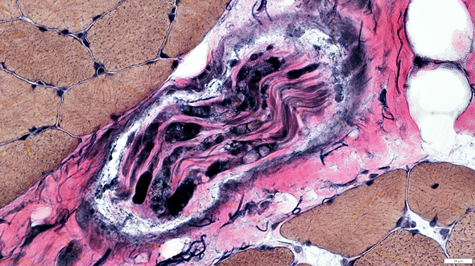
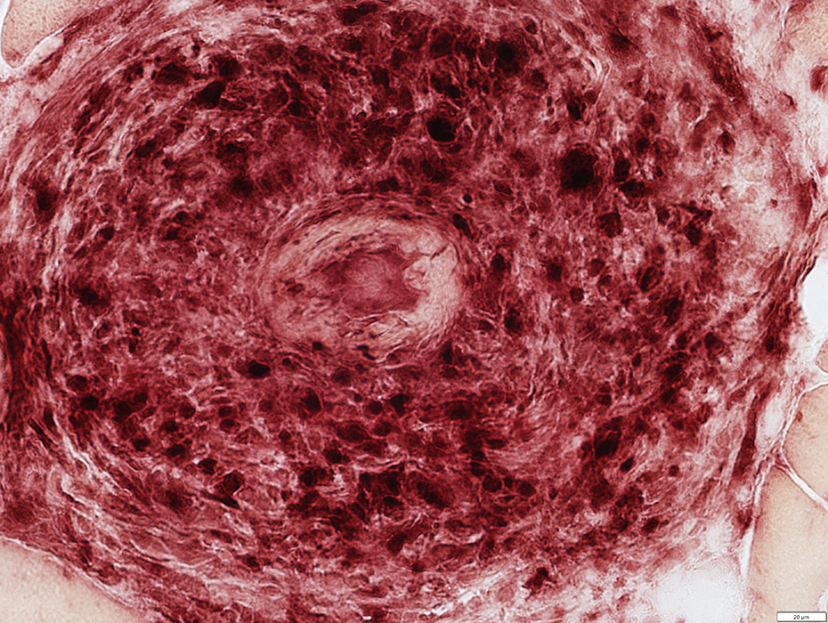
 VvG stain |
 VvG stain |
Within cell focus: No fibrils; Endothelial cells large
Vein: No fibrils
Vascular remnants: Clustered
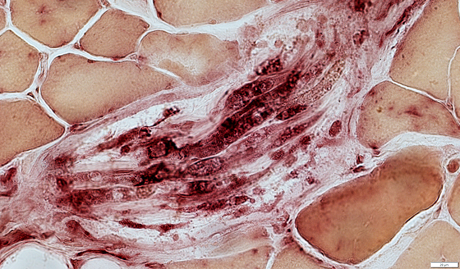
 Congo red stain |
Contains scattered eosinophils
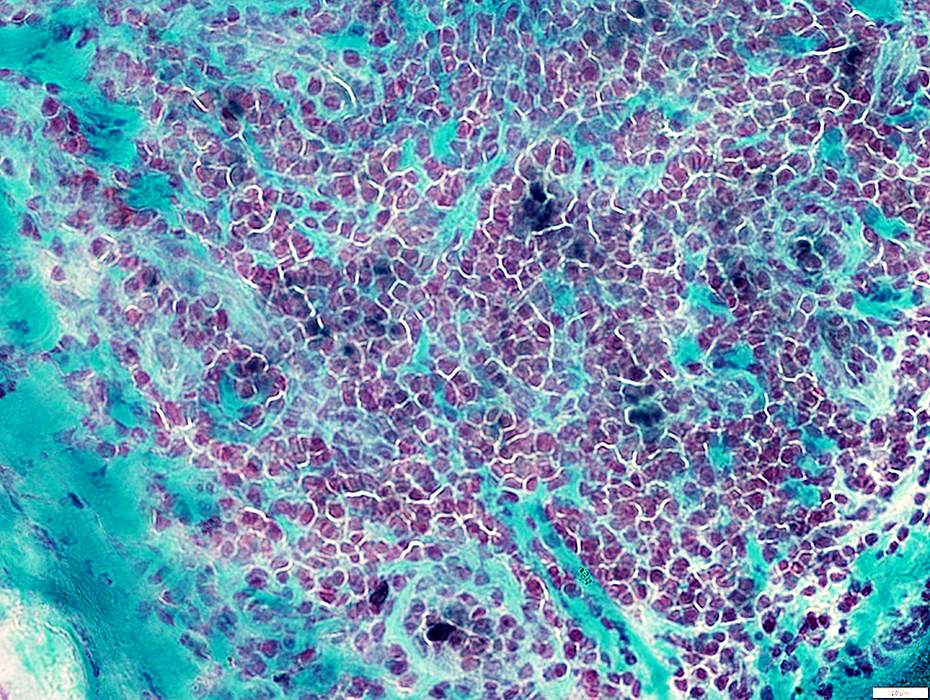 Gomori trichrome stain |
 VvG stain |
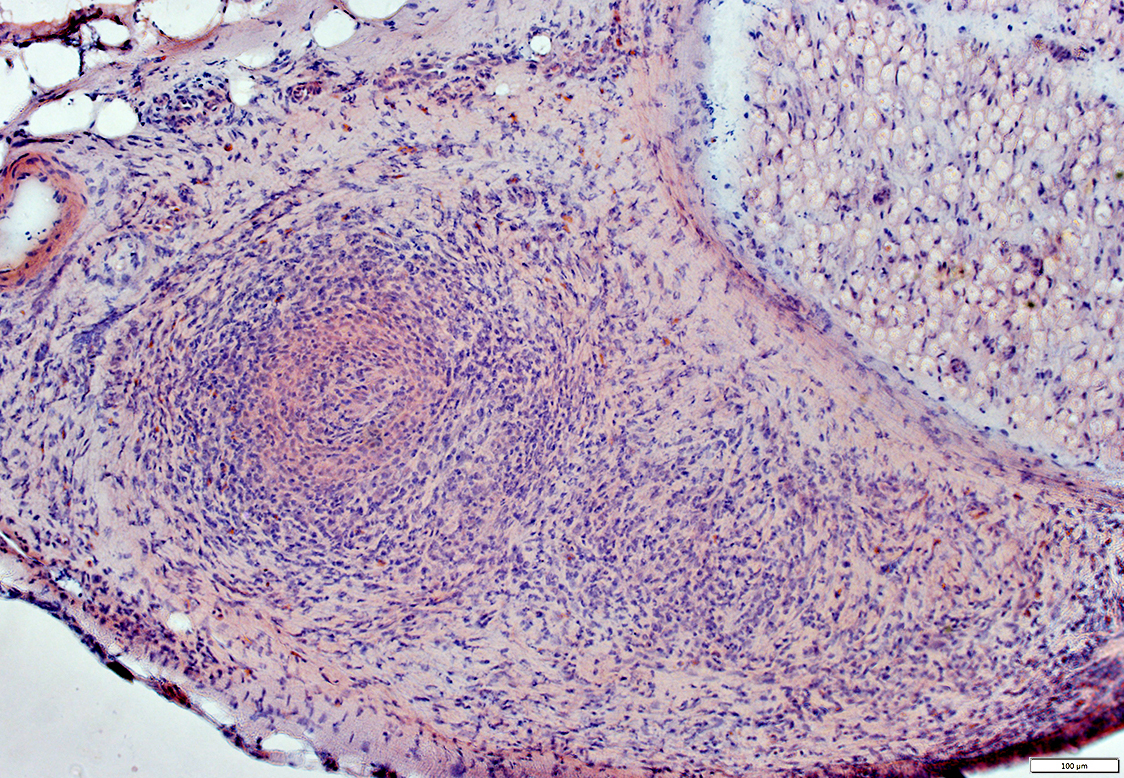
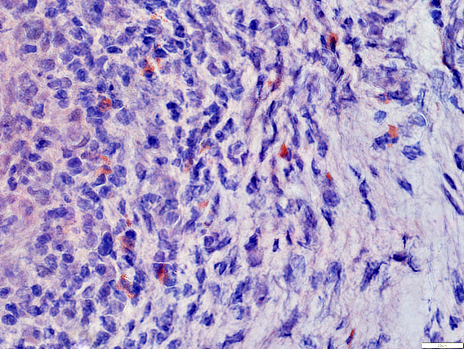
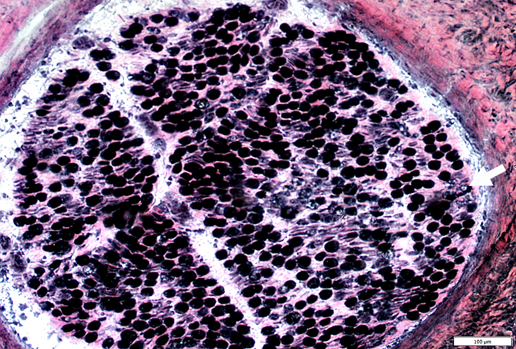
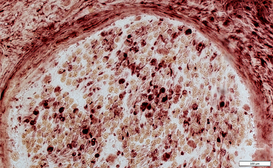
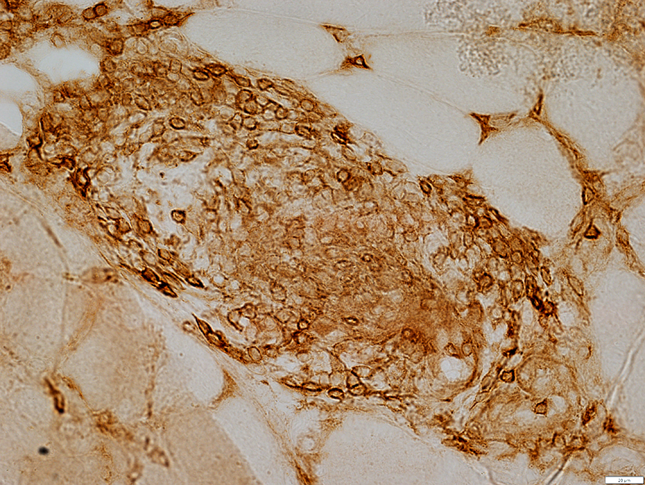
All cells in focus stain for acid phosphatase
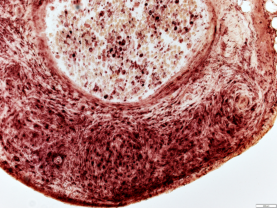 Acid phosphatase stain |
 Acid phosphatase stain |
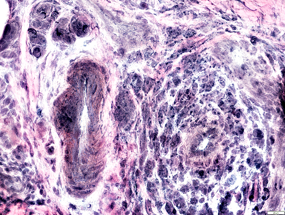
Vasculopathy: Smaller Epineurial Vessels
 VvG stain |
Surrounded by: Cells; Pale epineurial connective tissue
 H&E stain |
Epineurial Vessels: Neovascularization
Multiple small vessels within region of larger vessels
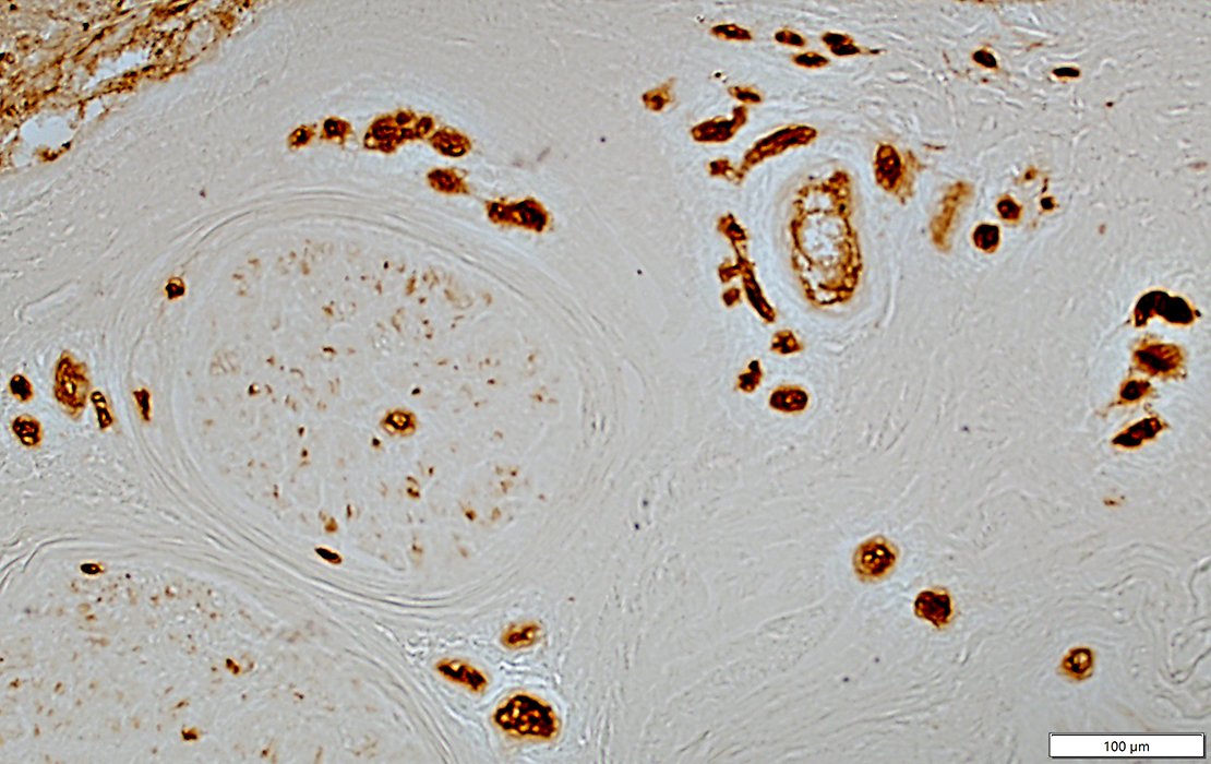 UEA I stain |
Histiocytes in several structures
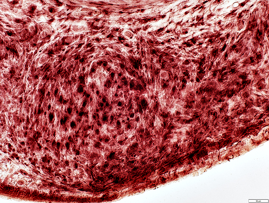
Epineurial Vessel: Acid phosphatase stained connective tissue inside fibril layer
Perineurium around small nerve fascicle (Arrow): Histiocytic cells
Epineurial connective tissue: Scattered histiocytic cells
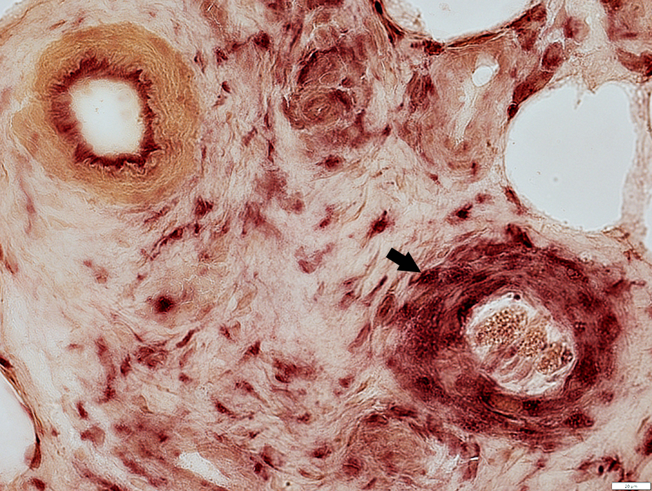 Acid phosphatase stain |
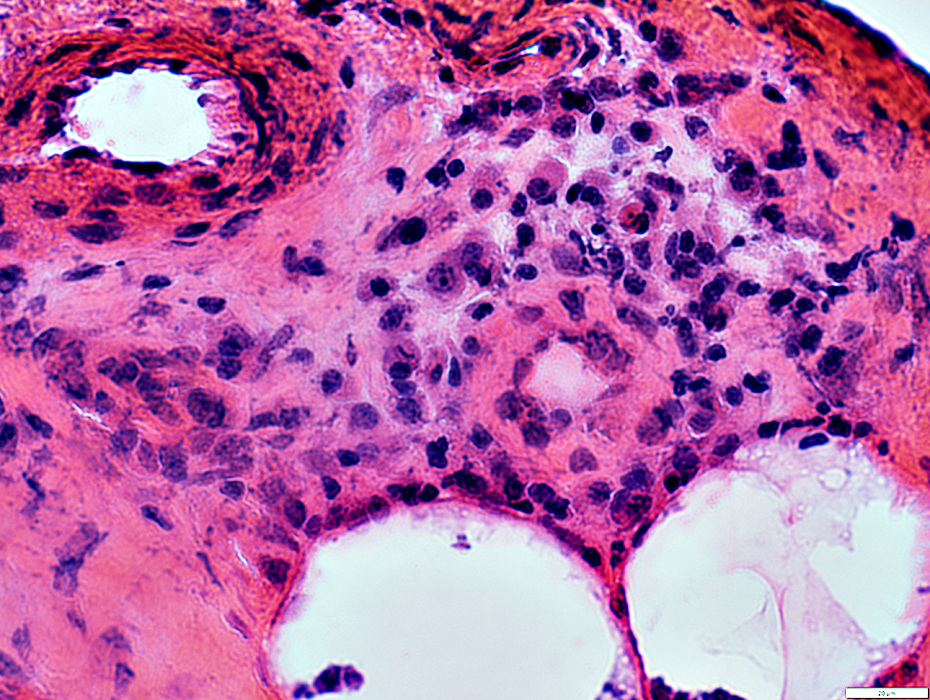
Fascicle Pathology: Subperineurial Edema
 H&E stain |
Pale-stained region under Perineurium
 Gomori trichrome stain |
Eosinophils: Within area of subperineurial edema
 H&E stain |
Wallerian Degeneration
 VvG stain |
Myelin sheath
Pale; Vesicular; Irregular (Arrow, Above)
Acid phosphatase stain (Below)
See: Intramuscular nerve
 Acid phosphatase stain |
Wallerian Degeneration: Intramuscular Nerve
 VvG stain |
 VvG stain |
Myelin sheath
Pale; Vesicular; Irregular (Arrow, Above)
Acid phosphatase stain (Below)
 Acid phosphatase stain |
Axon Loss
Differential: Intrafasciclar
More loss of large (Green) axons in some regions
NCAM-stained Schwann cells: Empty (Red) without associated axons
Active: Punctate or Irregular neurfilament staining
NCAM(r).jpg) Neurofilament (Green) + NCAM (Red) |
Axon Loss: Acute
Loss of large axons within pale areas (Myelin)
Punctate or Irregular neurofilament staining
 Neurofilament stain |
Axon Loss: Acute
Regions of myelin abnormally express NCAM
NCAM stain |
MBP(r).jpg) Neurofilament (Green) + MBP (Red) |
Regions of myelin (MBP above or P0 below) have no axons
P0(r).jpg) Neurofilament (Green) + P0 (Red) |
Eosinophilic Vasculitis: Muscle features
|
Myopathy Vasculitis |
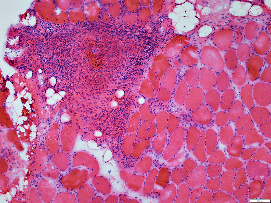 H&E stain |
Vessel location: Perimysium
Perivascular cells: Mostly histiocytes
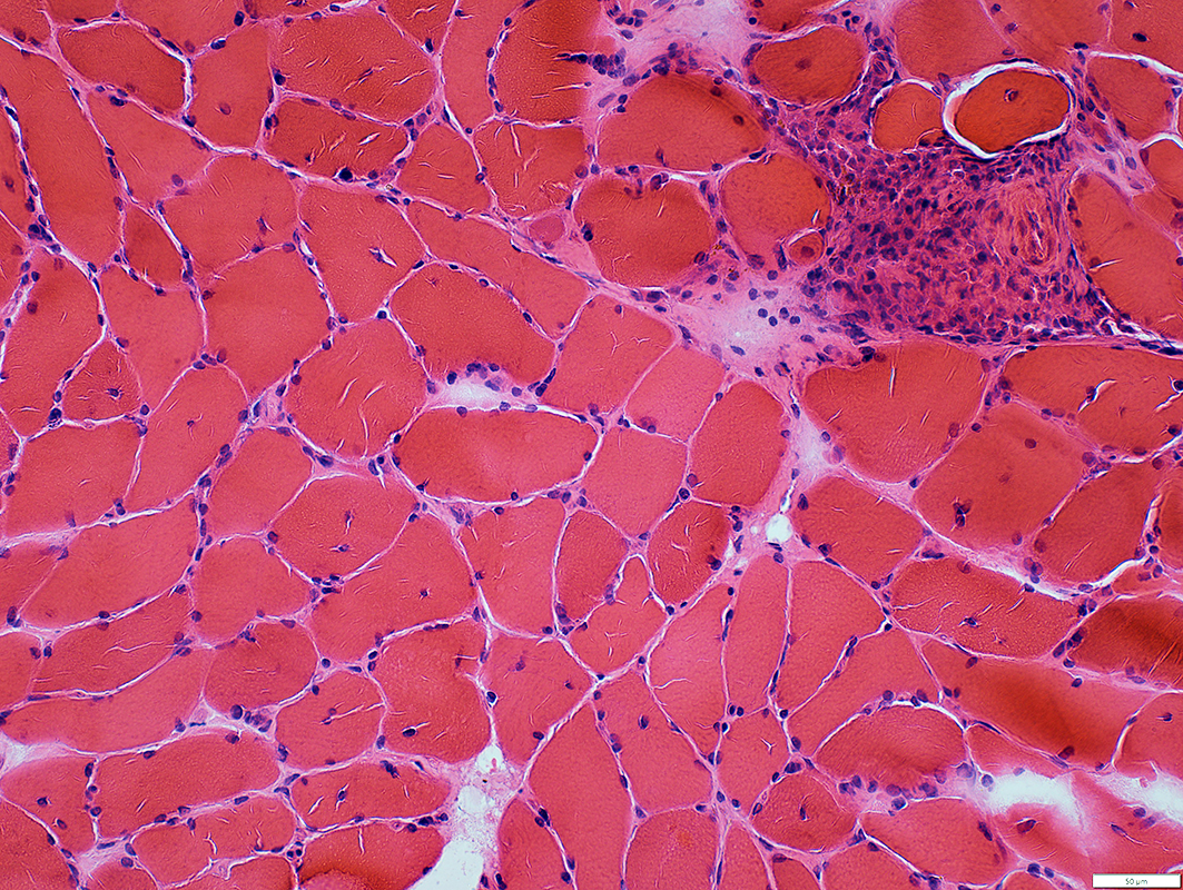 H&E stain |
Eosinophilic Vasculitis
Fibrinoid Necrosis: Inside fibril layer
Fibril layer: Irregular damaged
Surrounding vessel wall: Contains many histiocytes; Little other structure
 VvG stain |
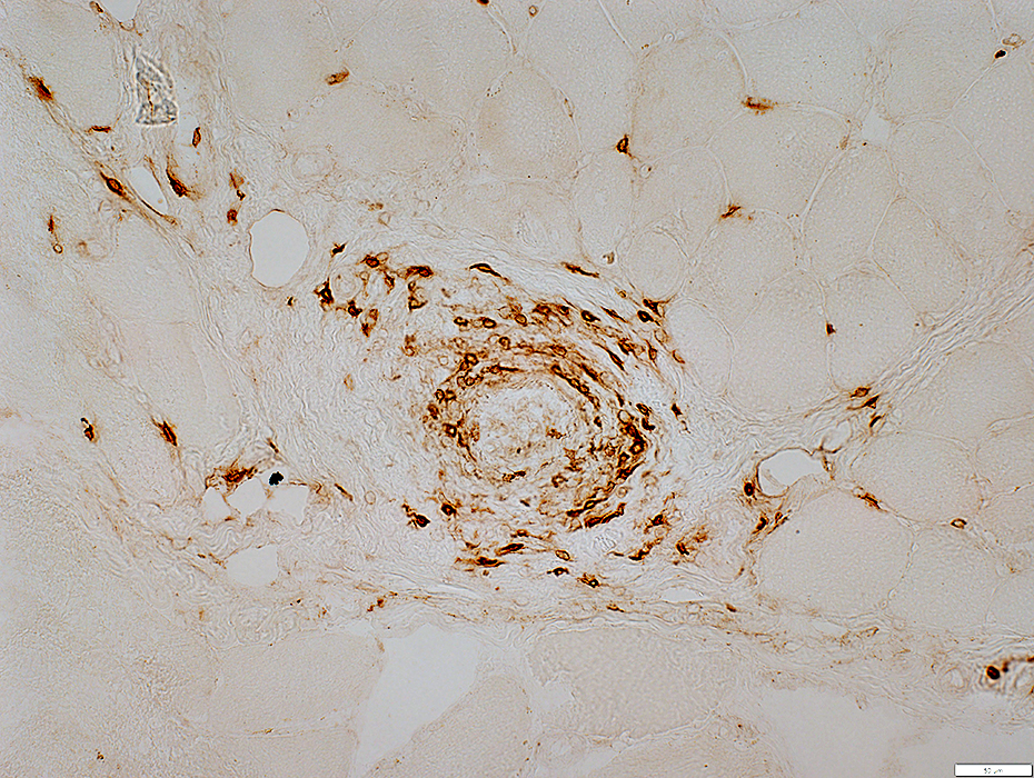
 Acid phosphatase stain |
Vessel location: Perimysium
Perivascular cells: Mostly histiocytes
 Acid phosphatase stain |
 CD4 stain |
Vessel location: Perimysium
Perivascular cells: Often stain for CD4 & MxA; Few stain for CD8
 MxA stain |
 CD8 stain |
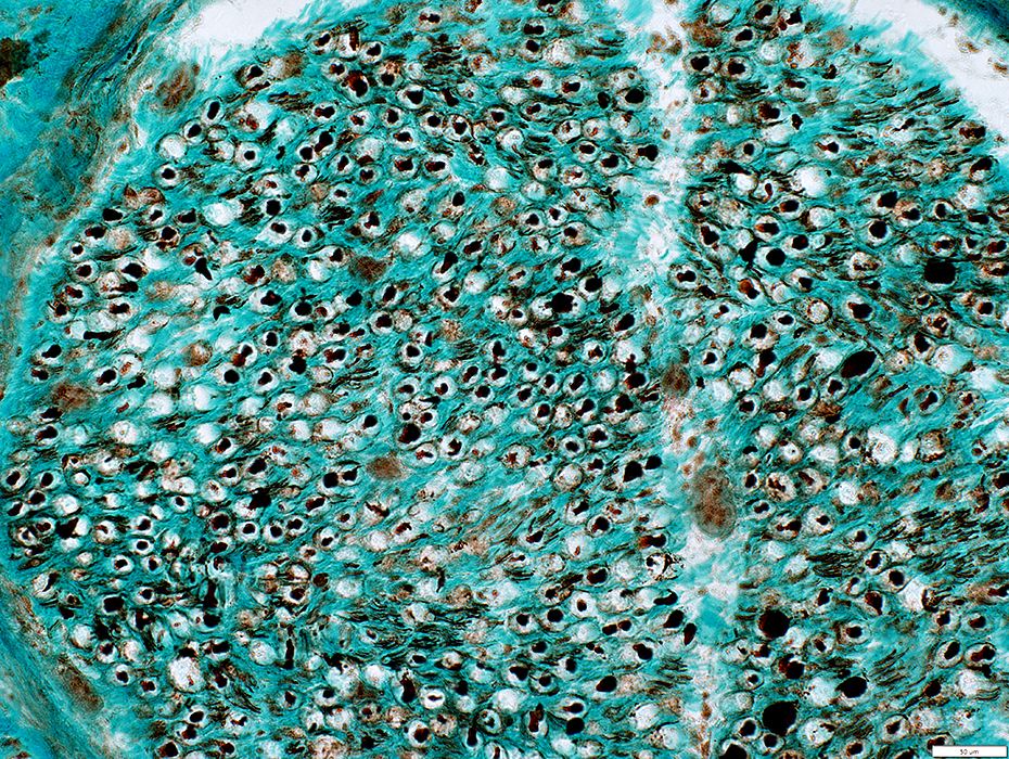
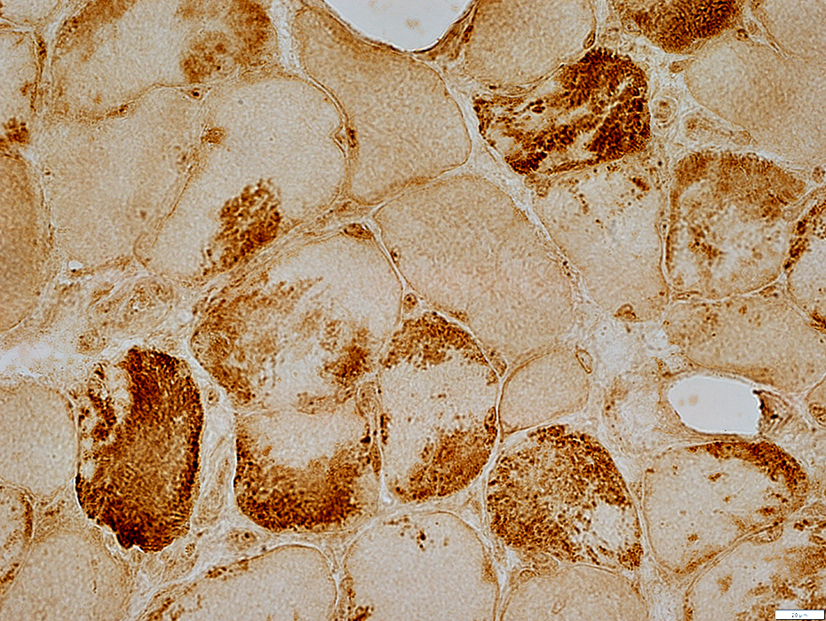
Eosinophilic Vasculitis: Myopathic features
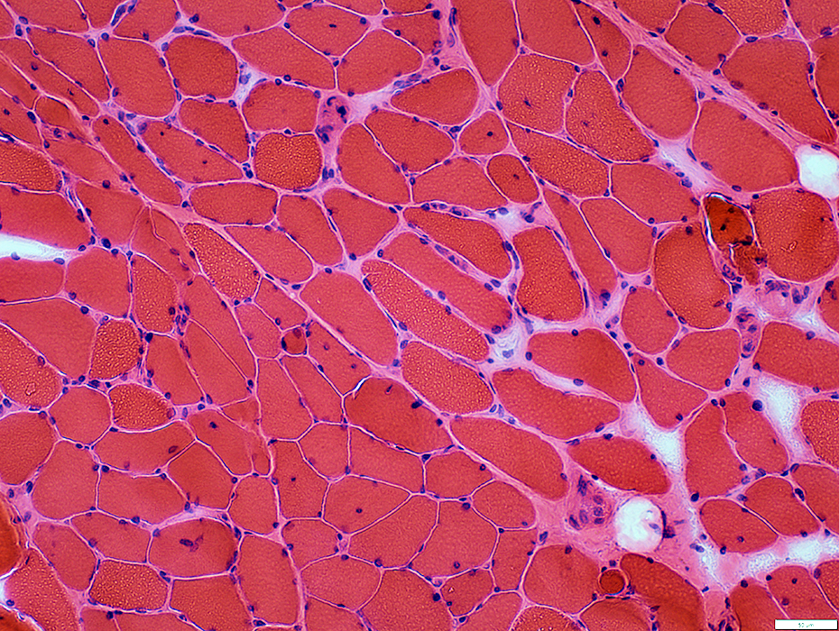 H&E stain |
Sizes: Moderately varied
Internal nuclei: Some fibers
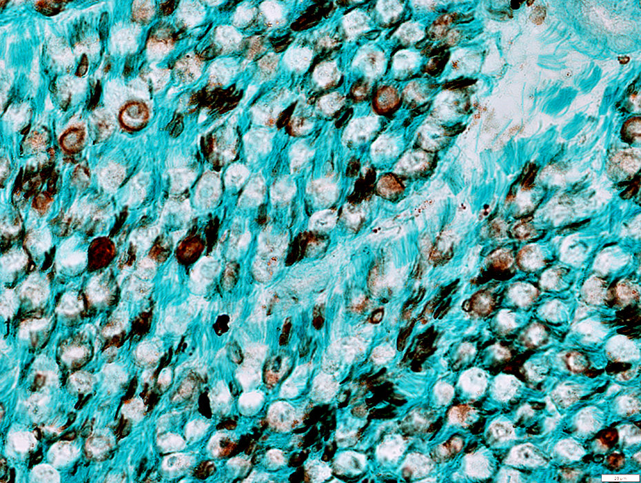
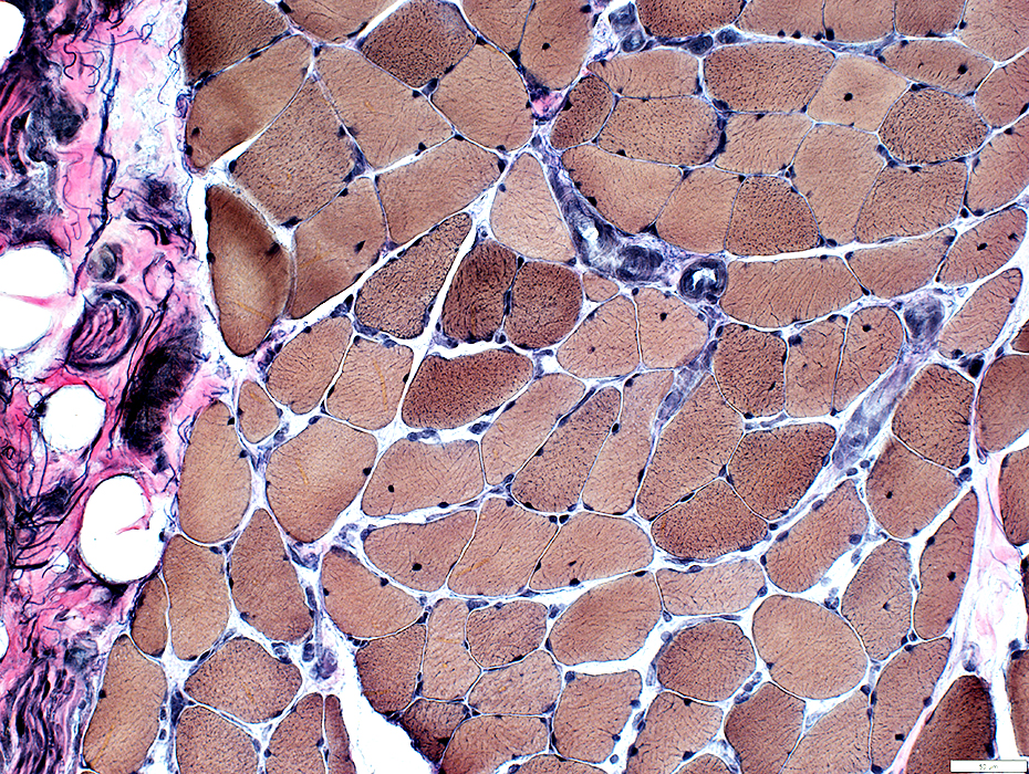 VvG stain |
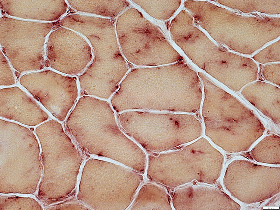 Acid phosphatase stain |
Cytoplasmic aggregates
Acid phosphatase positive (Above)
Contain LC3 (Autophagy marker; Below)
 LC3 stain |
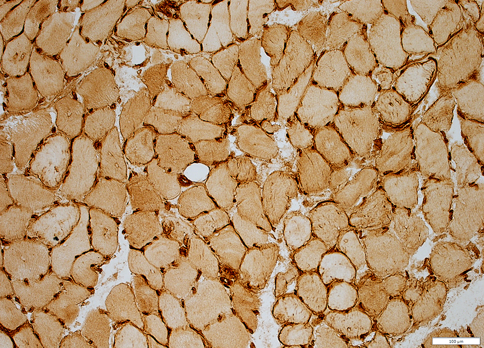 MHC Class I stain |
MHC1 upregulation
Perimysial vessels
Irregular fragments without lumens (Below; Arrow)
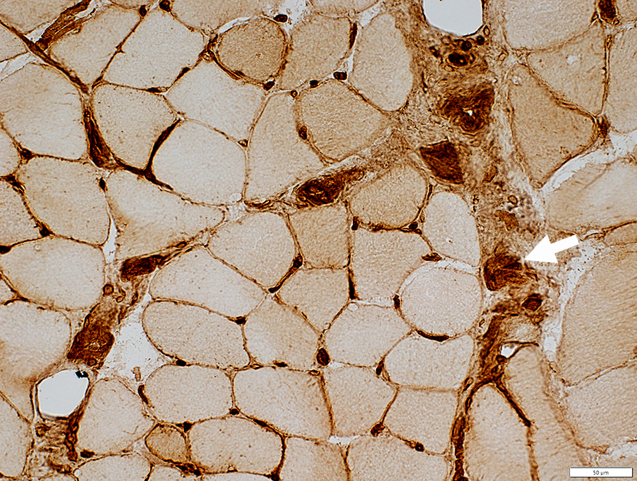 MHC Class I stain |
C5b-9 deposition
Location: Connective tissue, Endomysial & Perimysial
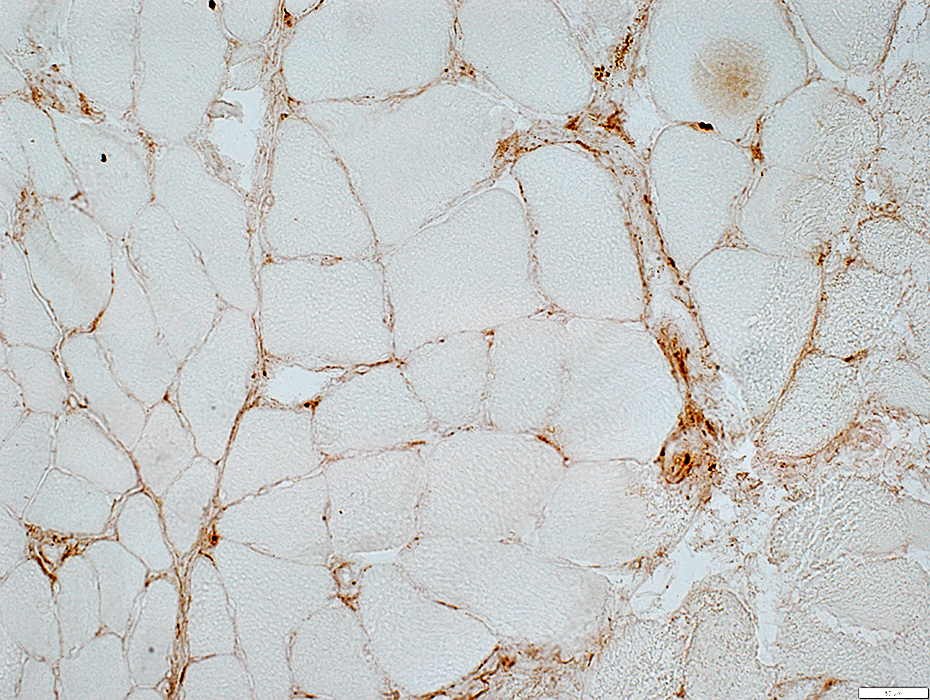 C5b-9 stain |
 CD163 stain |
CD163 histiocytes: Scattered in endomysium & perimysium (Above)
HAM56: Unusual distribution on endomysium & around surface of muscle fibers (Below)
 HAM56 stain |
Return to: Eosinophilic Vasculitis
Return to: Neuromuscular Home Page
7/3/2023