Neuromuscular

Home, Search, Index, Links, Pathology, Molecules, Syndromes,
Muscle, NMJ, Nerve, Spinal, Ataxia, Antibody & Biopsy, Patient Info

MYASTHENIA GRAVIS: DIAGNOSTIC TESTS

Antibodies
  Anti-AChR
  MuSK
  Striational
  Other
General principles
Electrodiagnostic
  Oculo-Vestibular EMP
  Repetitive CMAP
  Repetitive nerve stimulation
  Single fiber EMG
Tensilon test
Other diagnostic tests
Decrement of CMAP at 3 Hertz RNS
Decremental response to RNS in Myasthenia Gravis


General Principles of Diagnostic Testing for MG


Edrophonium (Tensilon) Testing

General
  • Drug properties
    • Action
      • Inhibits acetylcholinesterase
      • Prolongs presence of neurotransmitter, acetylcholine, in the NMJ
      • Results in enhanced muscle strength
        • Duration: Lasts for a few minutes
        • Response
          • In patients with NMJ dysfunction
          • Not specific for MG
    • Time course: Minutes; Rapid-onset; Short-acting
Method
  • Initially
    • Dosing: 2 mg of edrophonium is administered intravenously as a test dose
    • Monitoring heart rate: Bradycardia or ventricular fibrillation may develop
  • Follow-up
    • After observing for about 2 minutes, if no clear response develops
    • Up to 8 additional mg of edrophonium is injected
  • A double-blind protocol with a saline injection as placebo has been advocated
  • Testing performed with patient free of all cholinesterase-inhibitor medications
  • Cholinergic side effects of edrophonium
    • May include
      • Salivation & Lacrimation: Increased
      • Sweating & Flushing
      • Bladder urgency
      • Fasciculations, perioral
    • Atropine should be readily available
      • To reverse effects of edrophonium if hemodynamic instability
    • Extra precautions: Especially important in elderly patients
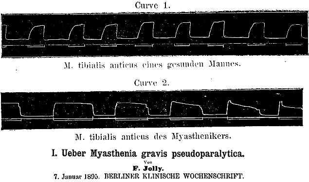
Positive test
  • Most myasthenic muscles respond in 30 to 45 seconds after injection
  • Improvement in strength that may persist for up to 5 minutes
  • Requires objective improvement in muscle strength.
  • Do not over interpret
    • Subjective or minor responses: Reduction of sense of fatigue
Utility of Tensilon test
  • Only useful in patients with objective, preferably measurable, findings on physical examination
  • Rarely helpful in the diagnostic evaluation of equivocal cases of MG
  • Sensitivity for MG is relatively low (60%) compared to other diagnostic tests
  • Tensilon testing should not be used to determine adjustments in the dose of pyridostigmine
  • False positive results
    • Can occur in patients with LES, ALS or even localized, intracranial mass lesions
    • Positive testing does not necessarily predict respose to a longer-acting anti-AChE drug
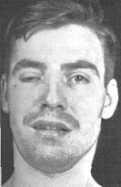
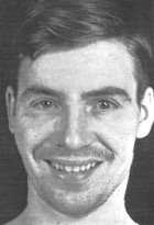
Cogan
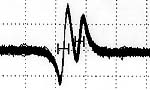
Tensilon test: Before (left); After (right)

Serum antibodies vs Acetylcholine Receptors

  • AChR antibodies: General testing method
  • Antibody properties & types
  • Antibody (IgG) targets in MG: Nicotinic AChRs
    • α1-subunit of AChR
      • Typical myasthenia gravis
      • Epitope location: Main immunogenic region on extracellular tip of α1-subunit
      • Main immunogenic region (MIR) features
        • Cluster of overlapping epitopes
        • Conformation dependent
        • Also commonly a target of anti-AChR antibodies in Experimental autoimmune MG (EAMG)
        • Cross-linking
          • Single bivalent antibody cannot bind to both MIRs on same AChR
          • Antibodies can cross-link adjacent AChRs
        • Clinical correlation
    • e-subunit
      • Acquired slow channel syndrome
      • Binding to Adult AChRs (containing e-subunit) 1
        • 13% of serums with no IgG binding to fetal AChRs
        • Increases finding (sensitivity) of anti-AChR antibodies in MG by ~ 3%
        • Occur in patients with either ocular or generalized MG syndromes
    • γ-subunit
  • AChR binding antibodies: Measurement of serum IgG (& IgM) antibodies that bind to AChRs
    • Antigenic target
      • AChRs from human skeletal muscle: Mixed innervated & denervated, or
      • Myogenic cell line expressing both adult & fetal AChRs
    • Clinical relevance
      • Relatively specific & sensitive test for MG
      • Other types of AChR testing (Modulating & Blocking): Low specificity
    • Anti-AChR antibodies occur in
      • Adults with generalized MG: 85 to 90%
      • Childhood MG: 50%
      • Ocular MG: 50% to 70%
        • Lower titers
        • Bind best to adult AChRs with e subunit
      • MG and thymoma: Nearly 100%
      • Some patients taking penicillamine with or without MG
      • "False" positives
        • Thymoma without MG
        • Immune liver disorders
        • Lambert-Eaton syndrome (13%)
        • Primary lung cancer: 3%
        • Older patients (> 70 years): 1% to 3%
        • Neuromyotonia
        • Guillain-Barré (Ganglioside Ab+): May be transiently positive
  • Antibody effects on AChRs: Other
    • AChR Modulating Antibodies
      • Mechanism
        • Anti-AChR antibodies cross-link AChRs on the post-synaptic membrane
        • Endocytosis & degradation of AChRs are accelerated
      • Test results
        • May occur in a rare patient when anti-AChR antibody binding is negative.
        • MG: Usual loss = 20% to 90% of AChRs (Normal < 20%)
        • MG with thymoma: > 90% loss
        • False positives: Common
          • Low specificity for MG
          • Specific causes: Hemolysis; Muscle relaxant drugs; Serum heating
    • AChR blocking antibodies: Block binding site for ACh on AChR (positive = 26% to 100% blocking).
      • Prevalence: Repeated arthrogryposis; 52% of generalized MG; 30% of ocular
      • 1% of patients with no binding or blocking antibodies
    • Complement binding
  • Clinical correlations of MG & Anti-AChR antibodies
    • Absolute titer of AChR binding antibodies
      • Among patients: No relation with severity of MG
      • In individual patient: Improvement often seen with reduction in titer of > 50%
    • Titer of antibody binding to main immunogenic region (MIR) of AChRs 2
      • Correlates with disease severity: Ocular vs Generalized
      • MIR location: Extracellular end of 2 α subunits of pentameric AChR
      • > 50% of MG AChR Abs bind to MIR
    • Antibody blocking & modulation of AChRs
      • Some correlation with disease severity
      • Not diagnostically specific
    • Neonatal MG: Transient
      • High anti-fetal/anti-adult muscle anti-AChR antibody ratio
    • Recurrent arthrogryposis
      • IgG vs γ subunit of fetal AChR: Blocks AChR function
    • Slow channel syndrome: Acquired
      • IgG vs adult AChR
    • IgG vs Clustered AChRs 3
      • Present in
        • "Sero-Negative" Ocular or Generalized MG patients: 50%
        • AChR Ab+ patients by RIA: 98% positive
        • May be more specific for MG than RIA binding Ab
      • Complement fixing
      • Antibody subclass: IgG1
      • Associated with thymic pathology: Lymphocytic infiltrates; Germinal centers
    • Neuronal AChR: α3 subunit
    • Also see: AChR disorders
  • Patients with MG but no anti-AChR antibodies
    • Rule out hereditary MG
    • Low frequency of thymic pathology & thymoma
    • May have antibodies to other neuromuscular junction antigens

Muscle AChR
(Adult)

Repetitive Nerve Stimulation (RNS): 2 to 3 Hz

RNS: General
  • Most frequently used electrodiagnostic test for MG
  • Procedure
    • Nerve to be studied with RNS
      • Electrically stimulated six to ten times at 2 or 3 Hertz
    • Compound muscle action potential (CMAP)
      • Recorded with surface electrodes over muscle
  • Nerves tested
    • At least one proximal & one distal motor nerve
    • Nerves innervating weak muscles
  • If Decrement present
    • Ensure decrement is reproducible
      • Repeat RNS
    • Test for: Post-exercise facilitation (Repair of decrement)
    • Perform EMG
      • Rule out
        • Denervating, or other, disorder producing decrement
      • Changes supporting NMJ disorder
        • Motor unit action potentials: Small-short or Unstable
  • If NO Decrement at baseline
  • Original description
Decrement with RNS in MG: Orignial description
RNS in Myasthenia Gravis
  • Normal muscles
    • CMAP amplitudes: No change with repetitive nerve stimulation
  • Myasthenia gravis
    • CMAP amplitudes: Progressive decline during first 4 to 5 stimuli
      • Caused by failure (Block) of increasing number of NMJs
    • Positive RNS test features
      • Decrement in CMAP amplitude
        • Size: More than 10% in reduction in CMAP amplitude
          • Measure from 1st to 4th or 5th potential in train
        • Smallest CMAP is often 2nd or 3rd potential in train
      • Post-tetanic potentiation (Post-exercise facilitation)
        • Definition: Reduction in (Repair of) decrement after exercise
        • Stimulus: Isometric exercise, brief (10 to 15 sec)
        • Time course
          • Onset: Immediate
          • Duration: ~2 minutes
        • Degree of repair: Partial or Complete
      • Post-exercise exhaustion
        • Definition
          • Post exercise: Appearance, or Exacerbation, of decremental response
        • Protocol
          • Stimulus: Muscle exercise for 1 minute
          • Repeat RNS after 1, 2, 3 and 4 minutes
        • Time course
          • Onset: Maximal 3 to 5 minutes after exercise
          • Disappears by: 10 minutes after exercise
    • RNS is positive in about 75% of patients with generalized MG, if:
      • Proximal & Clinically involved muscles are tested
      • Muscle is warm: Cooling reduces size of decrement
      • More than one muscle is tested: Strong muscles often have less decrement
    • Diagnostic issues
      • Sensitivity of RNS for MG
        • Sensitivity in generalized MG
          • Overall: 75% to 80%
          • Increased
            • Proximal & Clinically involved muscles are tested
            • Muscle is warm
            • More than one muscle is tested
          • Reduced
            • Only distal muscles are tested
            • Cooling: Reduces size of decrement
            • Childhood MG
            • Acute severe generalized or bulbar MG (≤ 4 weeks of disease)
              • Decrement frequency: 11% to 40% positive 4
              • Often have abnormal jitter
            • Strong muscles: Often have less decrement
        • Ocular MG: 50%
      • Diagnostic specificity

Myasthenia Gravis
Repetitive Nerve Stimulation


From: M Al-Lozi
Also see

Single fiber electromyography (SFEMG)

  • Principle: Muscle fibers innervated by a single axon
    • Normal: Activated with consistent latencies
    • NMJ disorders
      • NMJ abnormal: Increased variability of latencies among muscle fibers in single motor unit
      • NMJ fails completely: Muscle fiber potential may be blocked if transmission at NMJ fails completely  
  • SFEMG
    • Simultaneously records potentials of two muscle fibers innervated by an individual axon
    • Measures
      • Variability = "Jitter"
      • Block: NMJ failure
    • SFEMG is the most sensitive test for MG
      • Sensitivity: > 95% positive in generalized & ocular MG
        • When the test site includes facial muscles
    • Abnormal jitter is not specific for MG
      • May occur in other neuromuscular disorders, including ALS, polymyositis or LEMS
      • More specific for MG if large degree of jitter occurs with mild or no other changes on EMG
    • MuSK MG: Jitter may be present in absence of RNS decrement

Normal SFEMG

Increased jitter: MG patient
From: M Al-Lozi

Ocular-Vestibular Evoked Myogenic Potentials 5

Return to Myasthenia gravis
Return to Myasthenic Syndromes

References
1. Autoimmunity 2003;36:151–154
2. JNNP 2012; Online July
3. Arch Neurol 2012; Online June, J Neuroimmunol 2019;332:69-72
4. Clin Neurophysiol 2016;127:3480-3484
5. Neurology 2016;86:660-668

7/23/2020