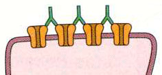
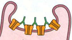
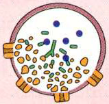
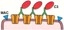
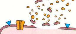
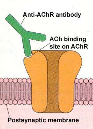
Anti-AChR antibodies: Mechanisms of action
Anti-AChR antibodies
AChR function: Altered
Post-synaptic membrane: Damage
AChR Degradation: Increased rate
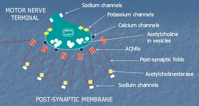
Neuromuscular junction: Normal
Neuromuscular Junction: Normal
From: M Al-Lozi
Normal NMJs
Many NMJs consist of several spot-like synaptic regions 1-5 μm across
Presynaptic terminal
Releases packets or quanta of acetylcholine
Spontaneous release rate: 0.1-10 Hz
Trigger: Increase in free Ca
++
in nerve terminal
Effect of release on post-synaptic membrane: Miniature endplate potentials (MEPPs)
Single nerve impulse
Causes release of 1-3 quanta from each presynaptic terminal bouton
Release event at a given
active zone
: Occurs in response to one of ten nerve impulses
Number of quanta released at a NMJ: Proportional to NMJ area
Frog NMJs: Large; Release 100-200 quanta per impulse
Human NMJs: 10x smaller; Release 20 quanta per impulse
Each quantum consists of 5,000 to 10,000 molecules of ACh
Post-synaptic membrane
Acetylcholine receptors
(AChRs): High concentration
AChRs are concentrated at peaks of, & 1/3 of way down, post synaptic folds
Density: ~10,000/μm
2
; Form near crystalline array
Voltage-gated sodium channels
High density of
Na
v
1.4
in folded postsynaptic membrane
Effect of transmission at human NMJs is amplified
Concentrated at base of folds
Structrure
Highly folded: Especially prominent in humans
Opening of folds: Opposite active zones on nerve terminal
Modulation of post-synaptic AChRs by anti-AChR antibodies
Increased AChR degradation
Anti-AChR antibodies cross-link post-synaptic AChRs
↓
Cross-linked AChRs are endocytosed more rapidly than normal
↓
Internalized AChRs are degraded
Fewer AChRs remain on the post-synaptic membrane
RESULT: Less efficient neuromuscular transmission
Damage to Post-synaptic Membrane
Complement binds to the Antibody-AChR complex
Membrane-attack complex (MAC) forms on the membrane
↓
The post-junctional membrane is damaged
Fewer post-synaptic membrane folds
Reduced numbers of AChRs
Widened synaptic cleft
RESULT: Less efficient neuromuscular transmission
Altered AChR function
Blockade of ACh binding site
Acquired immune MG
: No prominent blockade of ACh binding to AChRs
Recurrent arthrogryposis
: Blocking antibodies identified
Altered AChR channel function
Acquired immune MG
: No prominent change in AChR ion channel function
Acquired Slow Channel syndrome
: Antibodies to adult AChR alter AChR ion channel function
Return to
Neuromuscular Home Page
Return to
Myasthenia gravis
Return to
Anti-AChR antibodies
5/2/2003