|
Home, Search, Index, Links, Pathology, Molecules, Syndromes, Muscle, NMJ, Nerve, Spinal, Ataxia, Antibody & Biopsy, Patient Info |
Autophagic Vacuoles with Sarcolemmal Features (AVSF): Colchicine Treatment
|
Autophagy Description Differential diagnosis Pathology Ultrastructure Also see Desmin IBM Secretory XMEA |
AVSF: Pathology
|
|
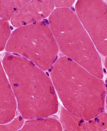
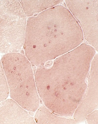
| Vacuoles: Small, Clear, Multiple (H&E stain) | |
|
|
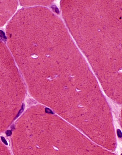
| Vacuoles: Magenta rim (GT stain) | |
|
|
| Vacuoles: May stain for esterase | |
|
|
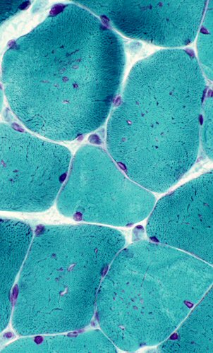
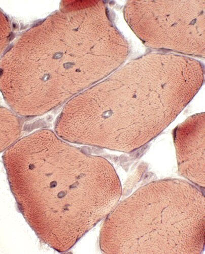
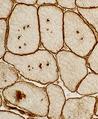
| Vacuoles: Staining of rim on VvG | Vacuoles: More common in type I fibers (ATP pH 9.4 stain) |
|
|
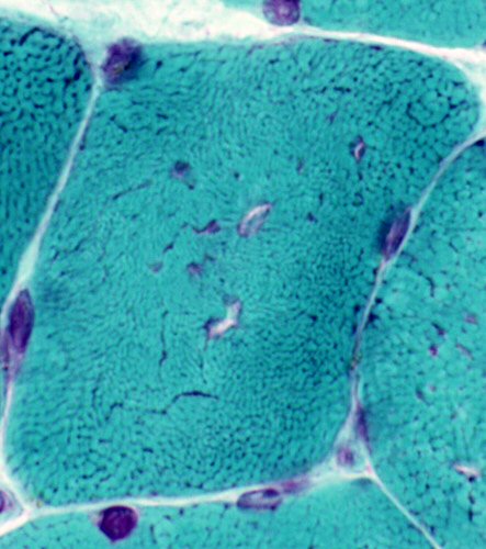
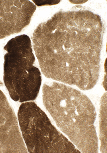
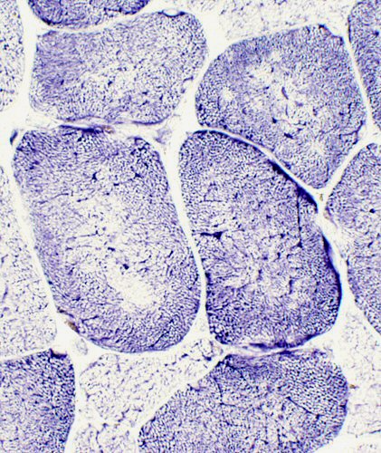
| Vacuoles: Staining of some vacuoles with acid phosphatase | Abnormal internal architecture (NADH stain) |
|
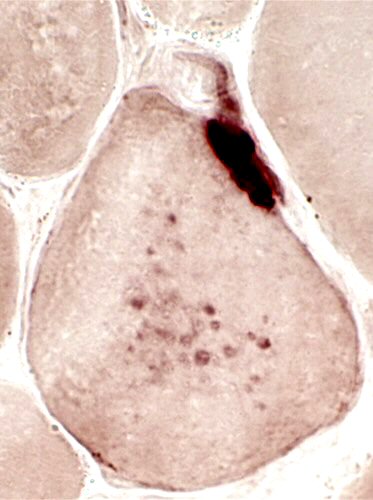
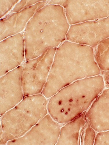
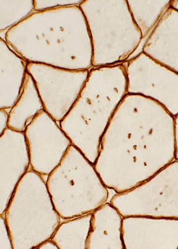
| Vacuoles: Caveolin-3 staining |
|
|
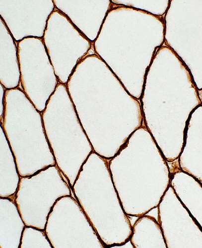
| Vacuoles: Caveolin-3; A caveolin aggregate is also present | Vacuoles: Dystrophin |
|
|
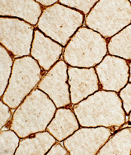
| Vacuoles: No staining with laminin-α2 | Vacuoles: Punctate staining with laminin B |
|
From: Robert Schmidt MD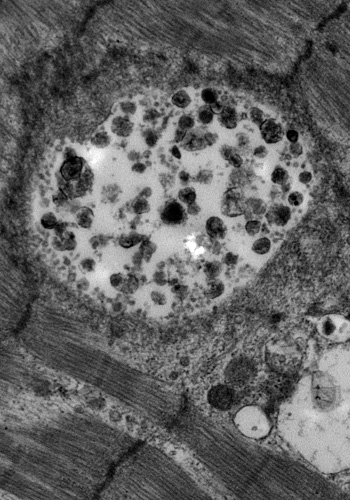
|
|
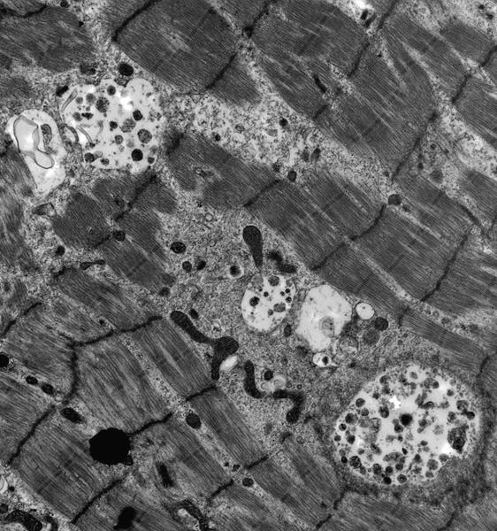
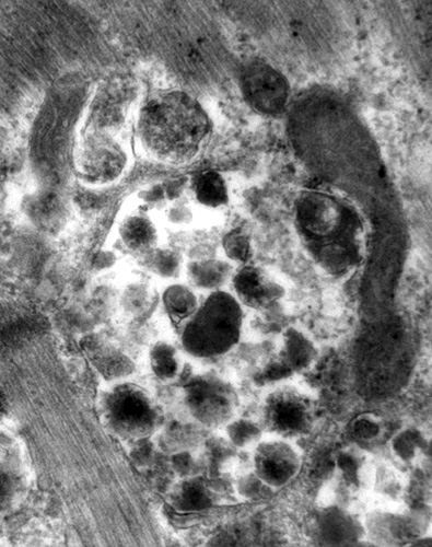
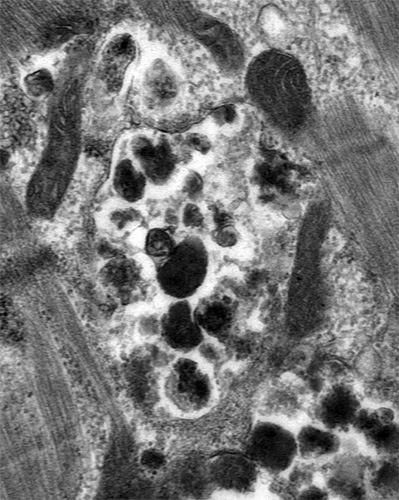
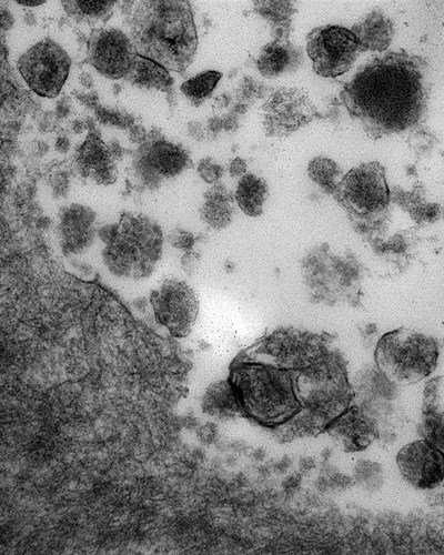
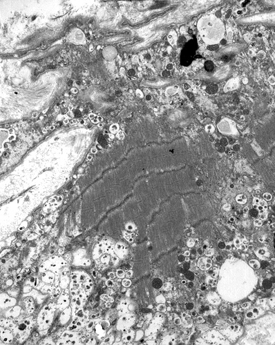
Vacuoles & Clusters Contain cytoplasmic debris, electron dense material & myeloid bodies. Some clusters are encircled by a membrane Abnormally shaped mitochondria are located near some clusters Also see: Autophagic vacuoles, Desmin myopathy |
|
|
|
|
|
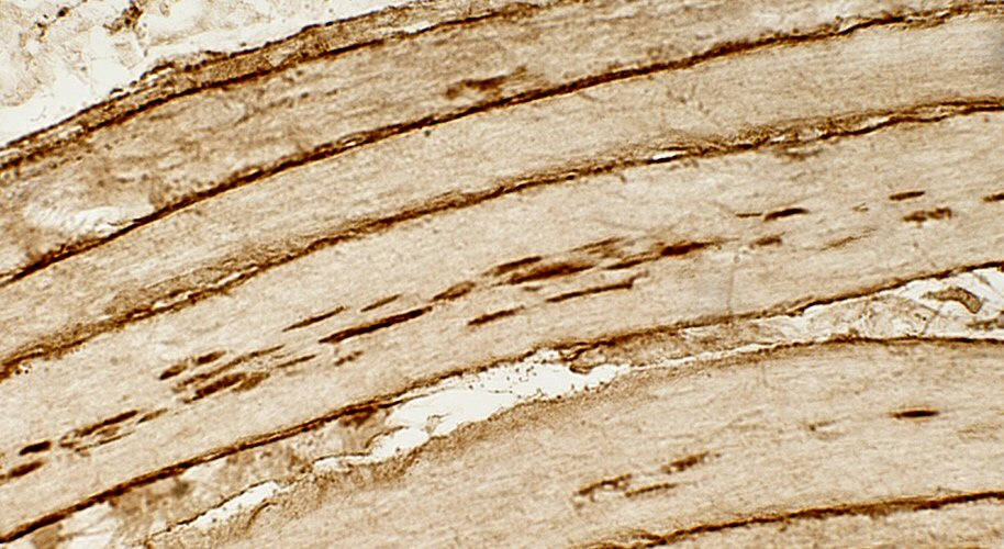
| Autophagic Vacuoles: Higher power view | Sarcolemma Structure: Disordered, Irregular Many associated vacuolar structures See: Secretory Autophagy |
Return to Pathology Index
Return to XMEA
3/18/2026