X-linked Myopathy with Excessive Autophagy
 H & E stain |
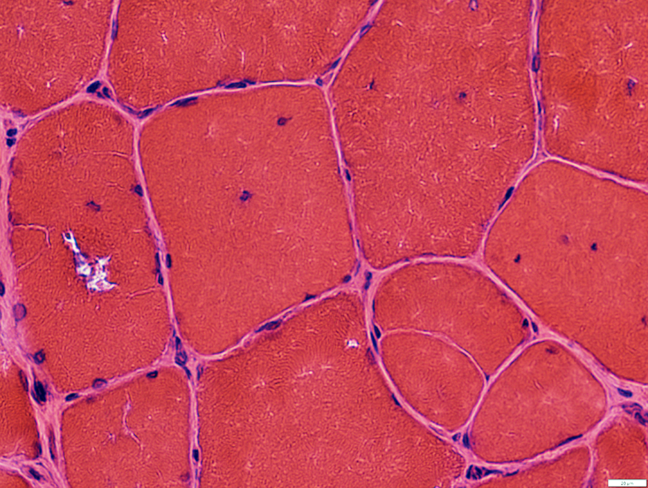
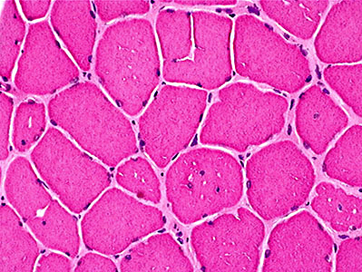 H & E stain From J Mandell Myopathic changes
|
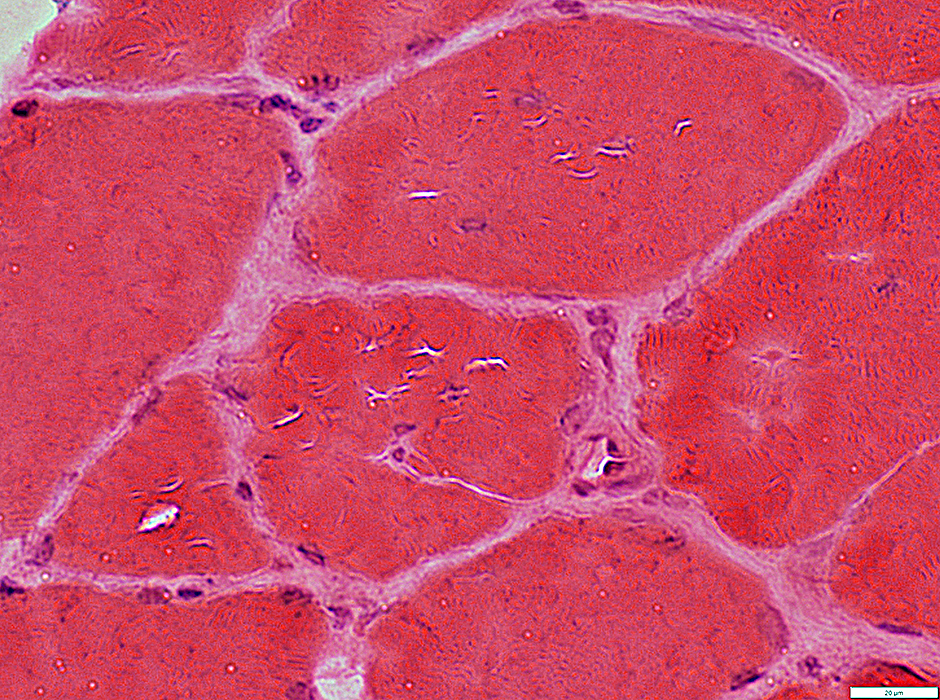
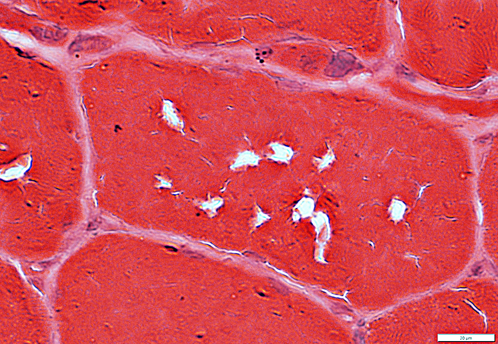
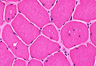 H & E stain From J Mandell Vacuoles: Small & Clear |
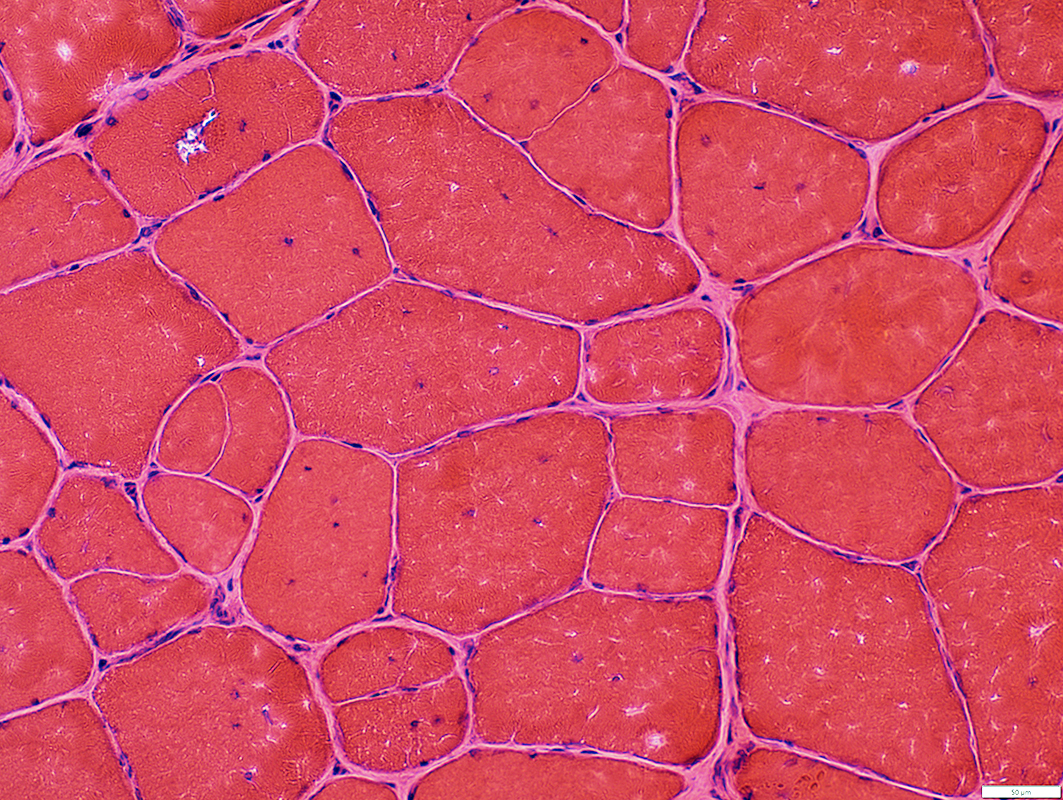
 H & E stain |
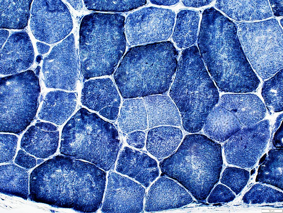
- Muscle fibers
- Size: Varied
- Split (Partially fused)
- Internal nuclei
- Vacuoles: Irregular shapes
- Endomysial connective tissue: Increased
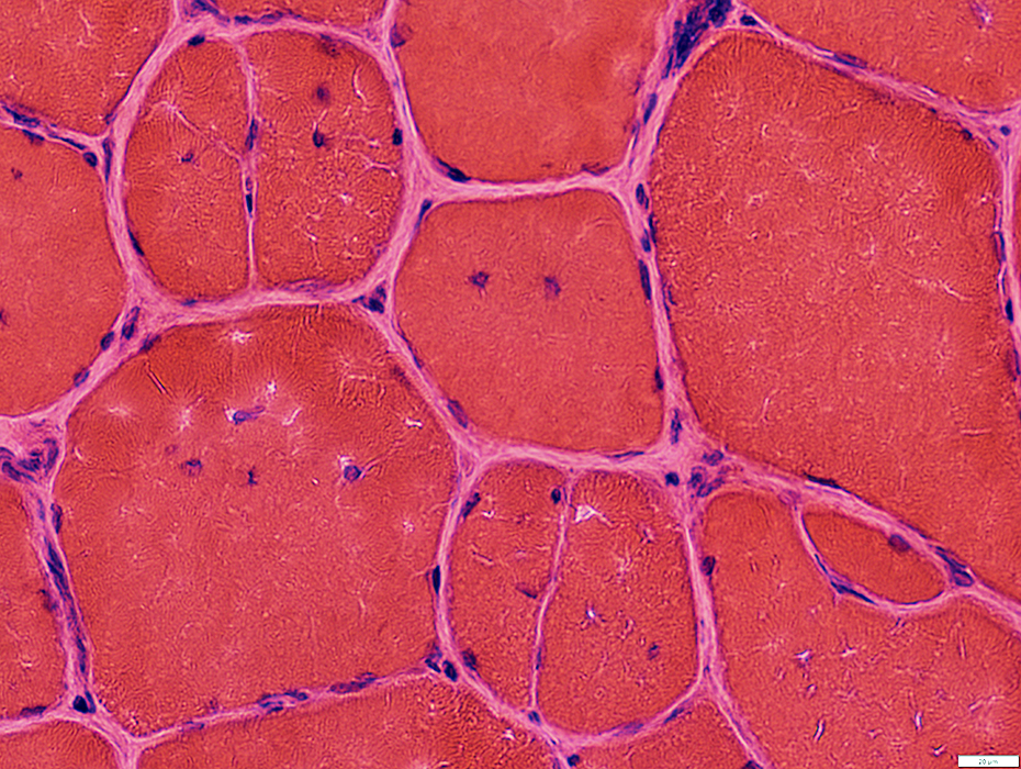
 H & E stain |
 H & E stain |
 H & E stain |
 VvG stain |
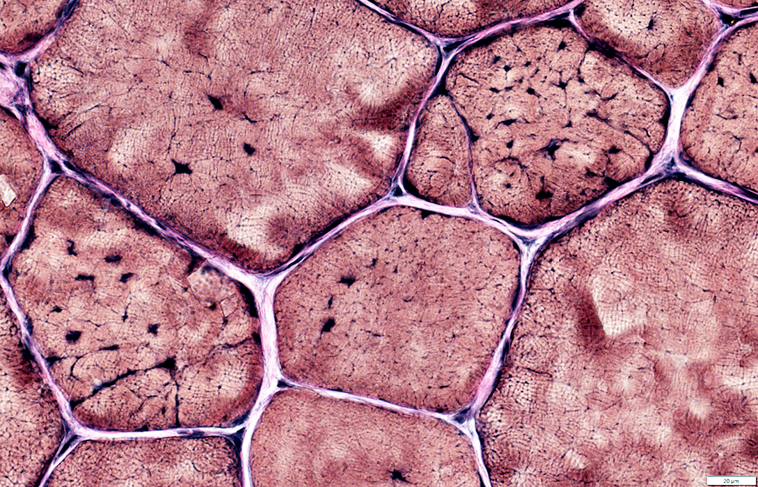
 NADH stain |
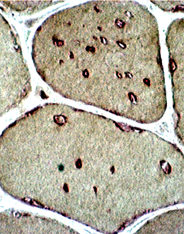 Esterase stain From J Mandell Vacuoles in Muscle fiber cytoplasm Many stain for Esterase |
|
 Esterase stain |
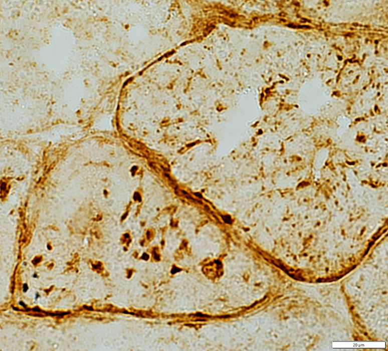
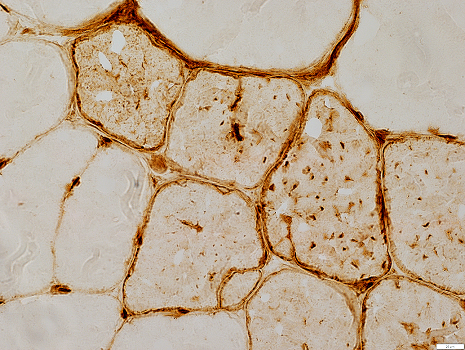
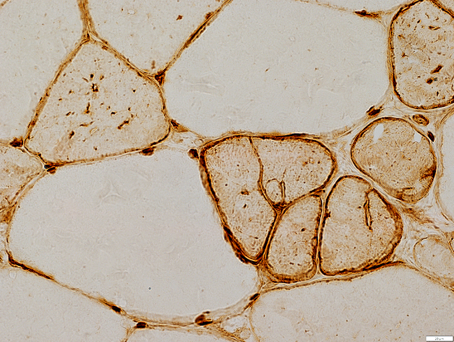
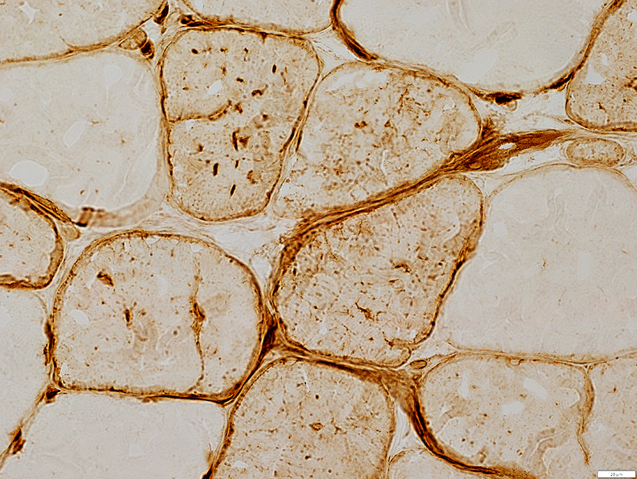
 Dystrophin stain Vacuoles: Stain for Dystrophin & other membrane proteins |
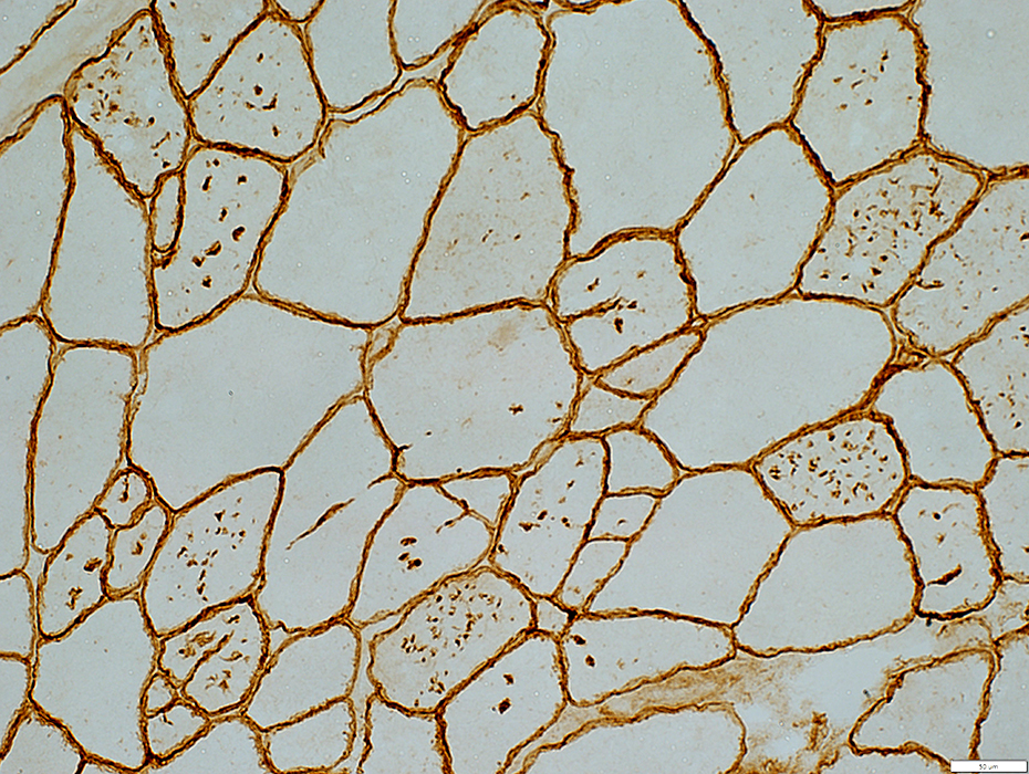 δ-Sarcoglycan stain Vacuoles: Stain for membrane proteins, including δ-Sarcoglycan |
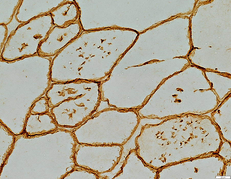 δ-Sarcoglycan stain |
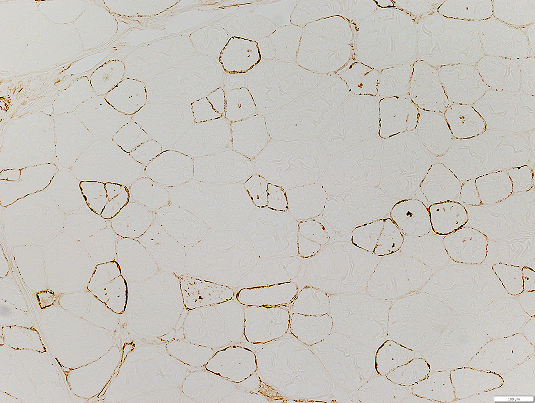
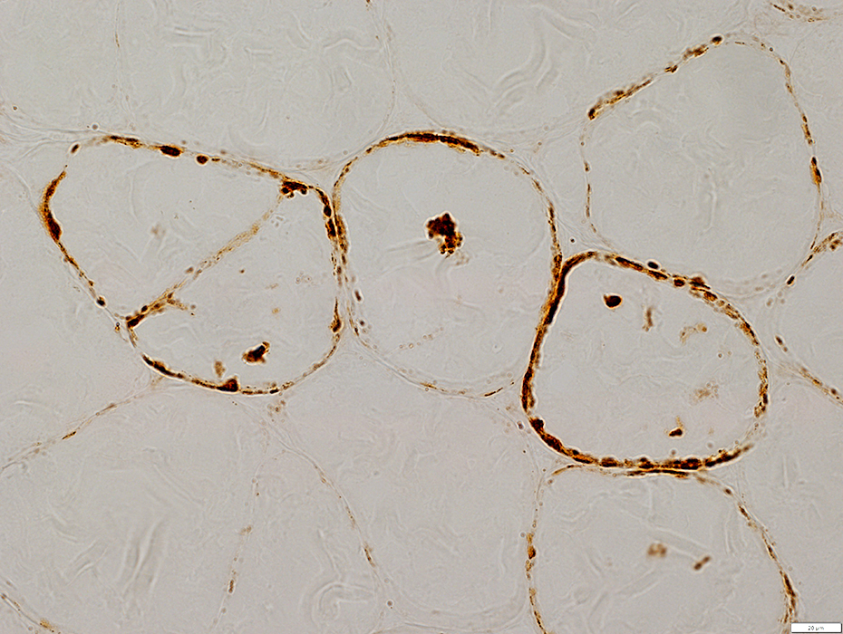
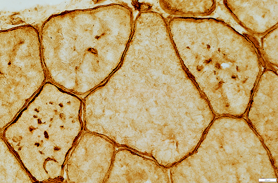 Caveolin-3 stain Vacuoles: Stain for membrane proteins, including Caveolin-3 |
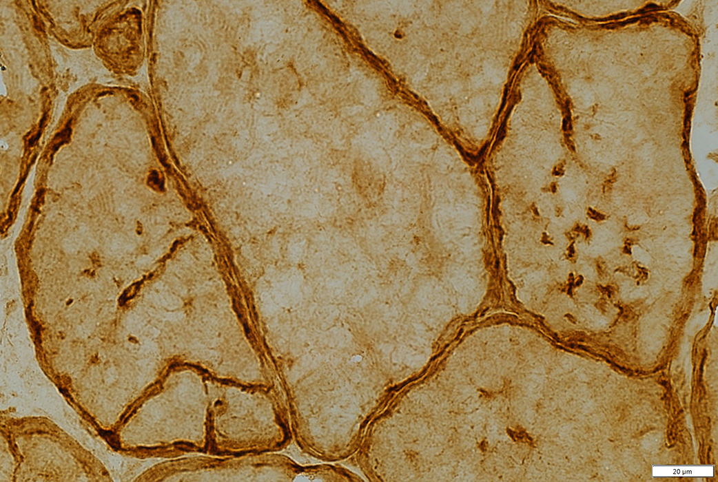 Caveolin-3 stain |
 LAMP2 stain |
XMEA (Above)
Control (Below)
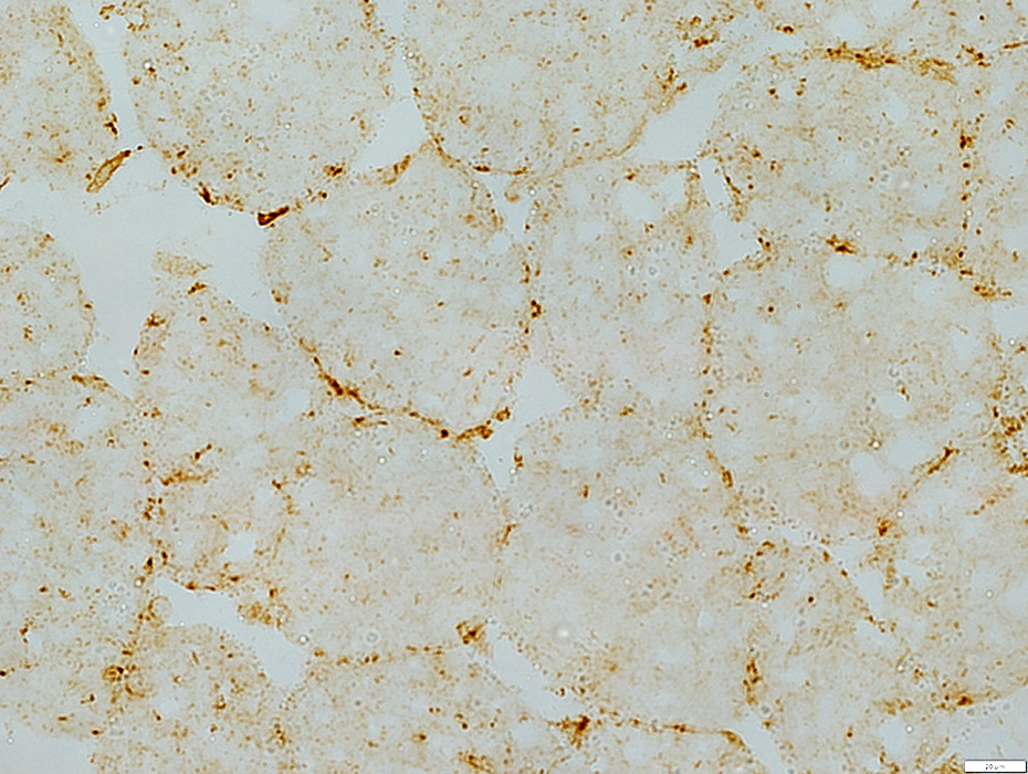 LAMP2 stain |
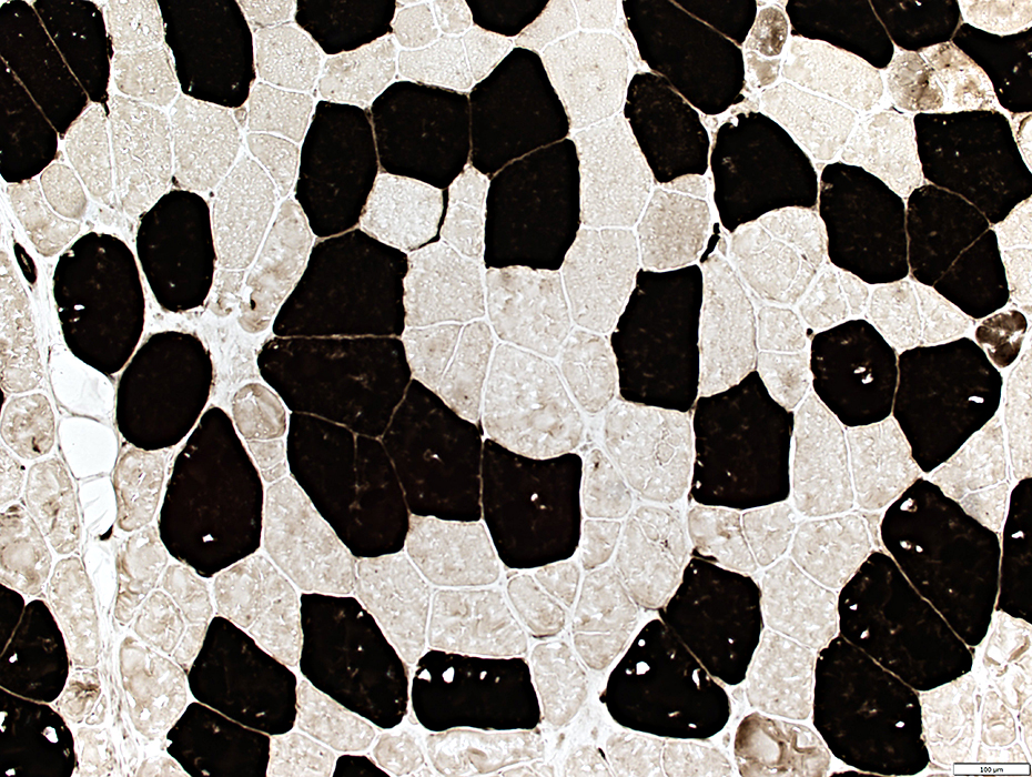 ATPase pH 4.3 stain |
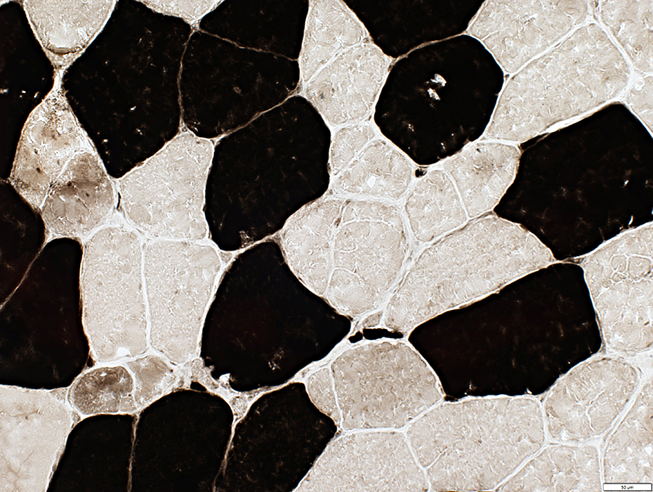 ATPase pH 4.3 stain |
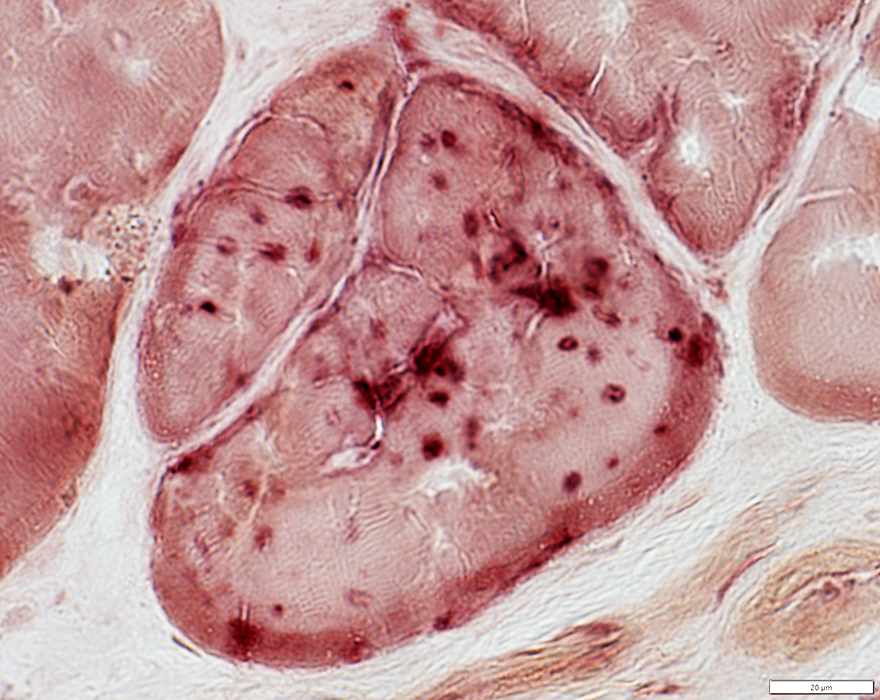
 C5b-9 complement stain |
 C5b-9 complement stain |
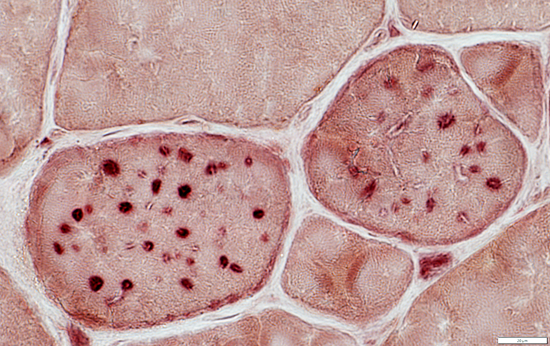
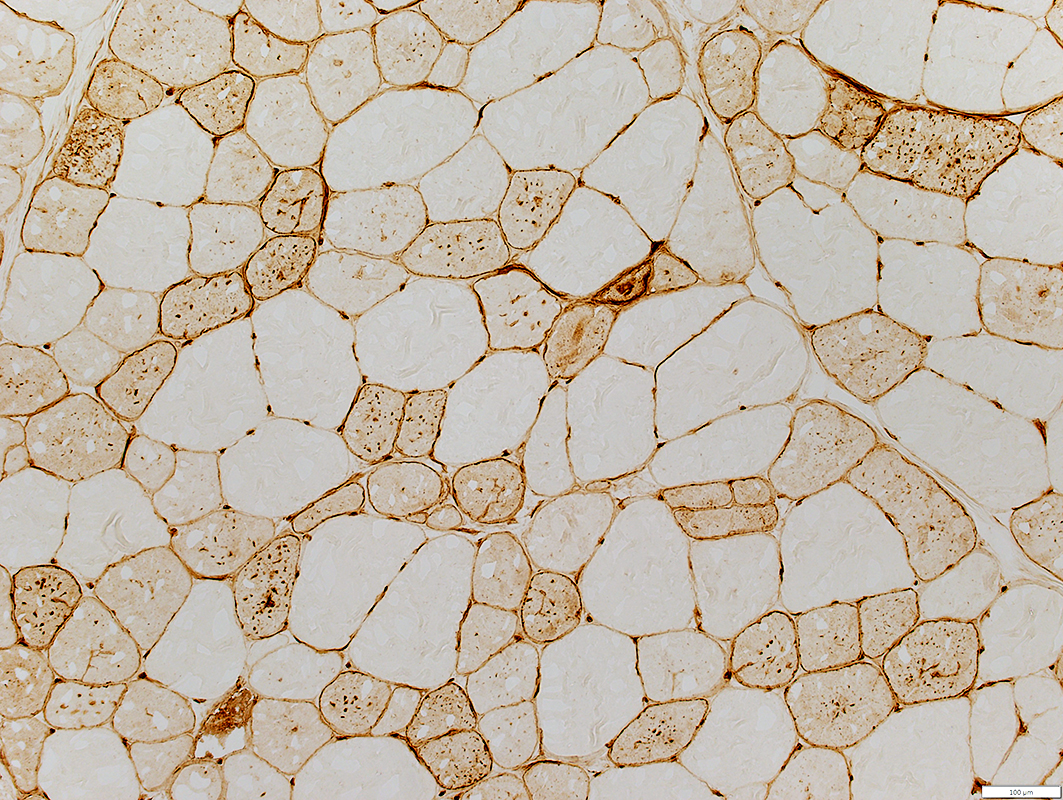 MHC Class I stain |
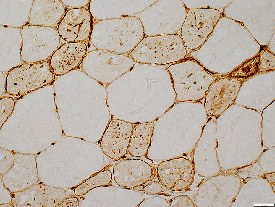 MHC Class I stain |
 MHC Class I stain |
 MHC Class I stain |
 MHC Class I stain |
Autophagic Vacuole
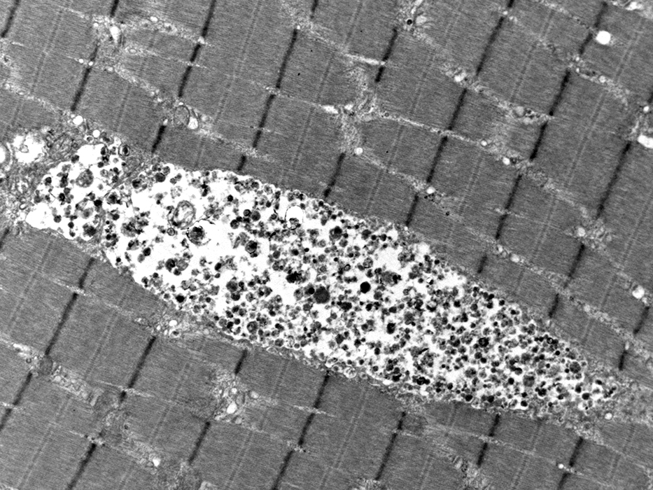
From: Steve Moore & Cameron Crockett |
Autophagic Vacuole
Extrusion
Basal lamina: Duplication
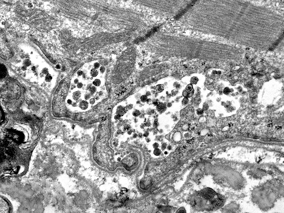 From: Steve Moore |
Return to: X-linked myopathy with excessive autophagy
Also see: Autophagic vacuoles with sarcolemmal features
Return to: Pathology index
Return to: Neuromuscular Home Page
5/14/2025