Chronic Immune Demyelinating Polyneuropathy (CIDP), Adult
CIDP: Nerve morphology
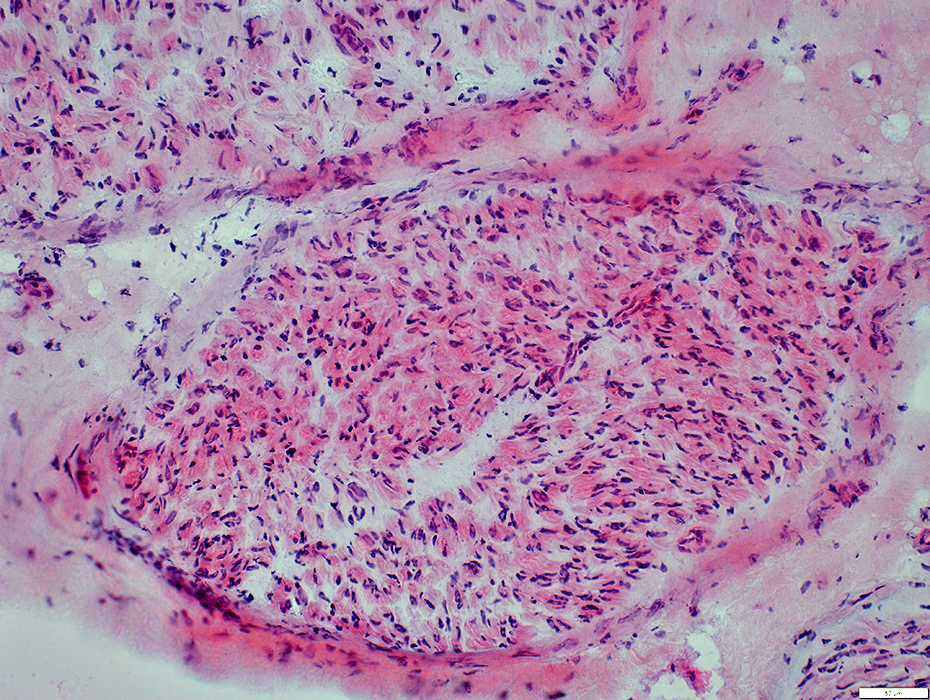 H&E stain |
Some regions are pale
Myelinated axons: Patchy loss
Perineurium: Thin & pale stained in regions
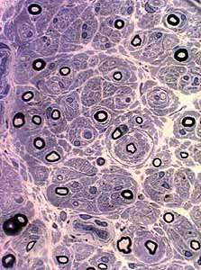
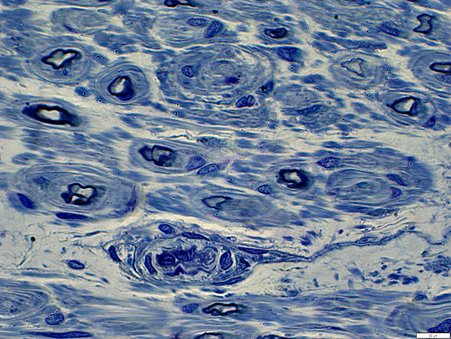
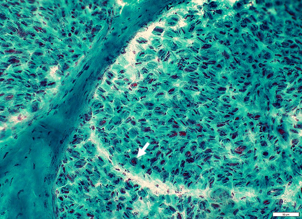 Gomori trichrome stain |
Small & Varied sizes
Scattered in endoneurium
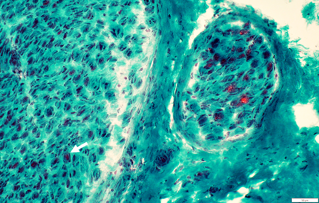 Gomori trichrome stain |
Onion bulbs, Small
Scattered in endoneurium
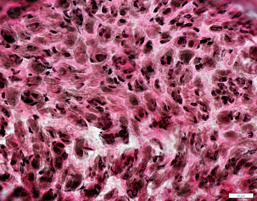 VvG stain |
CIDP
Myelinated axon loss There are no large myelinated axons
Remaining small axons are hypomyelinated
Subperineurial edema: Narrow; Surrounds much of fascicle
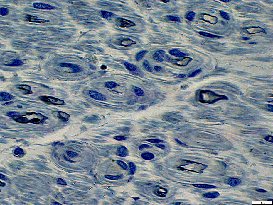
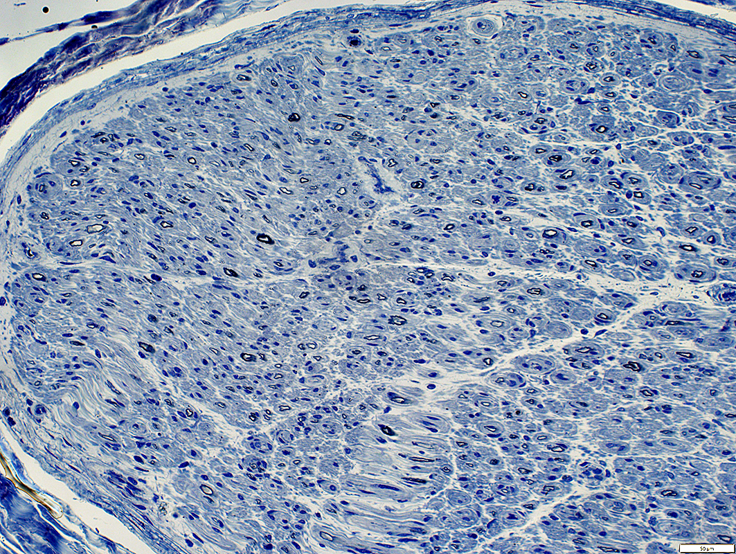 Toluidine blue stain |
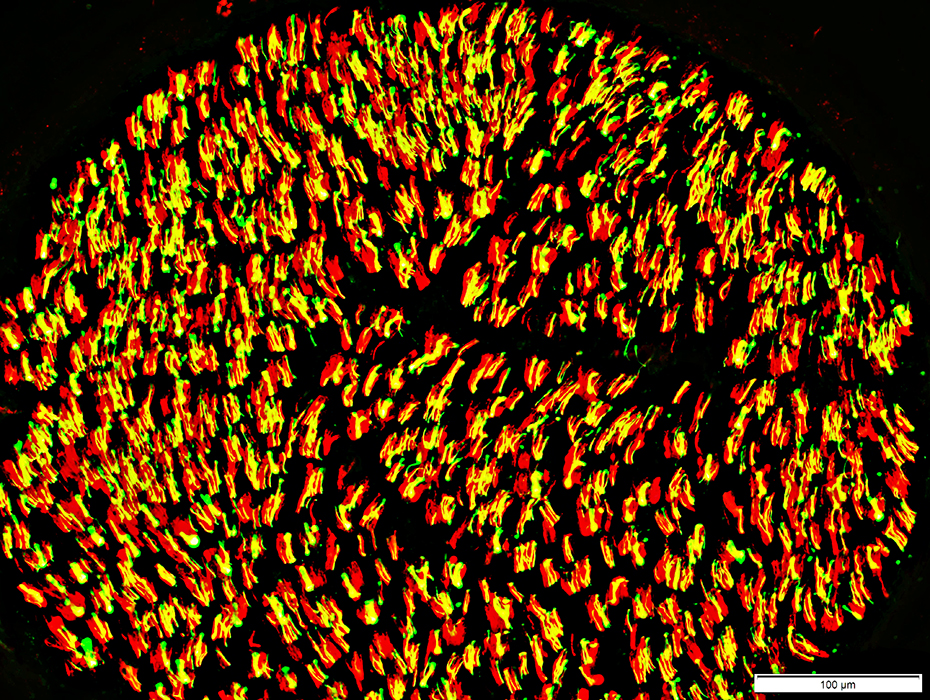
CIDP: Loss of the largest myelinated axons
Myelin containing MBP is mostly lost
With loss of large axons many non-myelinating Schwann cells express small amounts of MBP_
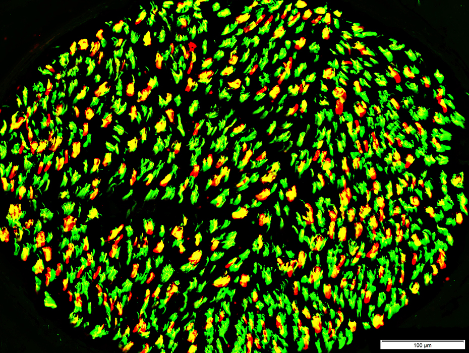
 Neurofilaments (Green) + MBP (Red) stain |
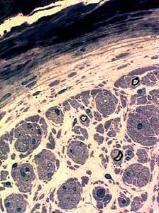
Myelin Pathology
Onion bulbs
Myelin sheath: Thin
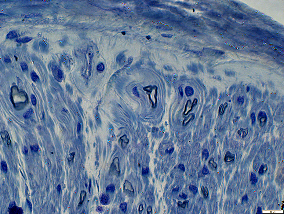
 Thinly myelinated axons. Axon loss. |
 Thinly myelinated axons. Axon loss: Patchy |
Onion bulbs: Small |
 Neurofilaments (Green) + NCAM (Red) stain |
Most axons, even intermediate sized ones, are surounded by NCAM-stained Schwann cells
NCAM normally surround only the smallest axons
Axon loss: The largest axons are completel lost
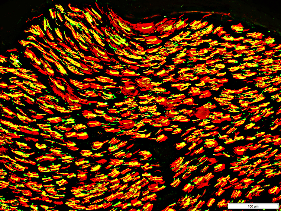 Neurofilaments (Green) + NCAM (Red) stain |
Onion bulbs: Many small
Many axons are surrounded by P0 even though myelinated axons are mostly lost
Schwann cells in onion bulbs: Similar to Büngner band Schwann cells (Yellow) with co-staining for NCAM & P0
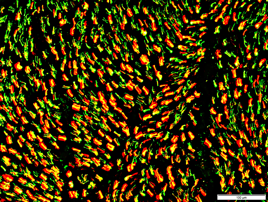 NCAM (Green) + P0 (Red) stain |
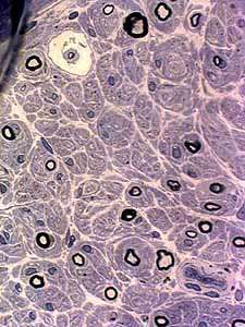
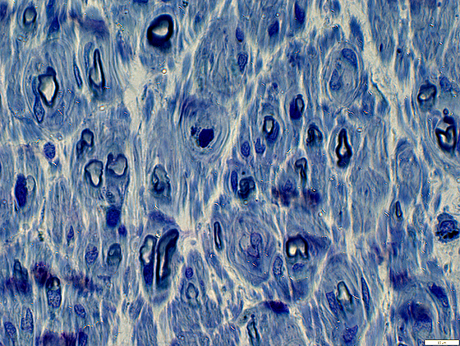 Toluidine blue stain |
Surround some axons
Varied sizes
Some contain no axon
Most axons in onion bulbs are hypomyelinated (Thin myelin sheath)
 Toluidine blue stain |
CIDP: Axon loss, Chronic: Schwann cells
Büngner band & Onion bulb Schwann cells (Yellow) co-stain for NCAM & P0
Normally unmyelinated axons (Green): Occur in small clusters
 NCAM (Green) + P0 (Red) stain |
Subperineurial edema
 H&E stain |
Subperineurial edema |
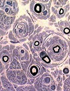
 Toluidine blue stain |
Onion bulbs
Endoneurial Microvessels: Mildly large
 Toluidine blue stain |
CIDP, Chronic: No inflammation
 Acid phosphatase stain |
CIDP: Muscle
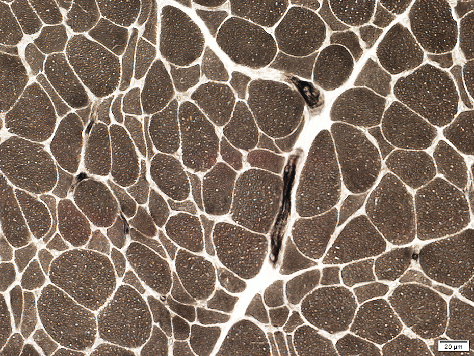 ATPase pH 9.4 stain Type 1 muscle fiber predominance |
Return to Normal nerve biosies
Return to Biopsy illustrations
Return to Neuromuscular Home Page
Return to Nerve biopsy
Return to Demyelinating neuropathies
Return to Active demyelination
4/16/2021