PERIPHERAL NERVE: Active Demyelination 1,2
Myelin stripping, Cell mediated
Cell Processes inside Schwann cell basal laminaPhagocytic cell process (White Arrow): Extends beneath a layer of myelin
Other, larger cell processes contain myelin breakdown products (Dark Arrow)
Multiple, very small, round Schwann cell processes are located immediately under the basal lamina
Axon within myelin sheath: Intact
Macrophage & Processes (Top, Left): Outside the basal lamina
Some macropahge processes extend near, but do not penetrate, the Schwann cell basal lamina
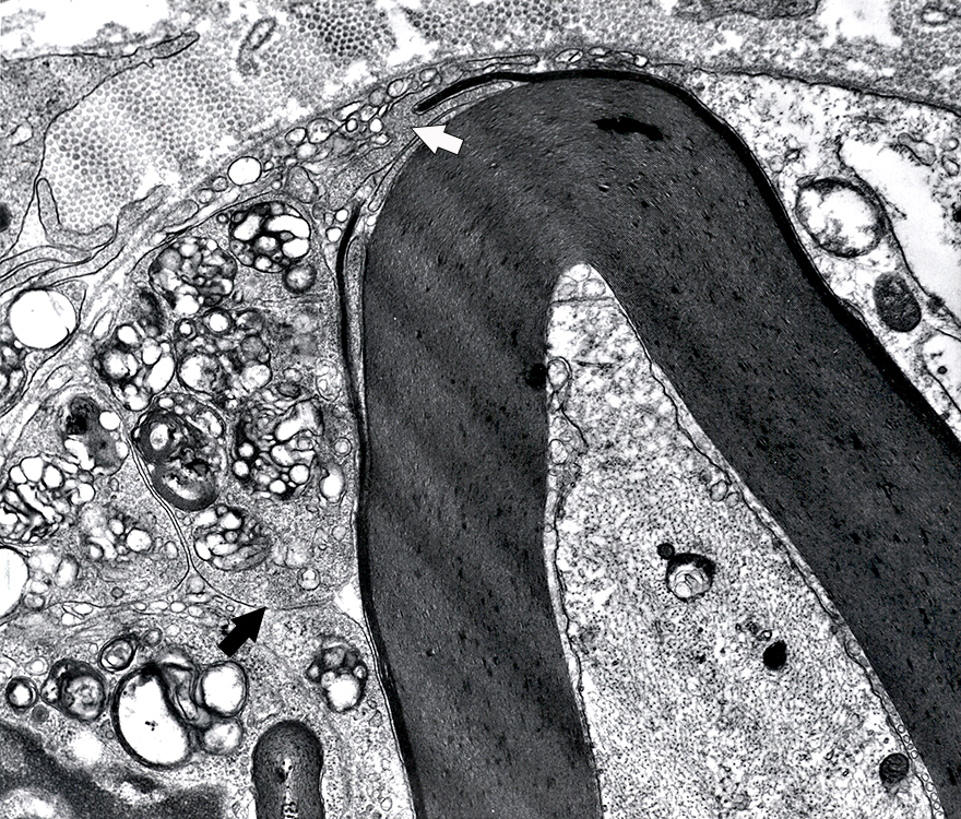 Electron micrograph: From Robert Schmidt MD |
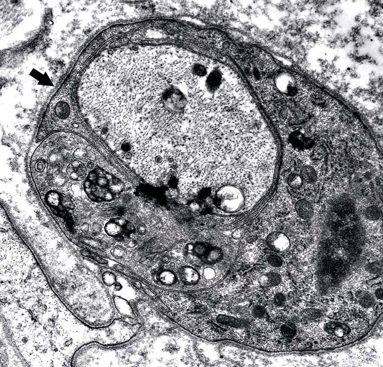
Demyelinated axons
Axon
Axoplasm is dark: Contains closely spaced neurofilaments.
Without surrounding myelin
Surrounded by cell processes: Contain inclusions
Entire structure is surrounded by a Schwann cell basal lamina (Arrow)
A pale Schwann cell process extends outward, but inside the Basal lamina (Bottom, Left)
2 small, round cell processes are apparent inside the basal lamina (Bottom, Left)
|
Demyelinated Axon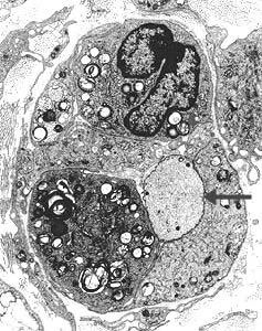 Electron micrographs: From Robert Schmidt MD |
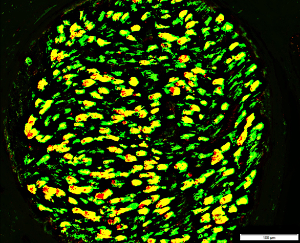
Active Demyelination: Histiocytic Schwann cells & Myelin
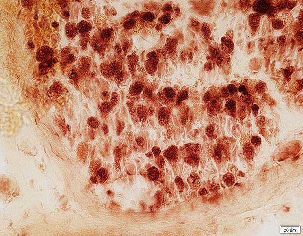 Acid phosphatase stain Most Myelin sheaths: Acid phosphatase+ Compare to: Wallerian degeneration |
 MBP = Myelin Basic Protein; P0 = P0 protein Myelin Structure: Irregular P0 & P0+MBP Sheaths |
See: AIDP
Return to Normal nerve biopsies
Return to Biopsy illustrations
Return to Neuromuscular Home Page
Return to Nerve biopsy
Return to Demyelinating neuropathies
Return to Chronic demyelination
References
1. Neurology 2018;91:1051-1060
2. J Neurol Neurosurg Psychiatry 2020;9:650-659
2/16/2024