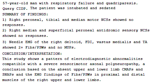
Chronic Immune Demyelinating Polyneuropathy, Relapsing, Severe
Relapsing CIDP, Severe: Electrodiagnostic summary
|
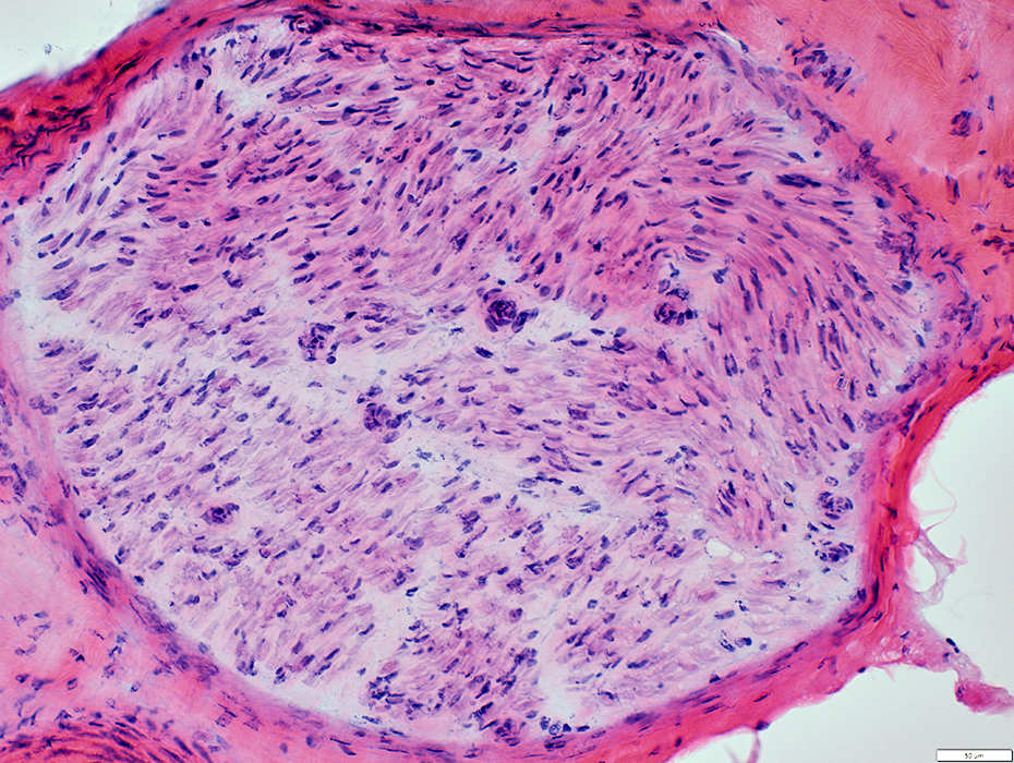
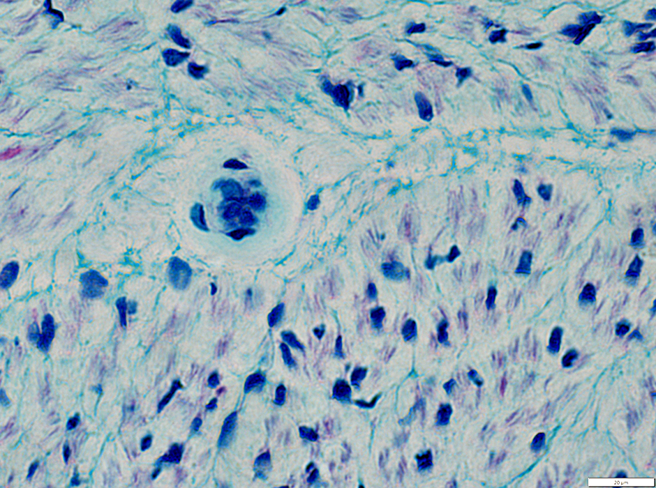
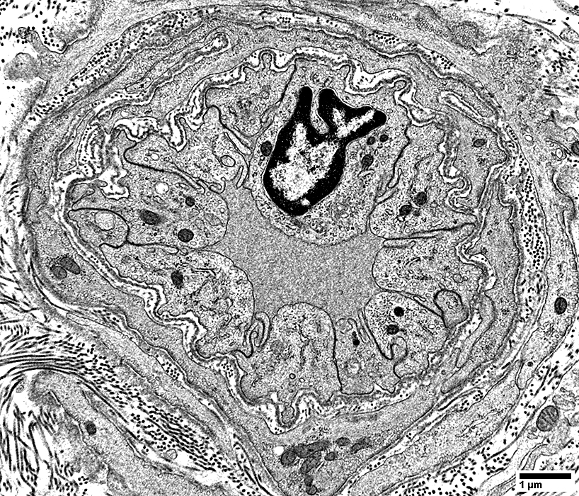
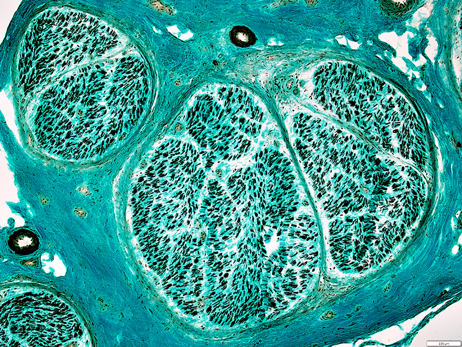
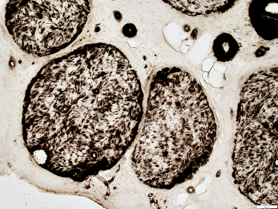
Relapsing CIDP: Endoneurial edema, No lymphocytic inflammation
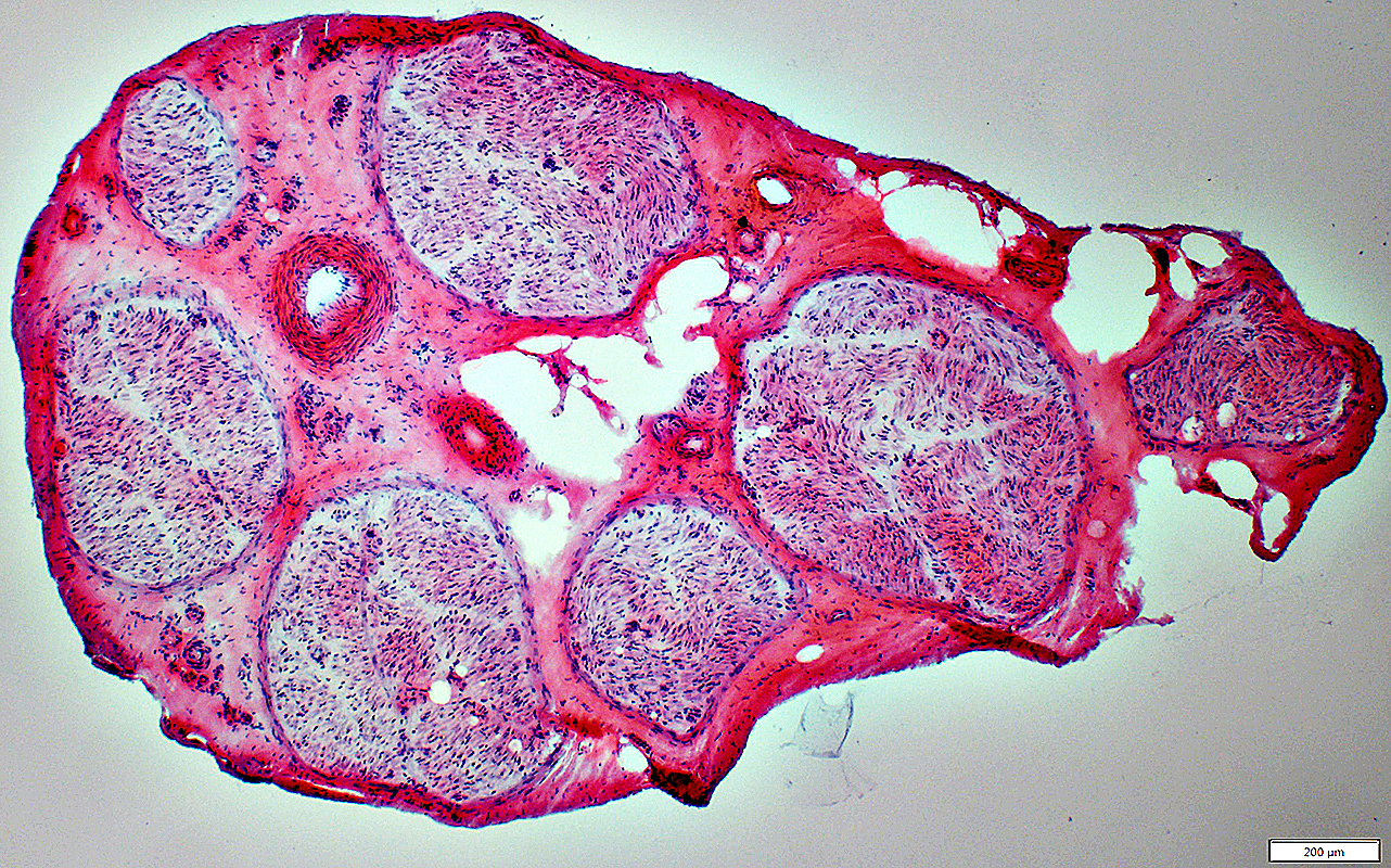 H&E stain |
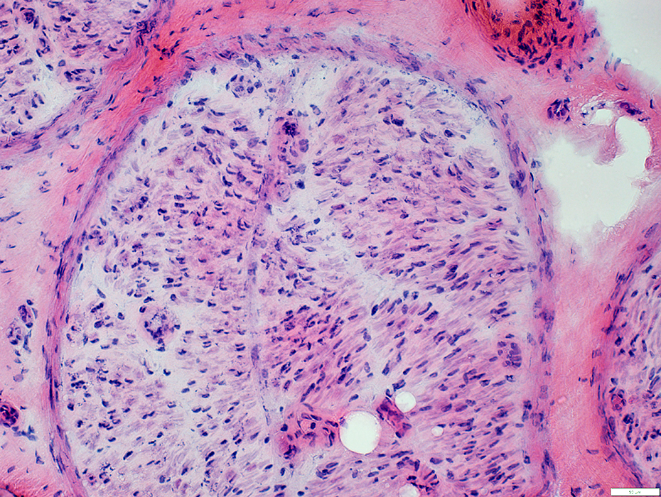
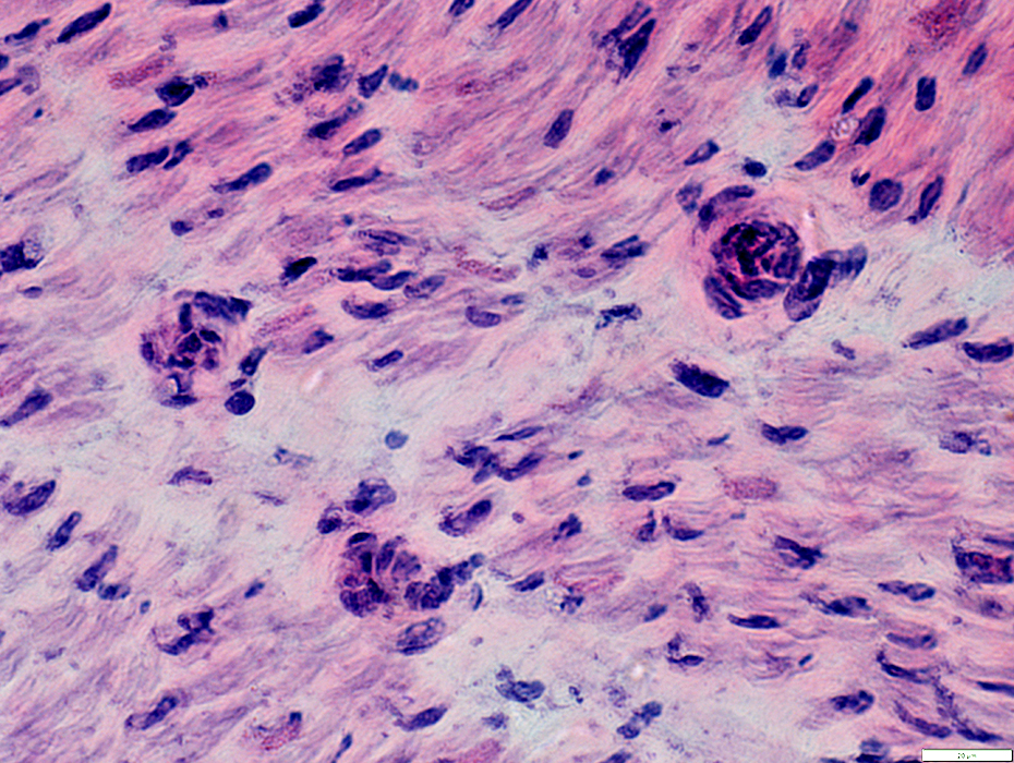
H&E stain |
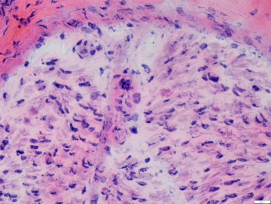 H&E stain |
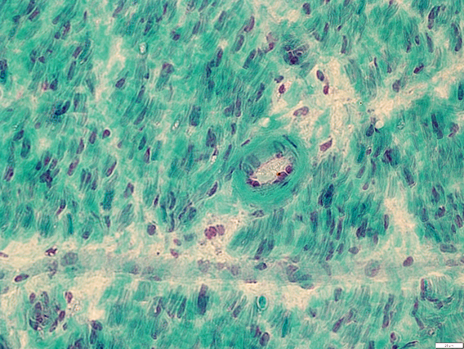
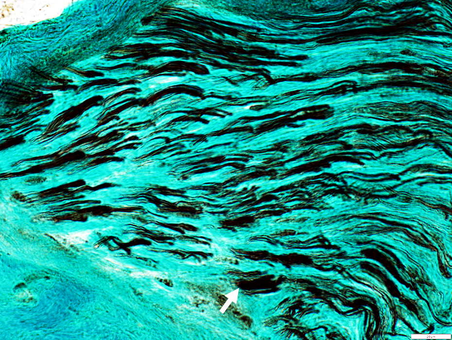
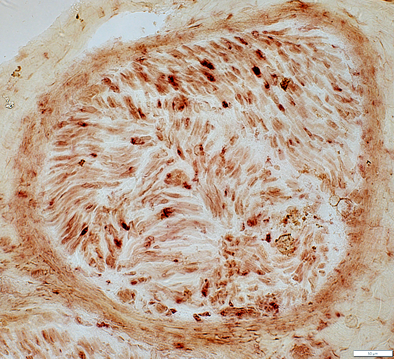
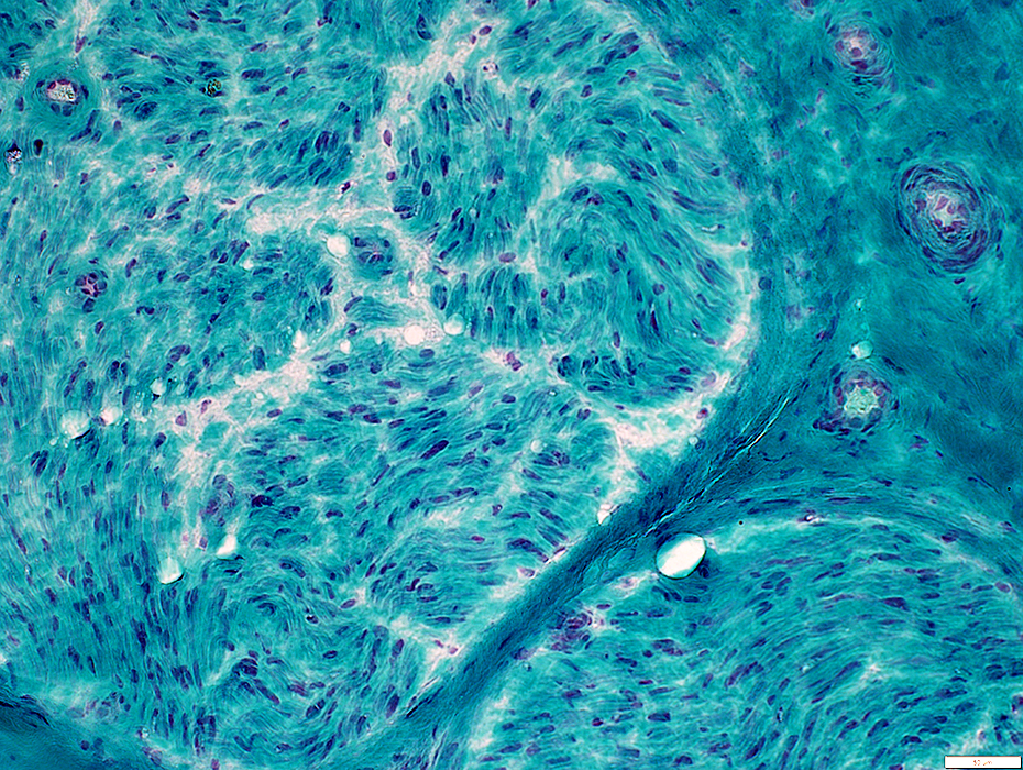 Gomori trichrome stain |
 VvG stain |
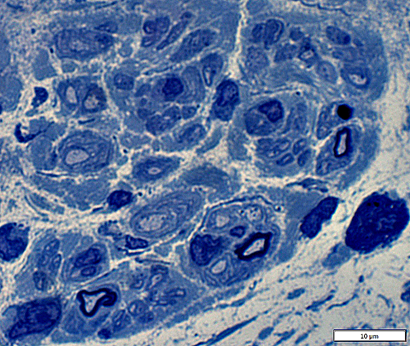
 Toluidine blue stain |
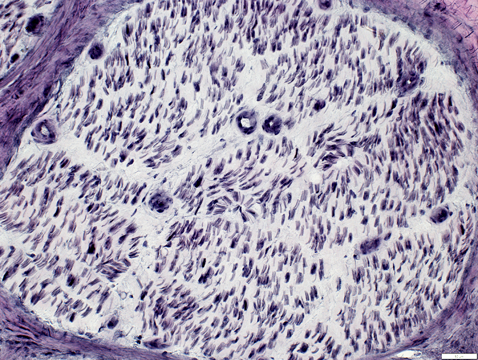
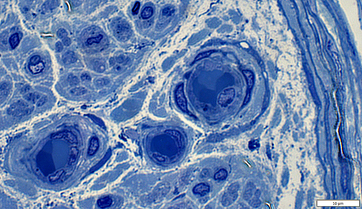
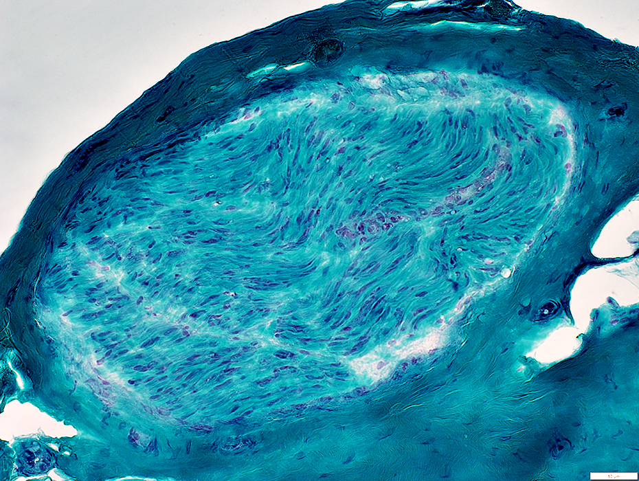
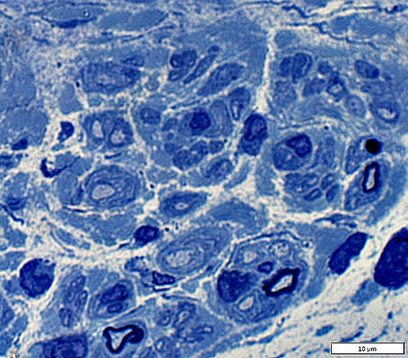
Loss of Myelinated Axons: Severe
 Toluidine blue stain |
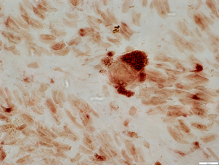
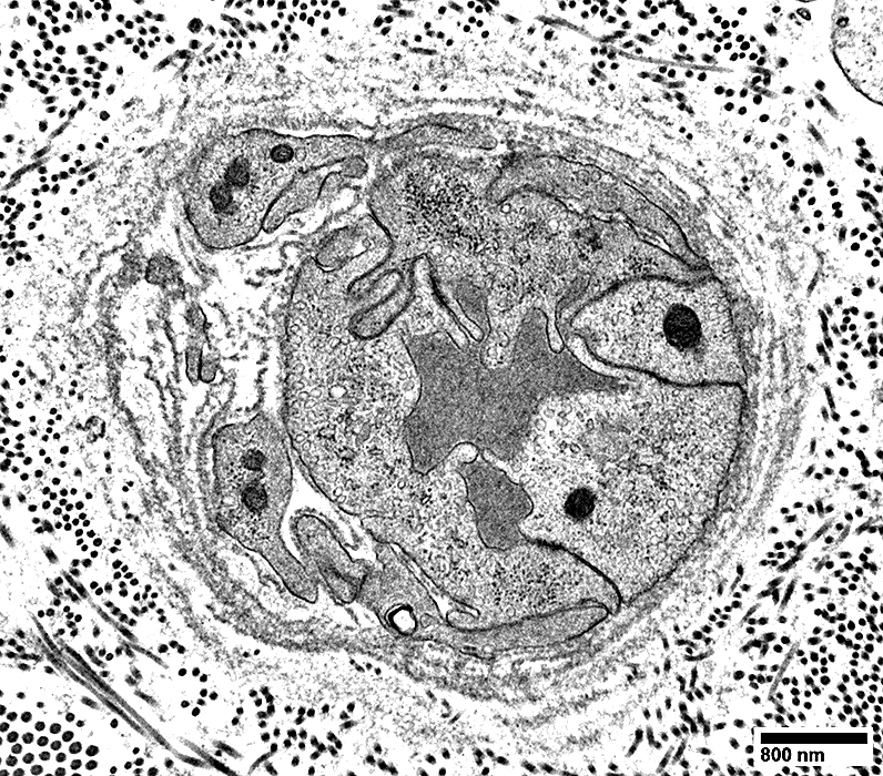
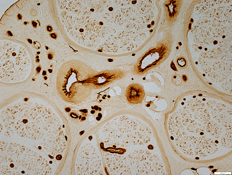
Endoneurial Microvessels: Endothelial cells prominent
 H&E stain |
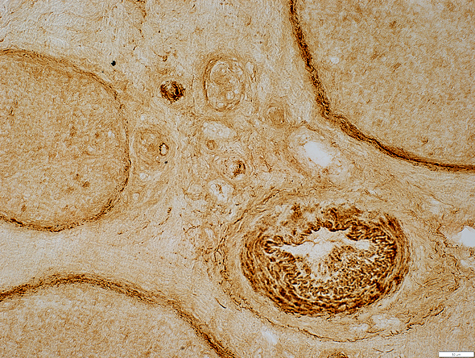
 H&E stain |
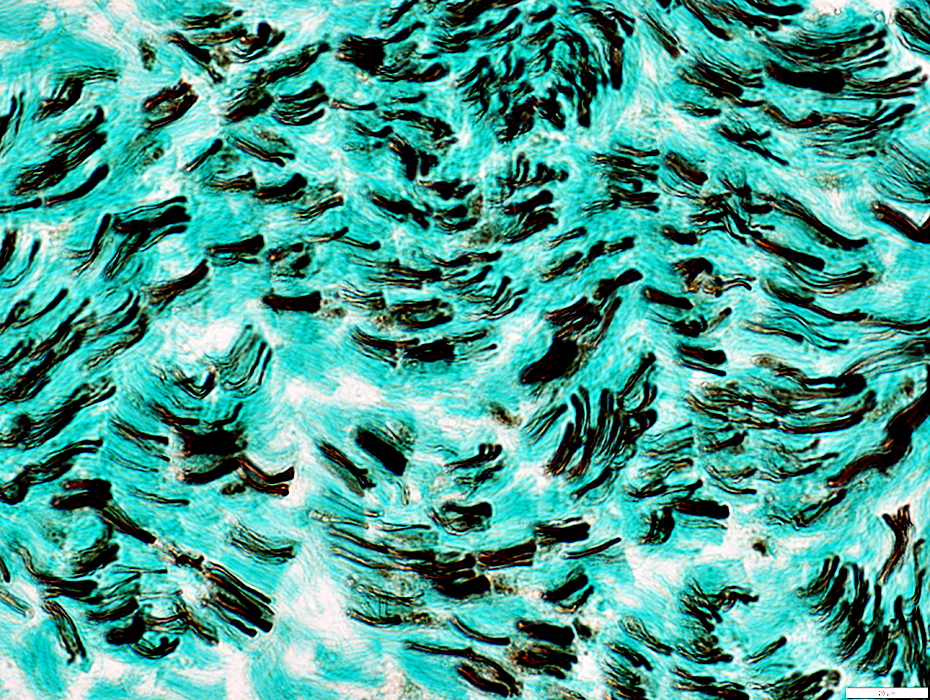
 Gomori trichrome stain |
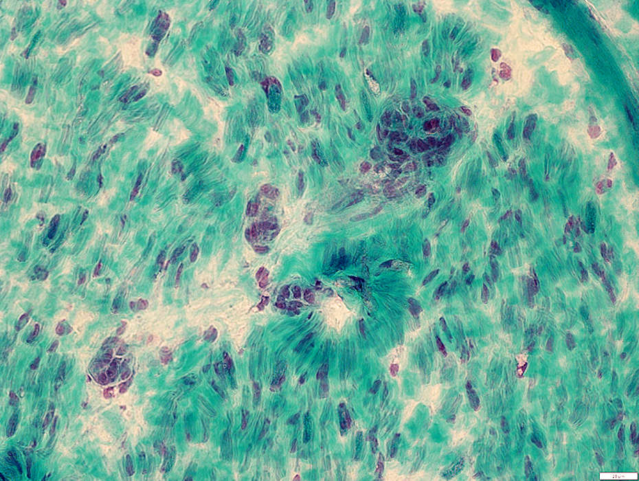 Gomori trichrome stain |
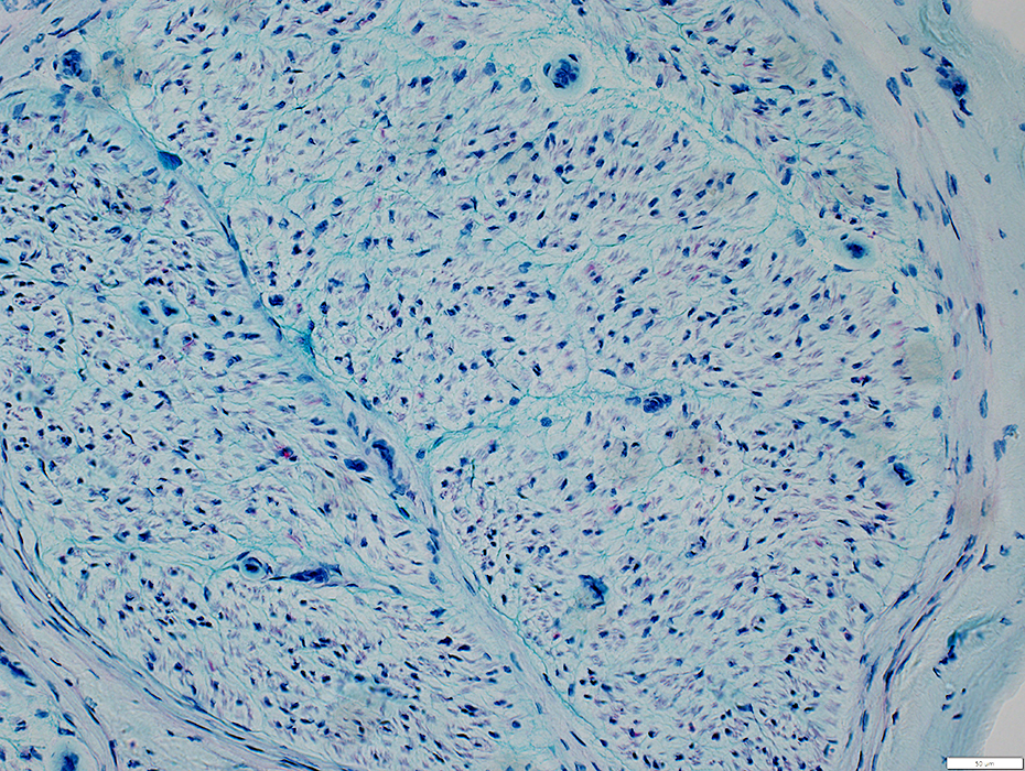
 Alcian blue stain |
 Acid phosphatase stain |
 VvG stain |
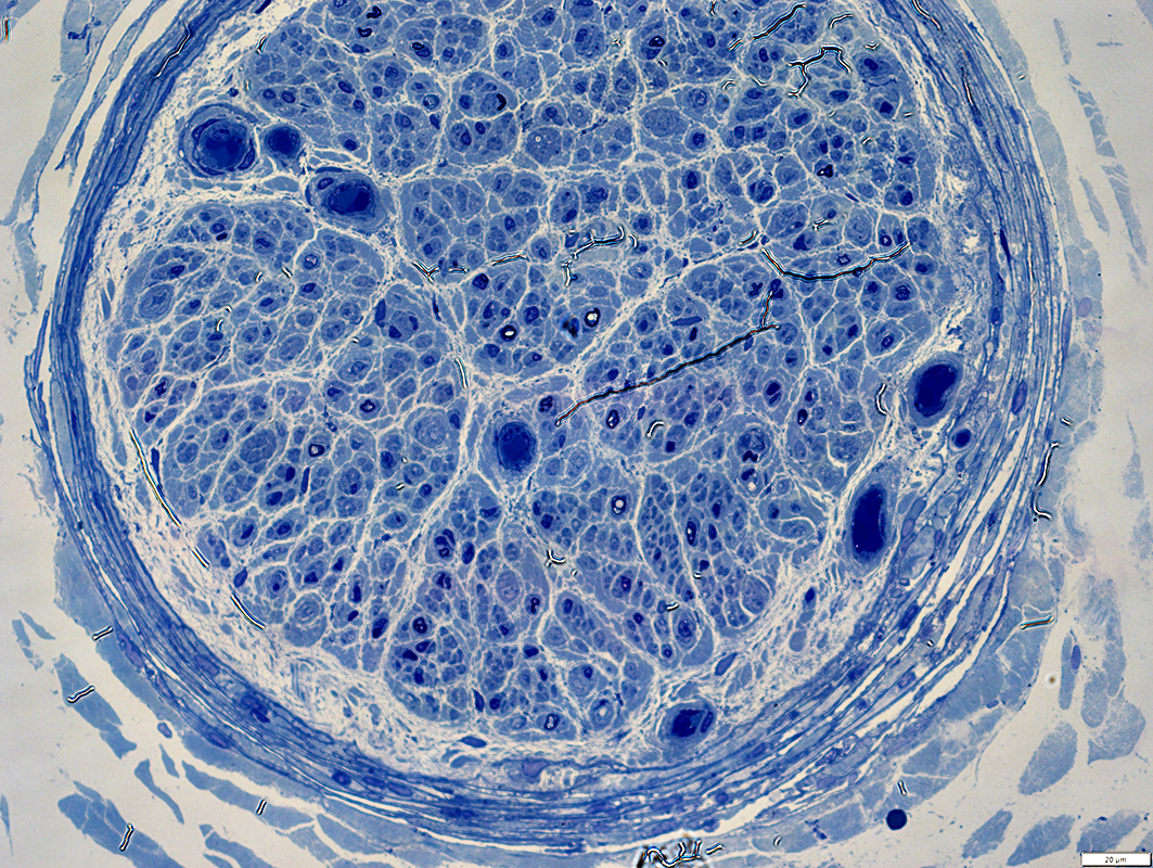
 Toluidine blue stain |
 From R Schmidt |
 From R Schmidt |
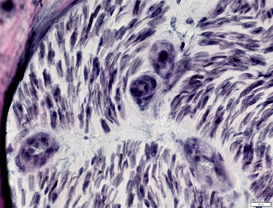
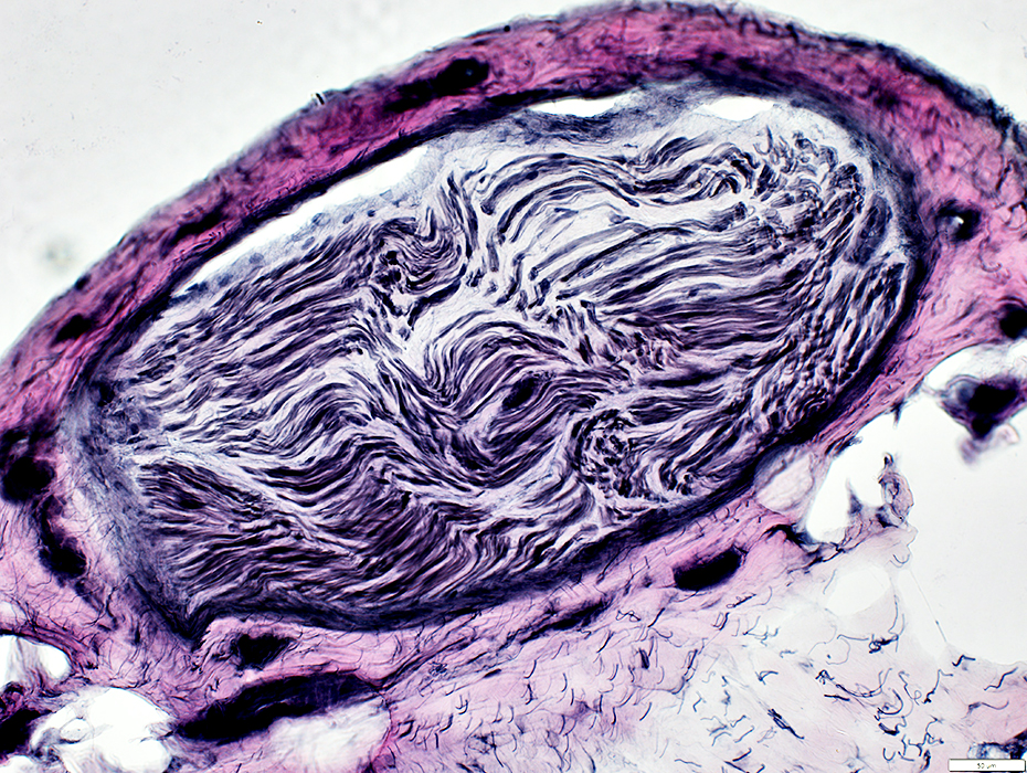
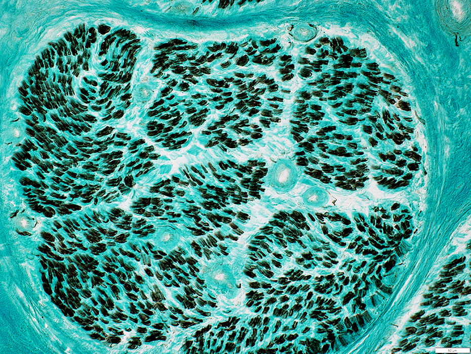
CIDP: Myelinated Axons: Markedly Reduced
 Gomori trichrome stain |
 VvG stain |
 Alcian blue stain |
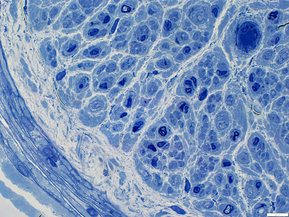
 Neurofilament stain |
Larger axons, that would normally be myelinated, are present (Arrow)
The largest axons have been lost
 Neurofilament stain |
 Toluidine blue stain |
Larger axons, that would normally be myelinated, are present
The largest axons have been lost
 Toluidine blue stain |
 Neurofilament (Green) + P0 (Red) stain |
The largest axons have been lost
Larger axons, that would normally be myelinated, are present but have no surrounding cells that stain for either NCAM or P0
Axons surrounded by P0 (Above) are often unusually small
 Neurofilament (Green) + NCAM (MBP) (Red) stain |
 Neurofilament (Green) + NCAM (MBP) (Red) stain |
 From R Schmidt |
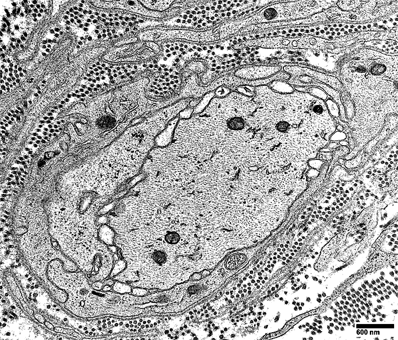
Larger (Intermediate-sized) axons, that would normally be myelinated, are present &
surrounded by Schwann cell processes but not myelin
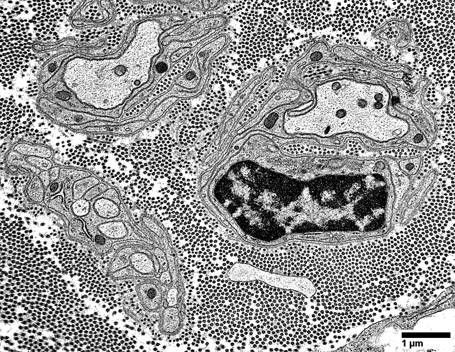 From R Schmidt |
 From R Schmidt |
Larger (Intermediate-sized) axons, that would normally be myelinated, are present &
surrounded by Schwann cell processes but not myelin
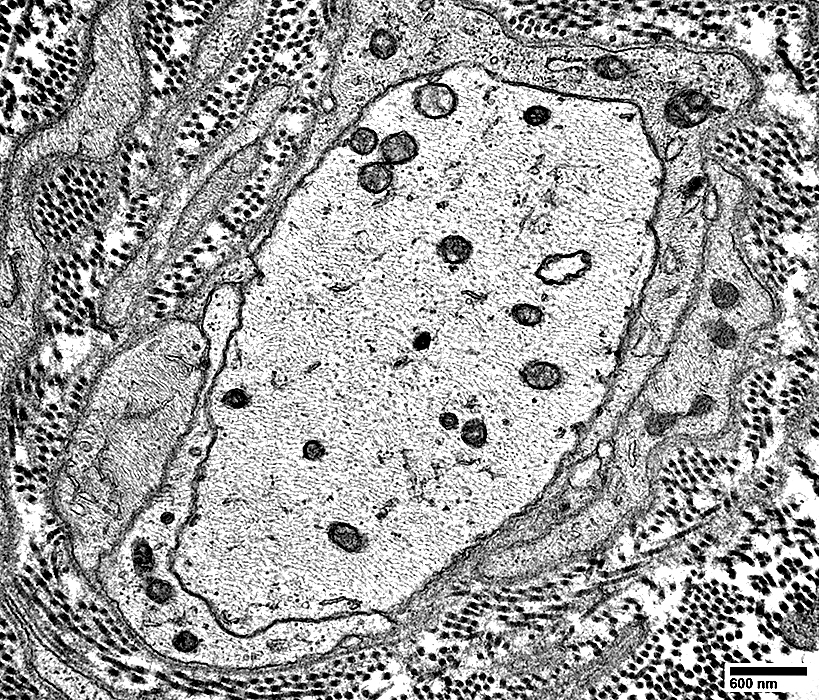 From R Schmidt |
 From R Schmidt |
 From R Schmidt |
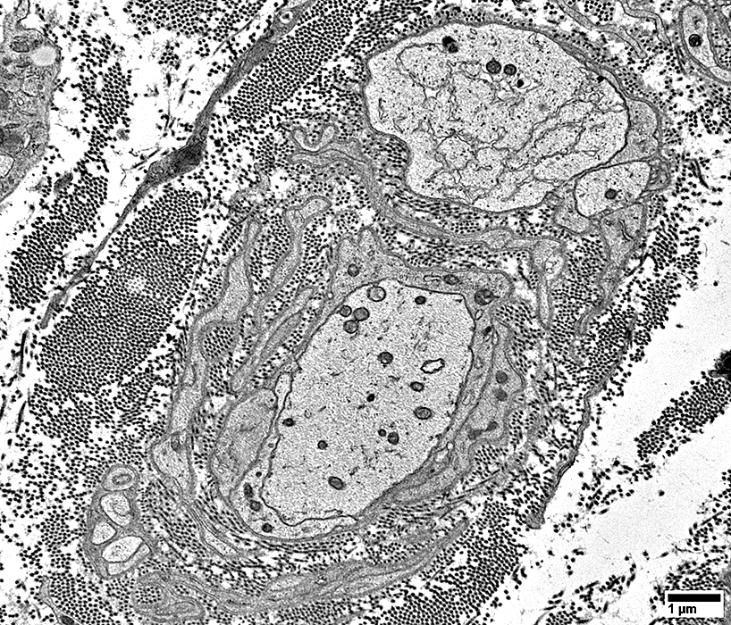
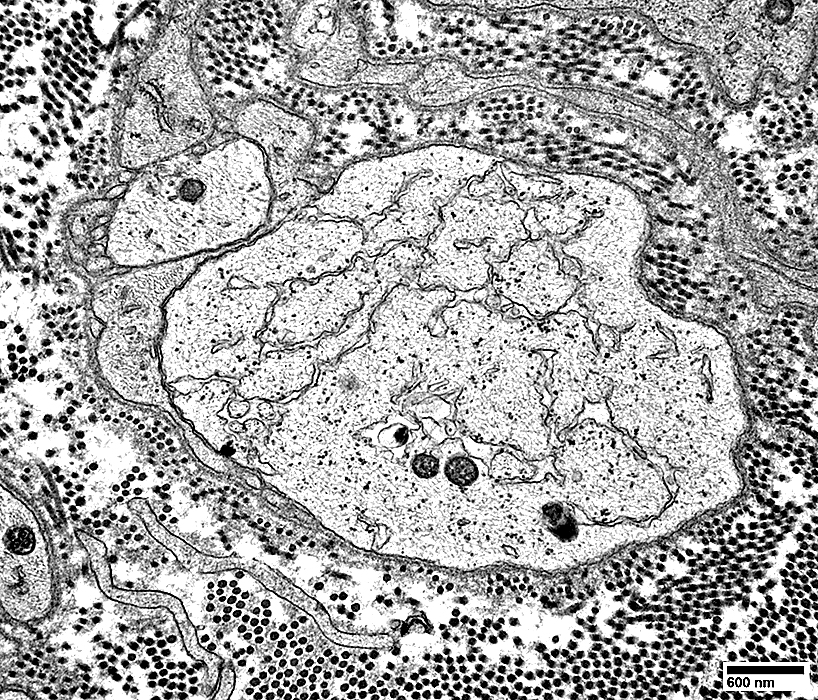
Axon, Small (Top left): Surrounded by several Schwann cell processes
Axon, Large (Center of image)
Contains serpentiginous membrane structures in cytoplasm
Surrounded by basal lamina: Possible withdrawal of Schwann cell process
 From R Schmidt |
 From R Schmidt |
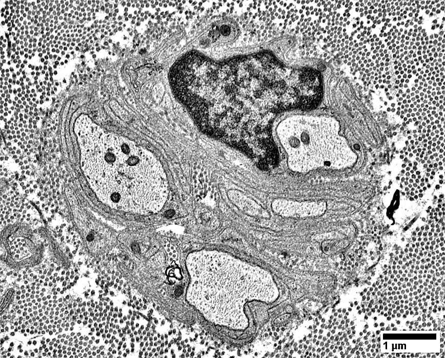
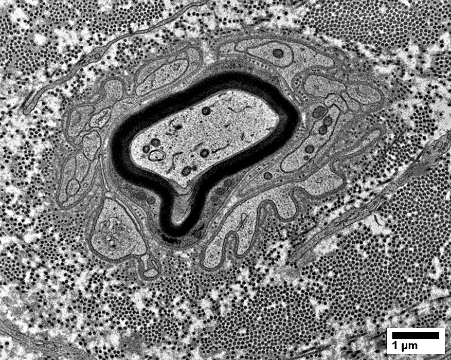
Myelinated axons surrounded by Schwann cell processes & Schwann cells
 From R Schmidt |
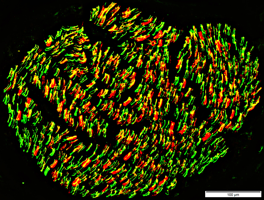
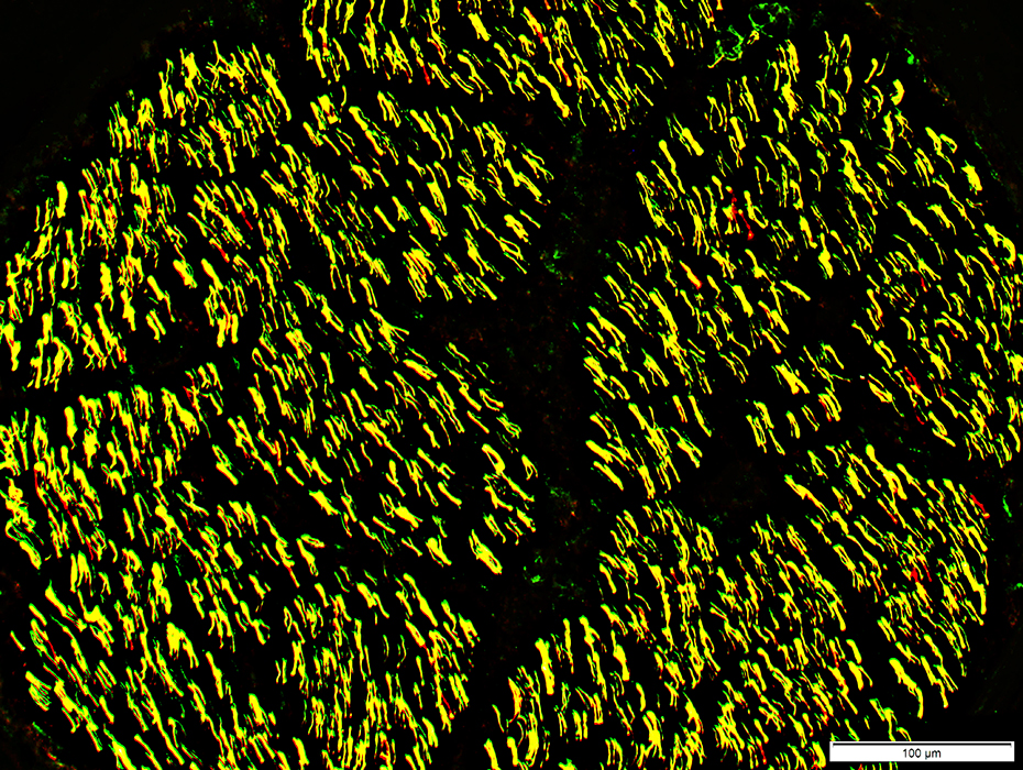
CIDP relapsing: Small axons are relatively preserved
 Neurofilament stain |
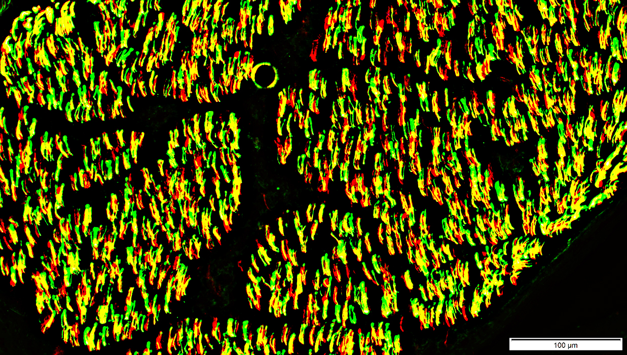
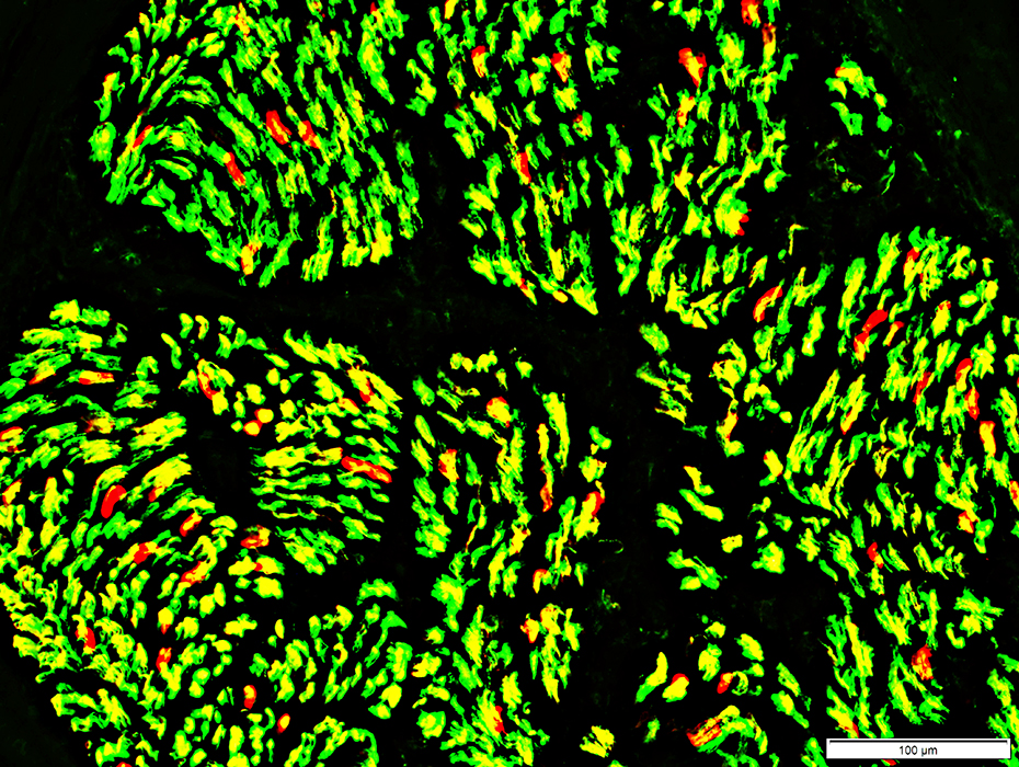
CIDP relapsing: Loss of large axons, Chronic
Schwann cells around remaining small axons stain abnormally for MBP
There are no MBP-containing myelin sheaths around axons
 Neurofilament (Green) + Myelin Basic Protein (MBP) (Red) stain |
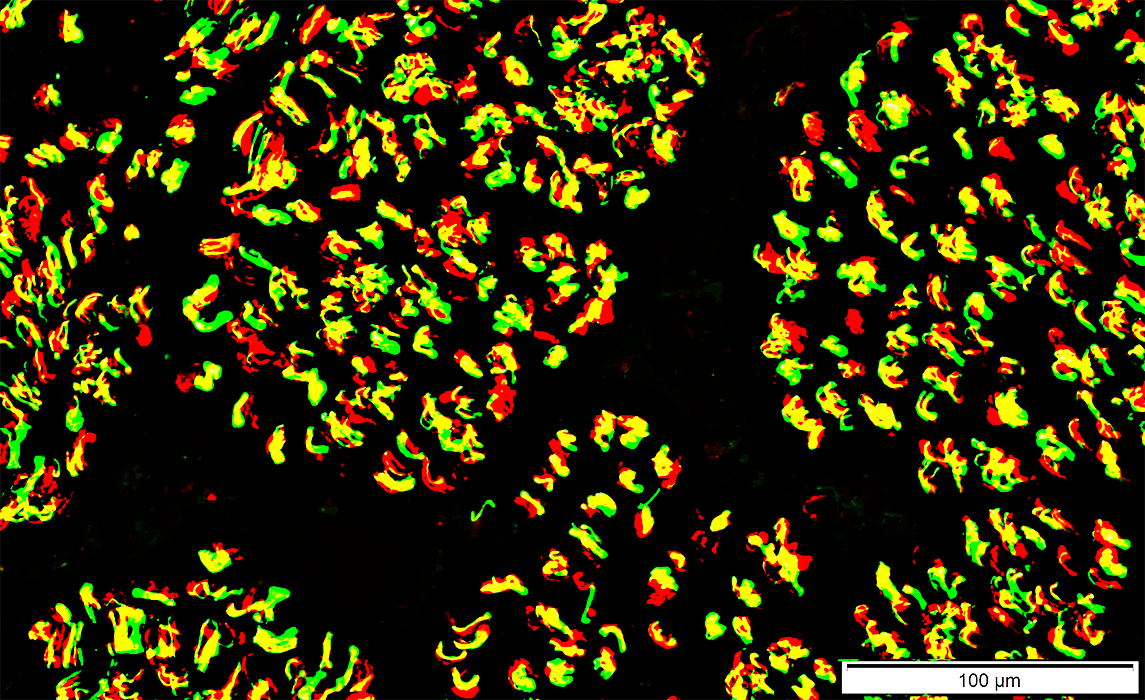
CIDP relapsing: Schwann cells
Büngner band Schwann cells (Yellow) co-stain for NCAM & P0
 NCAM (Green) + P0 (Red) stain |
CIDP relapsing, Schwann cells: Non-myelinating Schwann cells are diffusely distributed in endoneurium
 NCAM stain |
CIDP, Relapsing
Endoneurial cells: Darkly stained
Epineurial vessels: Normal smooth muscle
ATPase pH 4.3 stain |
Small histiocytic cells in Endoneurium
 Acid phosphatase stain |
Multiple small epineurial vessels
 UEA I stain |
 C5b-9 stain |
C5b-9 staining of perineurium is pale (Above, Left)
Perineurium: Damaged structure; Cellular invasion (Below)
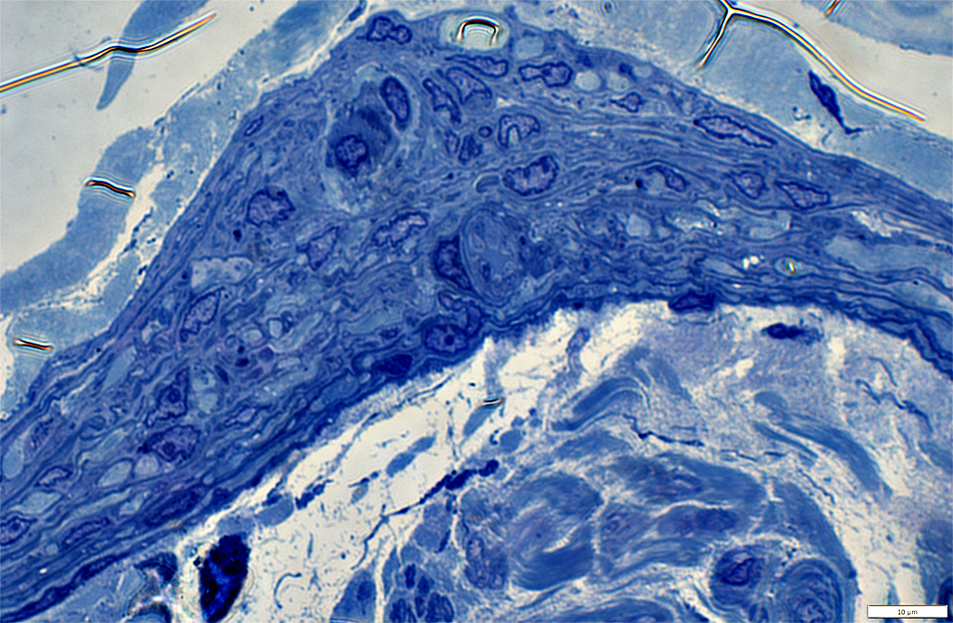 Toluidine blue stain |
Return to Normal nerve biosies
Return to Biopsy illustrations
Return to Neuromuscular Home Page
Return to Nerve biopsy
Return to Demyelinating neuropathies
Return to Active demyelination
4/15/2021