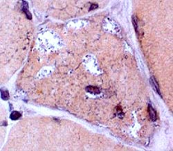
Examination shows asymmetric patchy weakness,
especially in the quadriceps and hand grip.
Serum CK is 300.
Image: Congo red stain highlights irregular vaculoes
Answers:
MHC Class I: Upregulated in sIBM but not hereditary disorders
SMI-31: Shows aggregates in muscle fibers
Lymphocyte stains: Shows inflammation in sIBM but not hereditary disorders
Likely diagnosis: Inclusion body myositis
Differential diagnosis of vacuoles with basophilic debris in muscle fibers:
Distal myopathies; HIBM2; Oculopharyngeal dystrophy
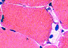
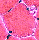
Examination shows normal strength, and a tendency to "give way".
Serum CK is 500. EMG is myopathic.
Image: H&E stain shows subsarcolemmal blebs. Some contain nuclei
Answers:
PAS: Increased in muscle fibers in some patients.
Phosphorylase: Absent in most muscle fibers; Present in vessels & regenerating fibers
Diagnosis: Phosphorylase deficiency (McArdle's)
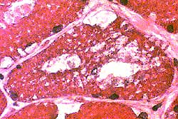
Exam shows a vital capacity of 50% of predicted,
and weakness of thigh adductors.
Serum CK is 1,465. EMG shows myopathy and fibrillations.
Image: H&E stain shows cytoplasmic & subsarcolemmal vacuoles.
Answers:
PAS: Increased in muscle fibers in children
Acid phosphatase: Positive staining granules in muscle fiber cytoplasm
Biochemical analysis for Acid maltase activity in muscle & blood
Diagnosis: Acid maltase deficiency
She was normal until 1.5 years of age.
Examination shows weakness in proximal & facial muscles.
Serum CK is 600.
Cardiac evaluation: Ventricular hypertrophy & R bundle branch block.
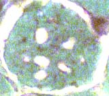
Image: Gomori trichrome shows multiple clear round regions in muscle fiber cytoplasm
Answer: Sudan Black, or Oil Red O, stains lipid in muscle fibers
Diagnosis: Lipid storage, often Carnitine disorders
Examination shows mild proximal weakness.
Serum CK is 300.
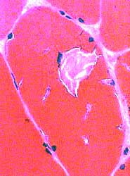
Answer: NADH stains tubular aggregates dark
Morphologic diagnosis: Tubular aggregates
NOTE: Tubular aggregates are not a final diagnosis and can occur in many disorders.
They are common in: Hypokalemic periodic paralysis
Now he is having difficulty arising from chairs.
Examination shows mild proximal weakness.
EMG is myopathic.
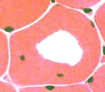
Image: H&E stain shows a clear region within scattered muscle fibers.
Answer: H&E evaluation of other muscle fibers
Diagnosis: Hypokalemic periodic paralysis
Differential diagnosis: Freeze artefact (Change would be present in many contiguous, not scattered, fibers)