NEMALINE RODS
|
General Description Characteristics Clinical Pathology Infant Diffuse Type 1 small Ultrastructure Congenital Child ACTA1 Adult RYR3 SLONM DDx Atypical Z-Disk streaming |
|
Nemaline Rod: Muscle Pathology
|
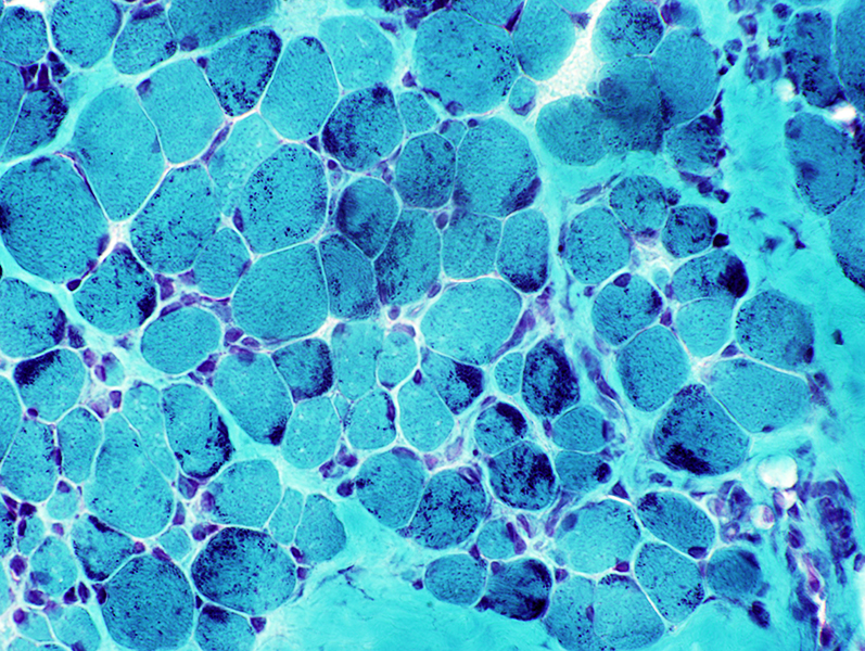
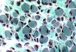
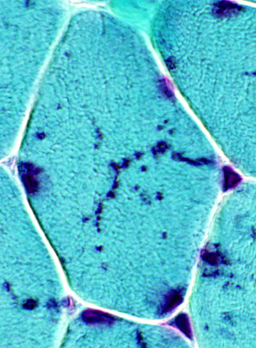
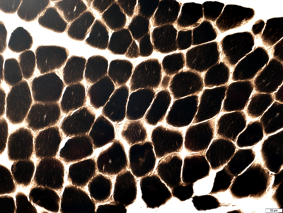
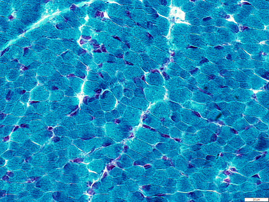
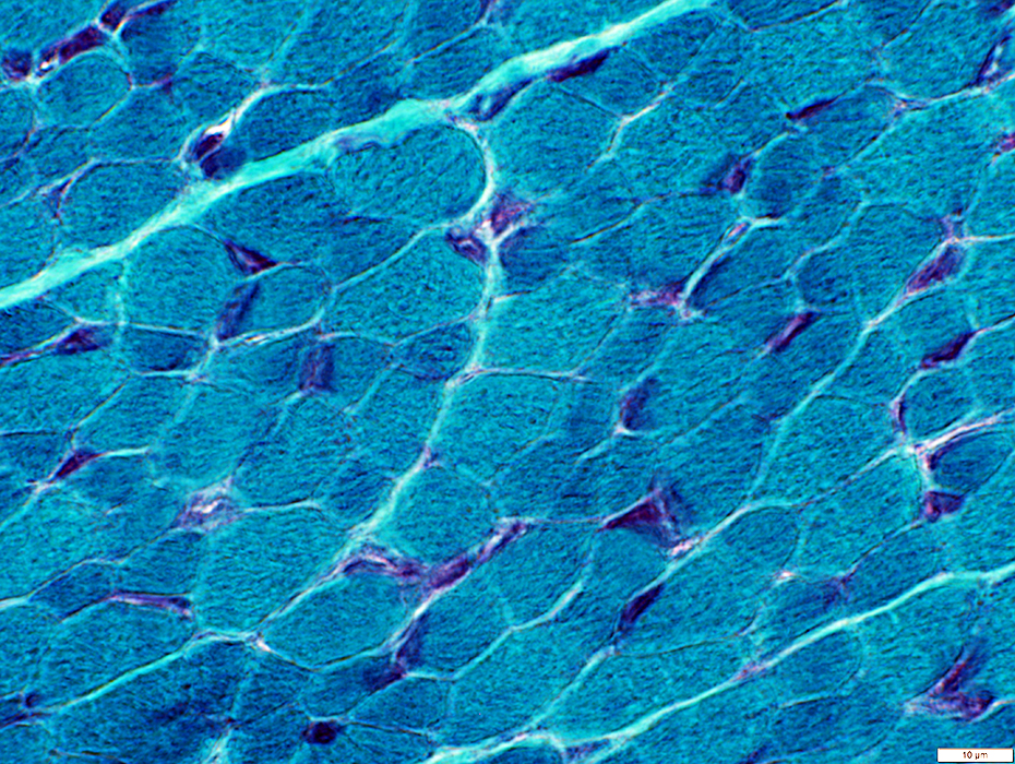
Nemaline rod myopathy: Infantile
 Gomori trichrome Nemaline rods in gastrocnemius muscle from 4 month old child |
|
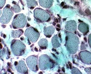 No nemaline rods in quadriceps muscle from same child biopsied at 2 months of age |
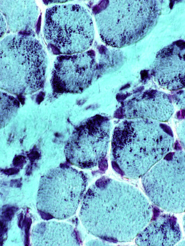
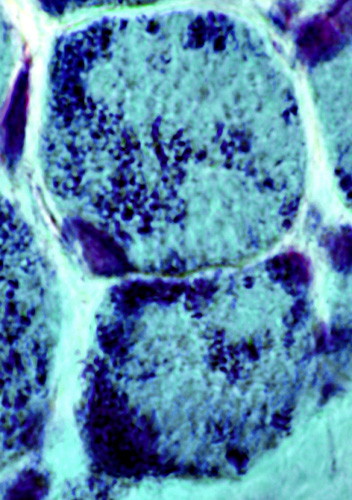
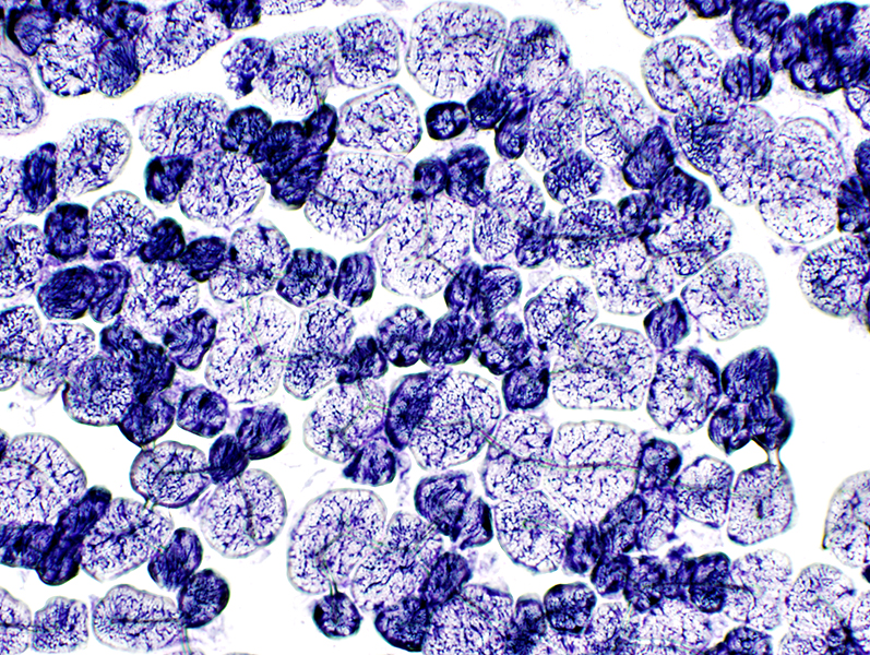
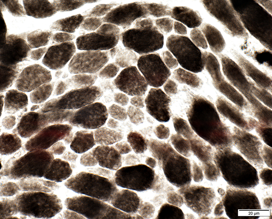
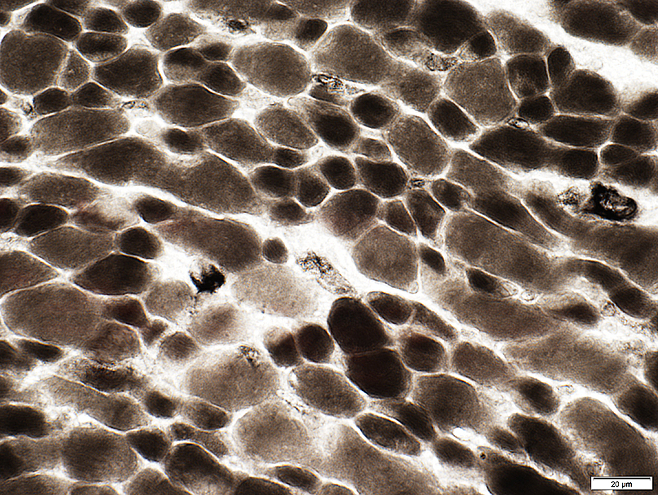
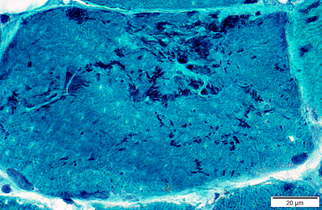
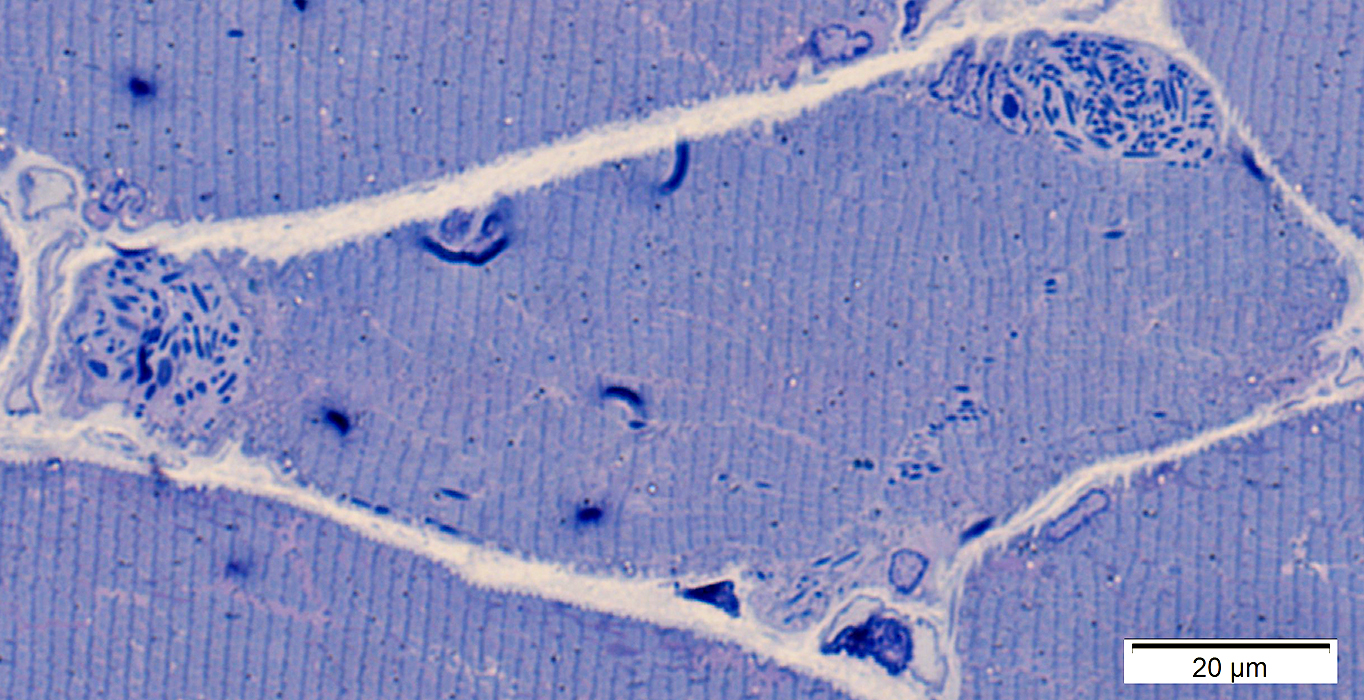
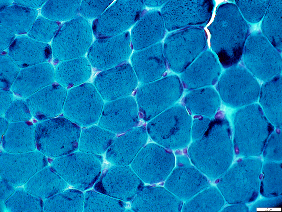
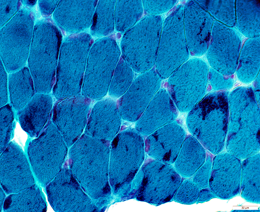
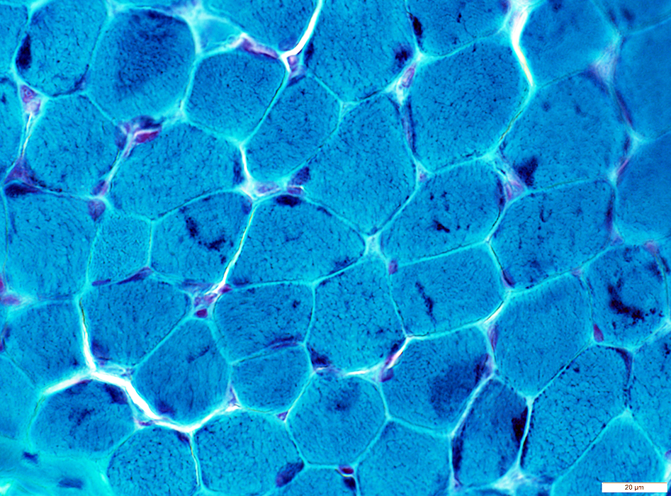
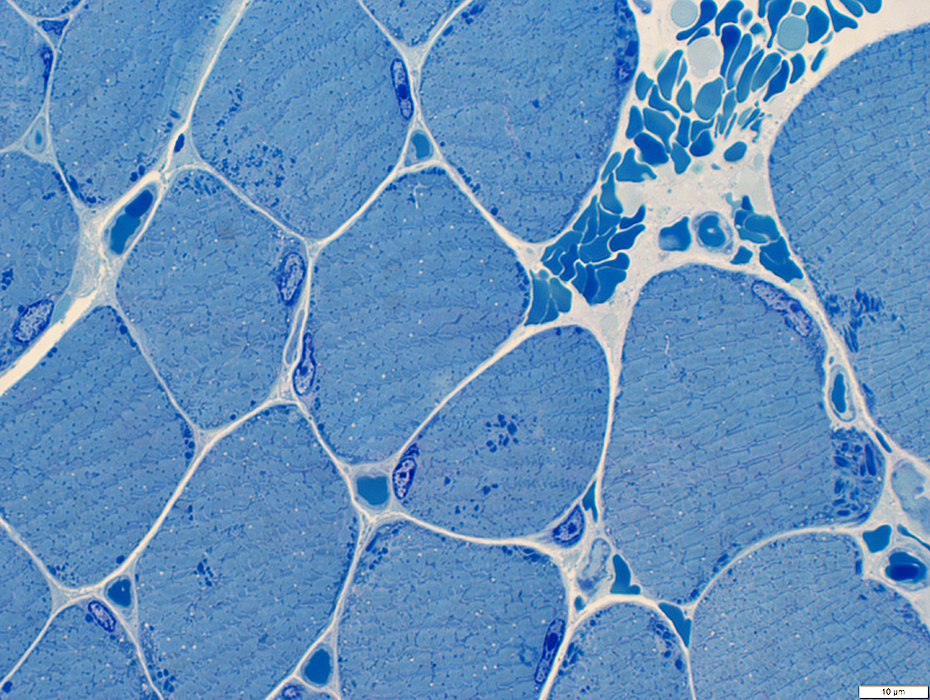
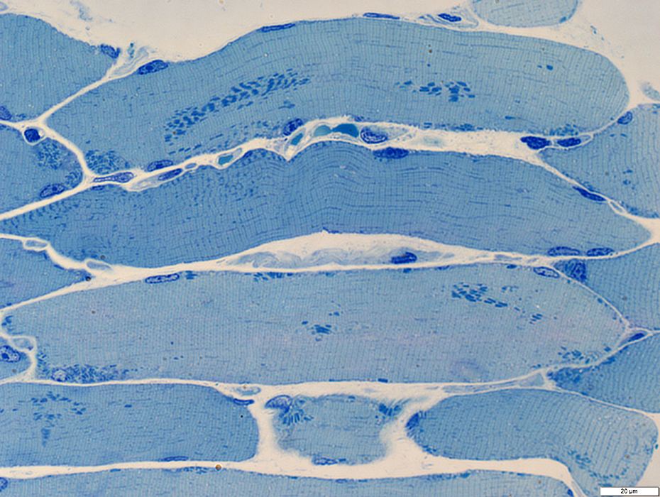
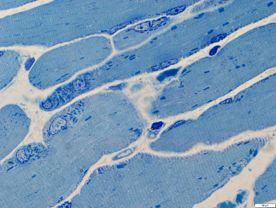
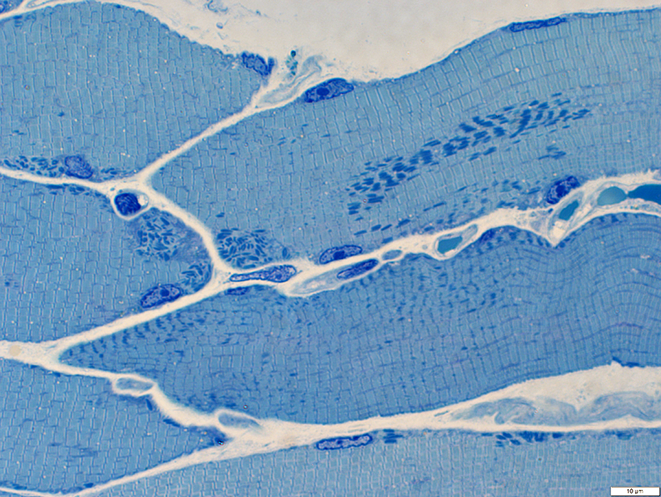
Nemaline rod myopathy, Infantile: Toluidine blue stains
|
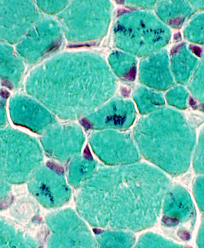
Nemaline rod myopathy, Infantile: Other stains
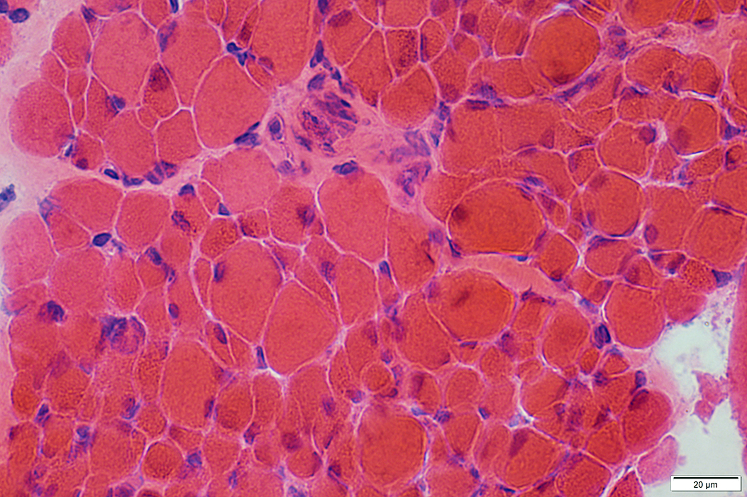
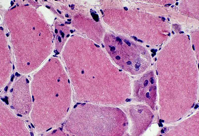
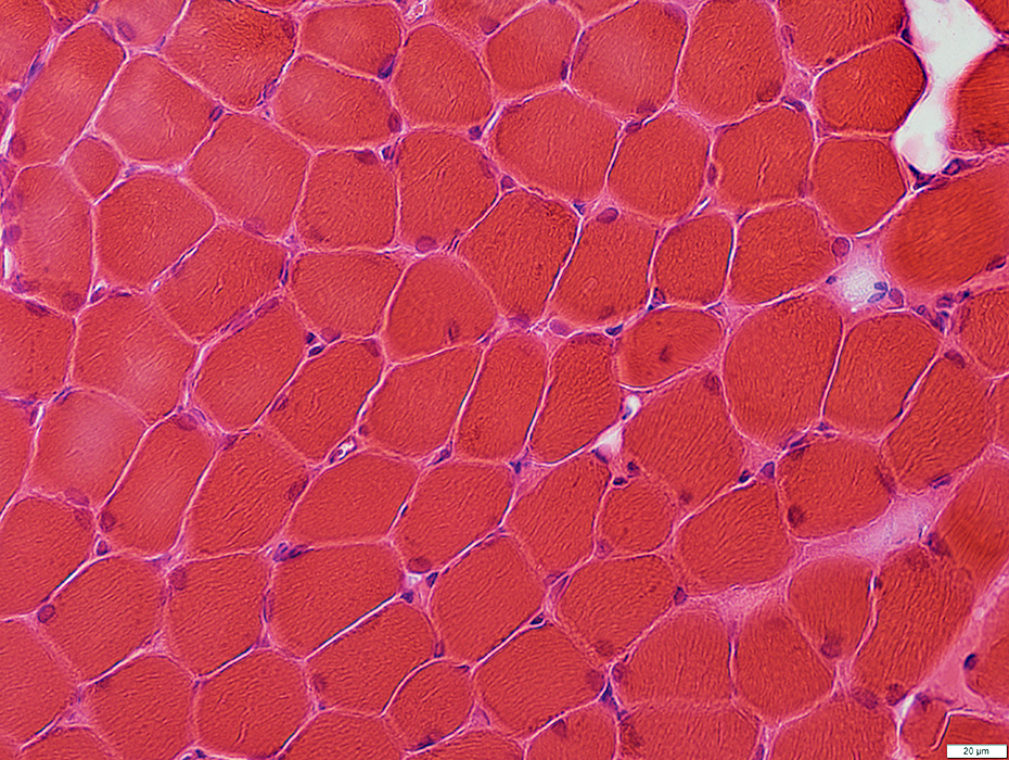
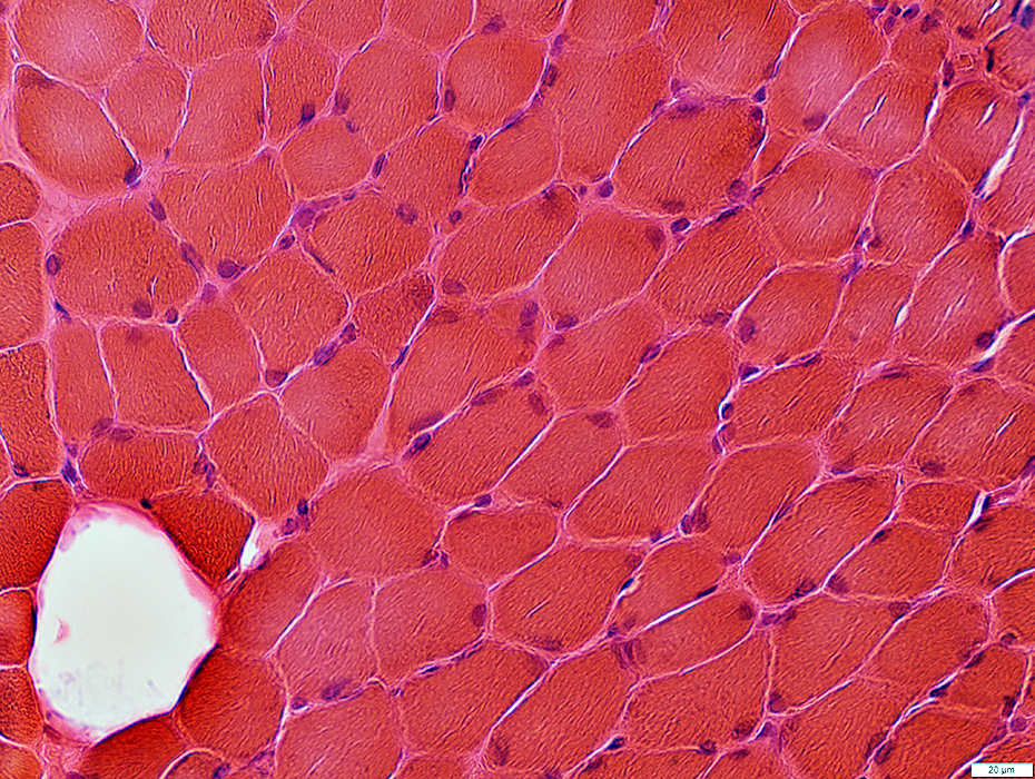
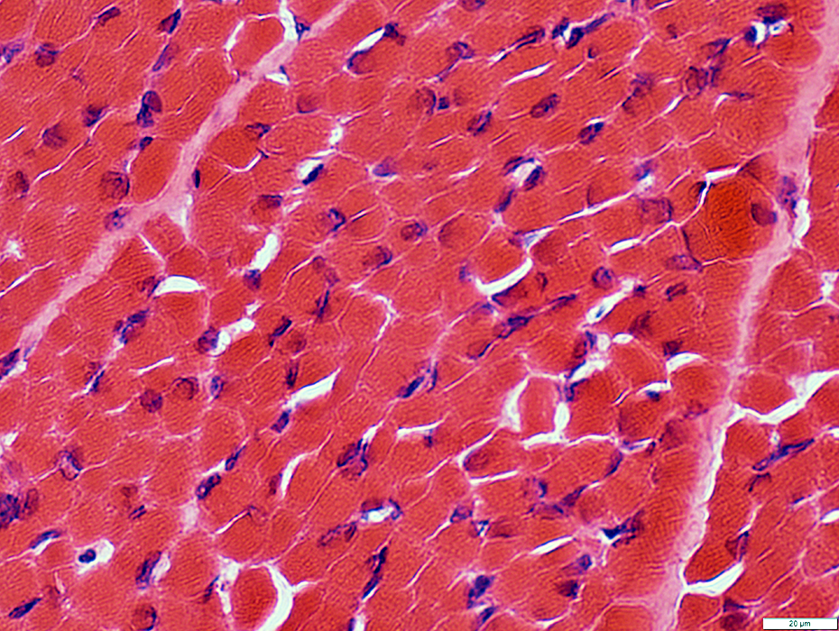
 H&E stain |
 H&E stain |
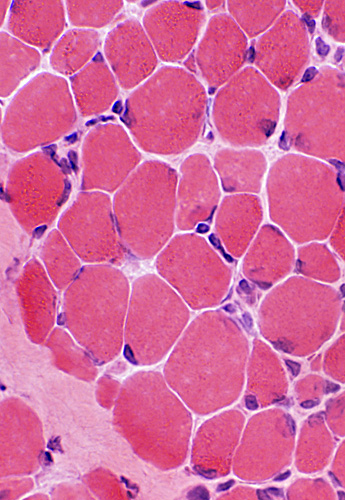 H& E stain Muscle fiber size: Varied Refractile rods can be seen |
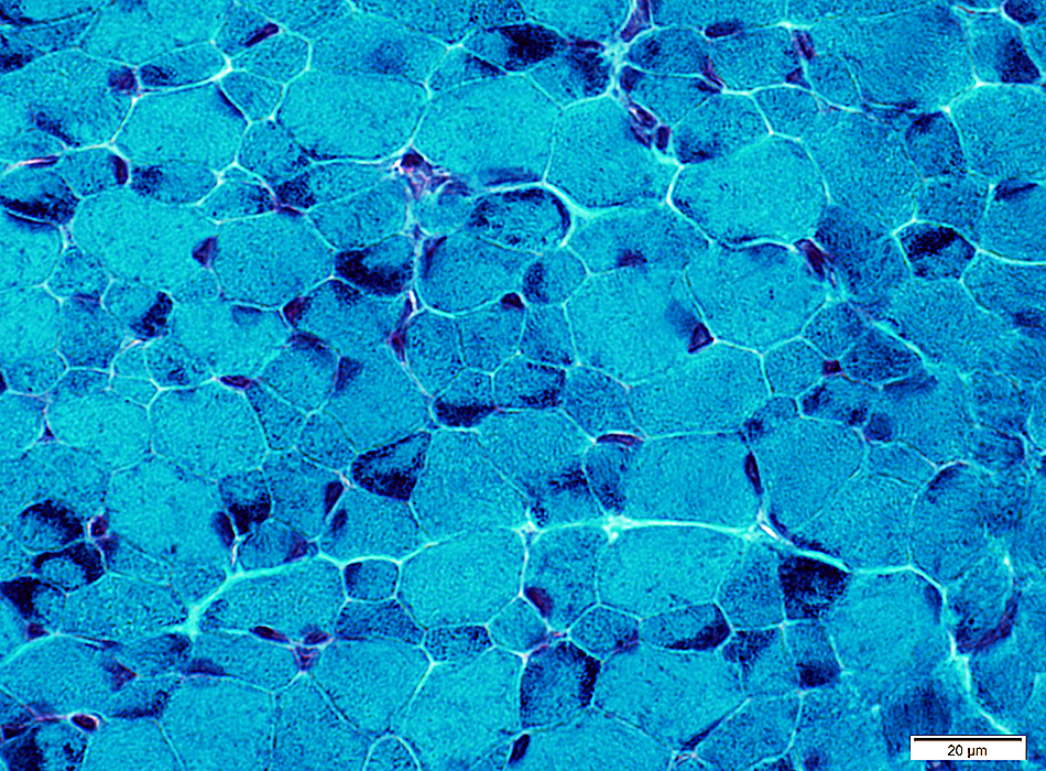
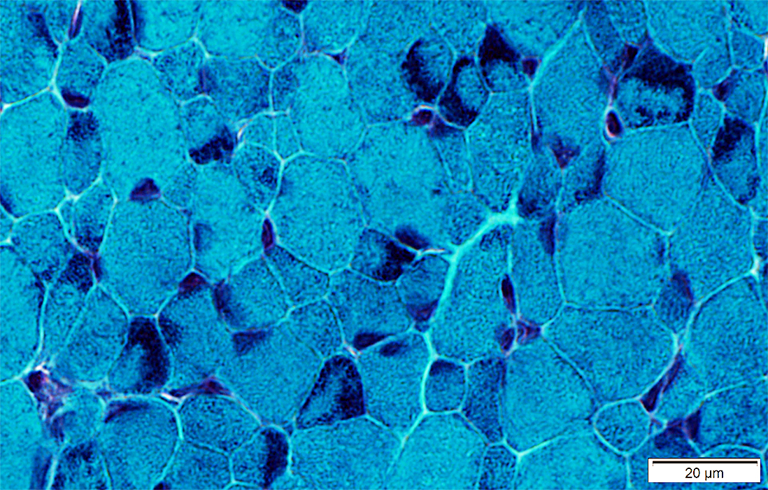
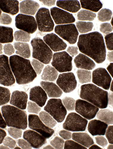
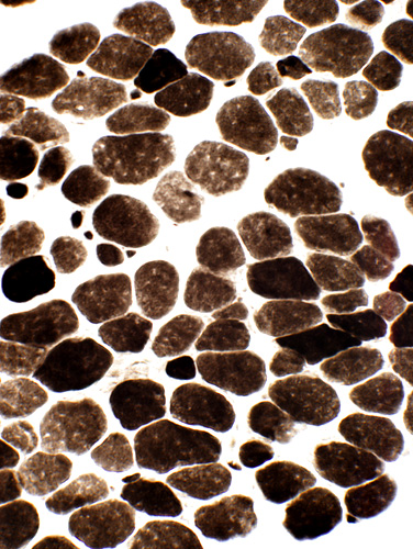 ATPase stain, pH 9.4 Type I muscle fiber predominance |
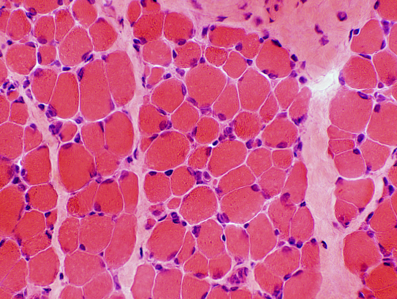 H& E stain |
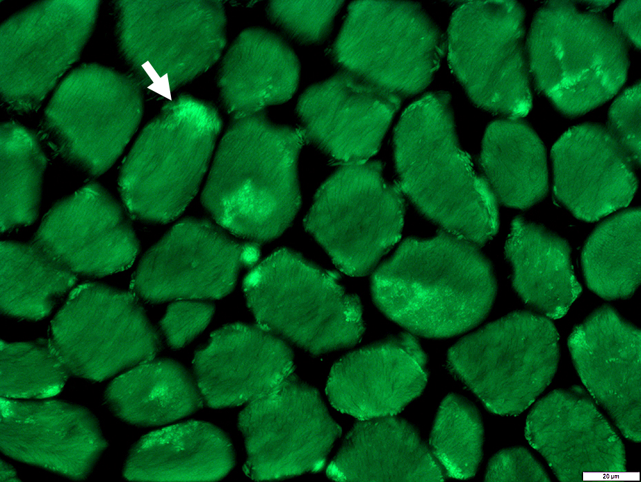
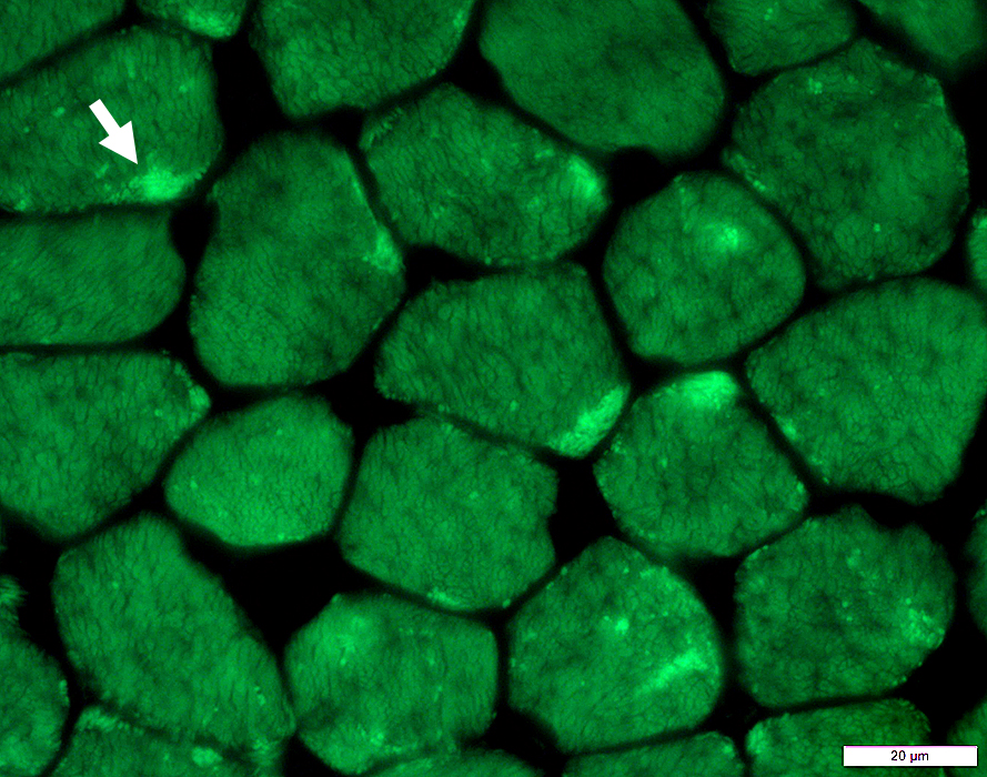
Rods: Actin stain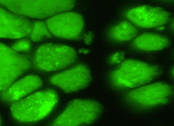 Phalloidin staining for actin |
| Focal (bright green), large and small aggregates of actin are present in some muscle fibers |
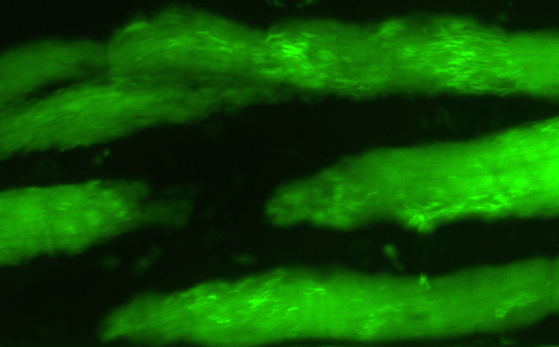 Phalloidin staining for actin |
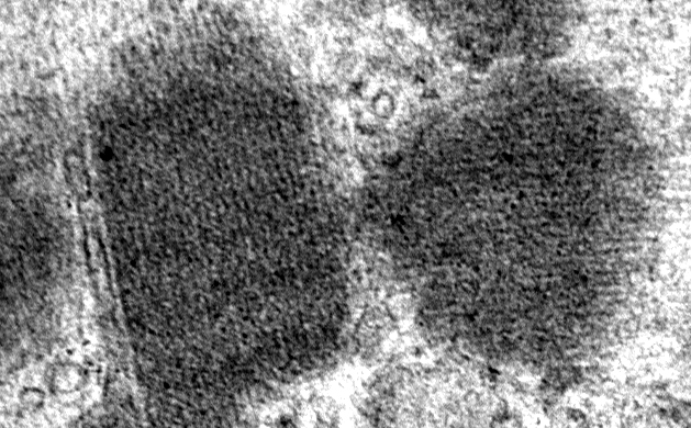
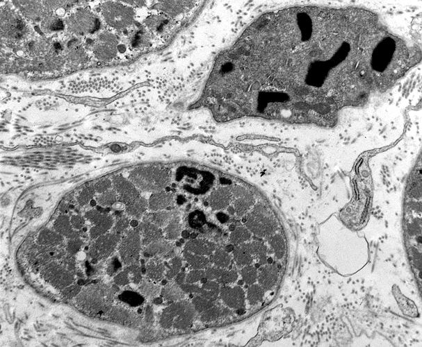
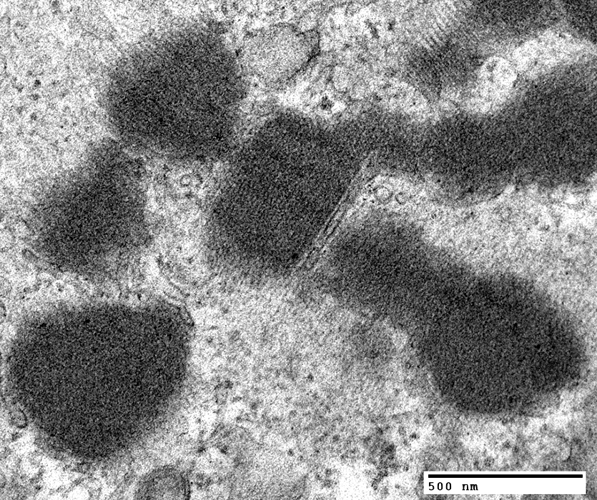
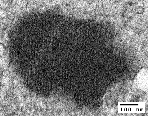
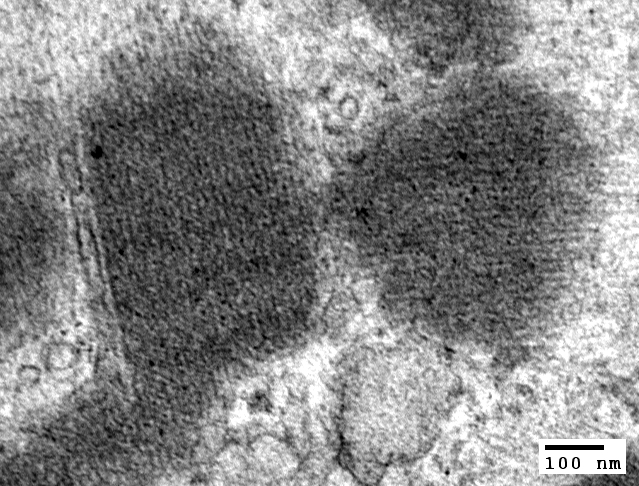
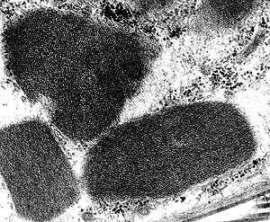
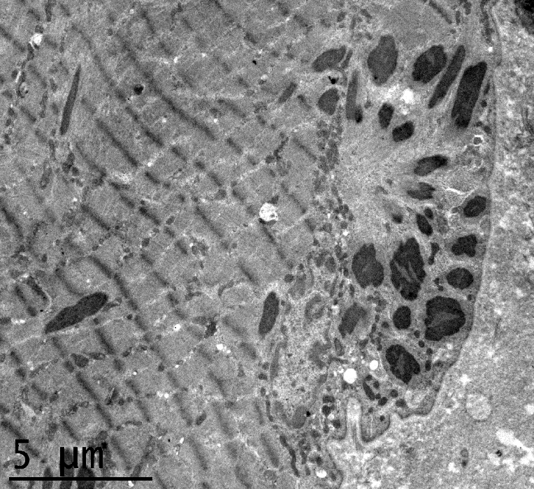
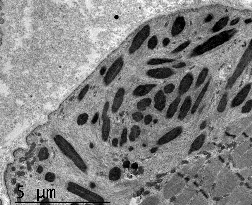
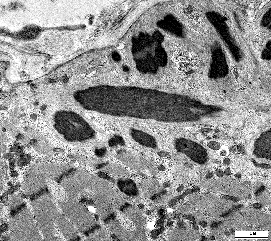
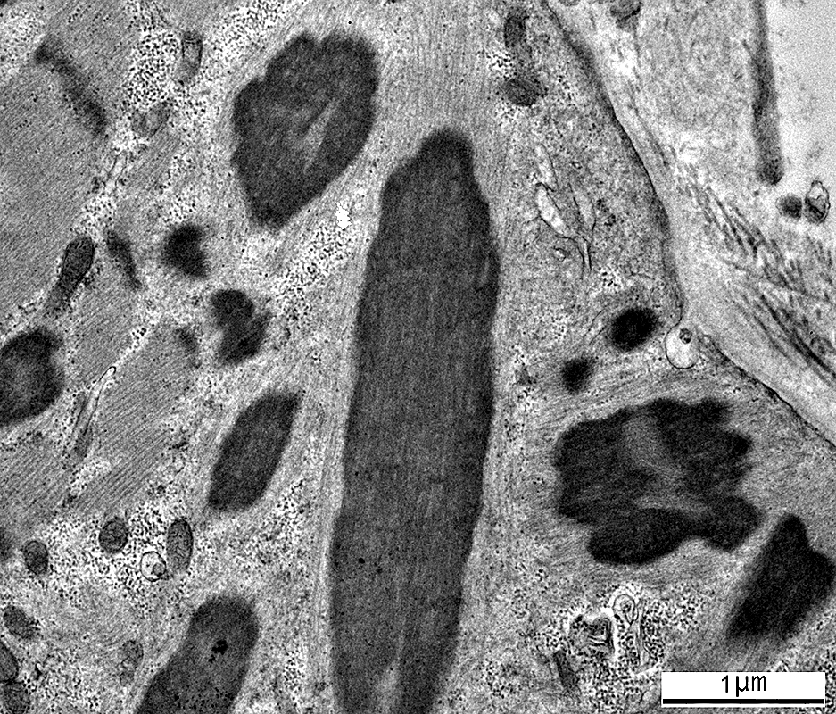
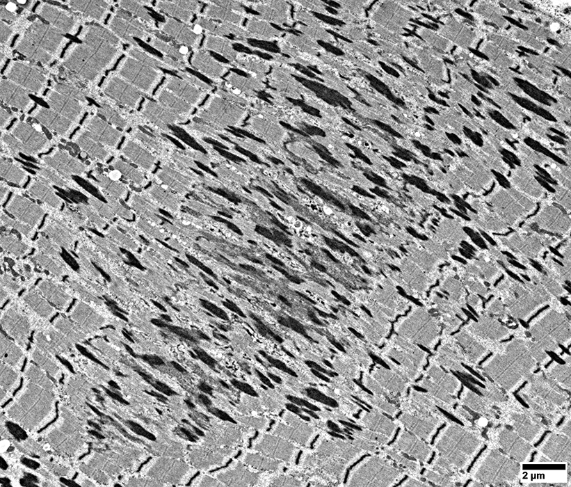
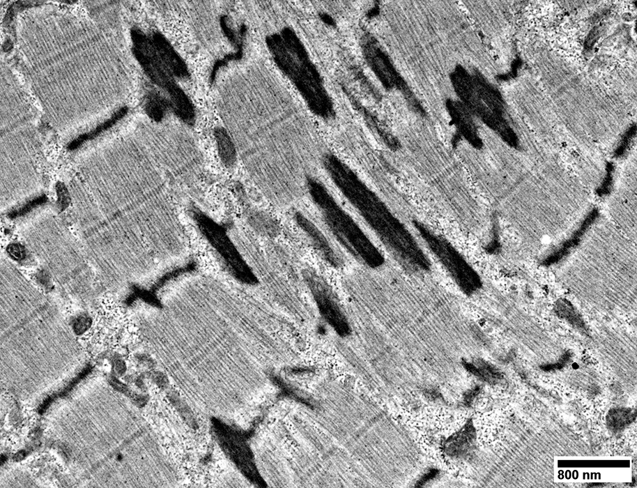
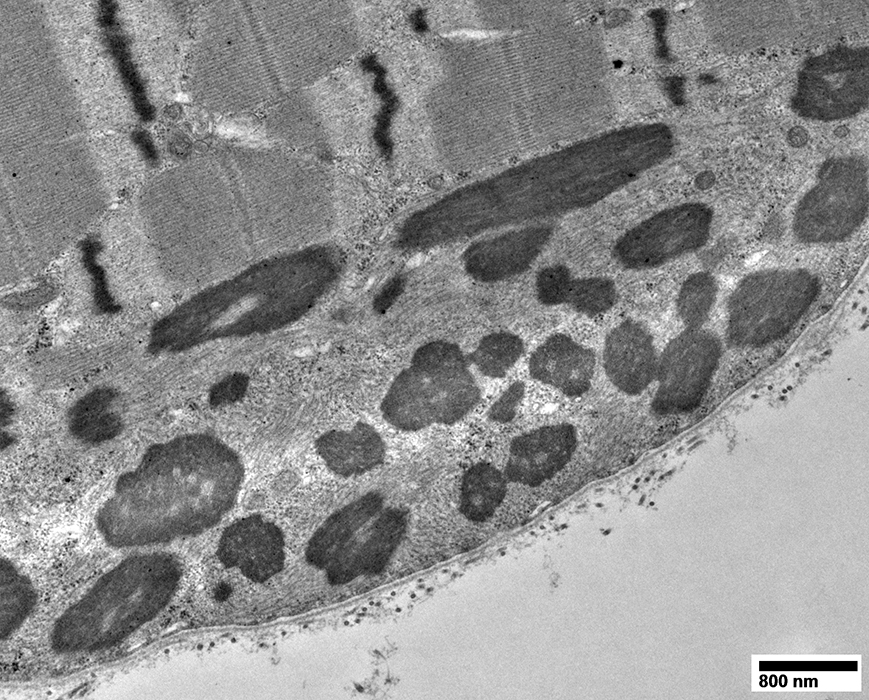
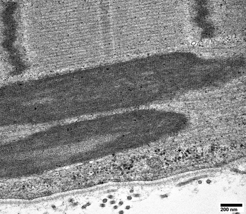
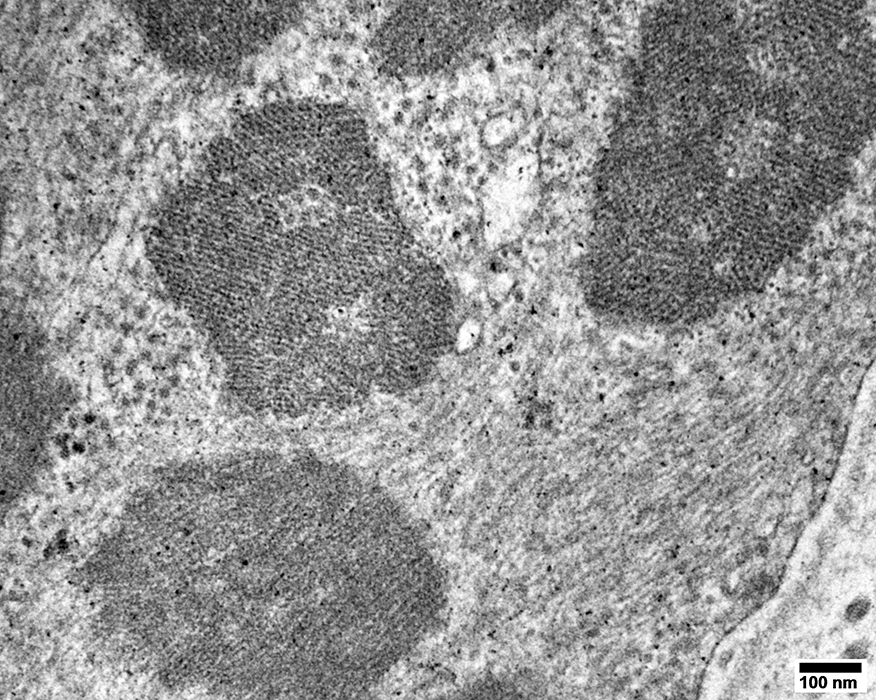
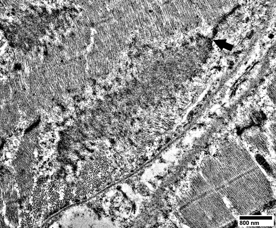
Nemaline Rods: Ultrastructure
Also see: SLONM| Infantile-onset Rod myopathy | |
|
|
|
 From: R Schmidt |
Nemaline Rods
 From: R Schmidt |
 From: R Schmidt |
Nemaline Rods: Mouse Model
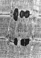 From E Hardeman |

|
|
| Electron microscopy of rods from Z-lines in Met9Arg αTMslow mouse | ||
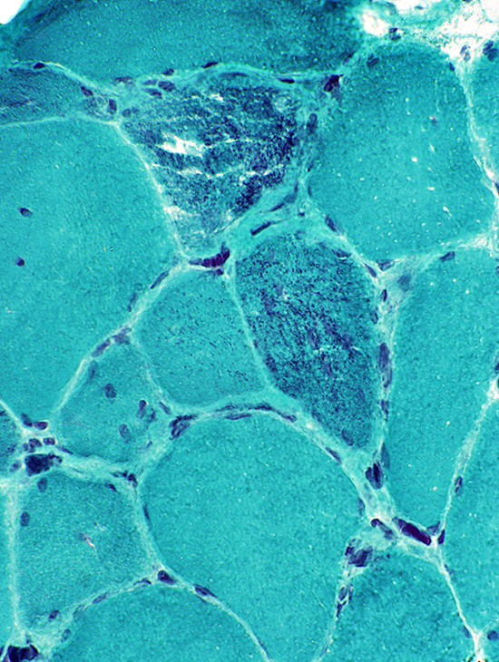
Nemaline rod myopathy, infant (8 months of age): Type 1 muscle fiber smallness
Rods: More prominent in smaller muscle fibers
 Gomori trichrome |
 ATPase pH 9.4 Type 1 muscle fibers: Small (Pale) |
 H&E Muscle fiber sizes: Bimodal |
 NADH Internal architecture of muscle fibers: Irregular |
Rod Myopathy: Congenital + Arthrogryposis
 H&E stain |
 Gomori trichrome stain |
Dark-stained: Punctate or Clustered regions, Often sub-sarcolemmal
More common in smaller muscle fibers
 Gomori trichrome stain |
 Gomori trichrome stain |
 ATPase pH 9.4 stain |
Type 1 fibers: Usually small
Type 2 fibers: Larger; Abnormal intermediate staining (2C) at ATPase pH 4.3
 ATPase pH 4.3 stain |
 NADH stain |
Rods: In clusters in muscle fibers
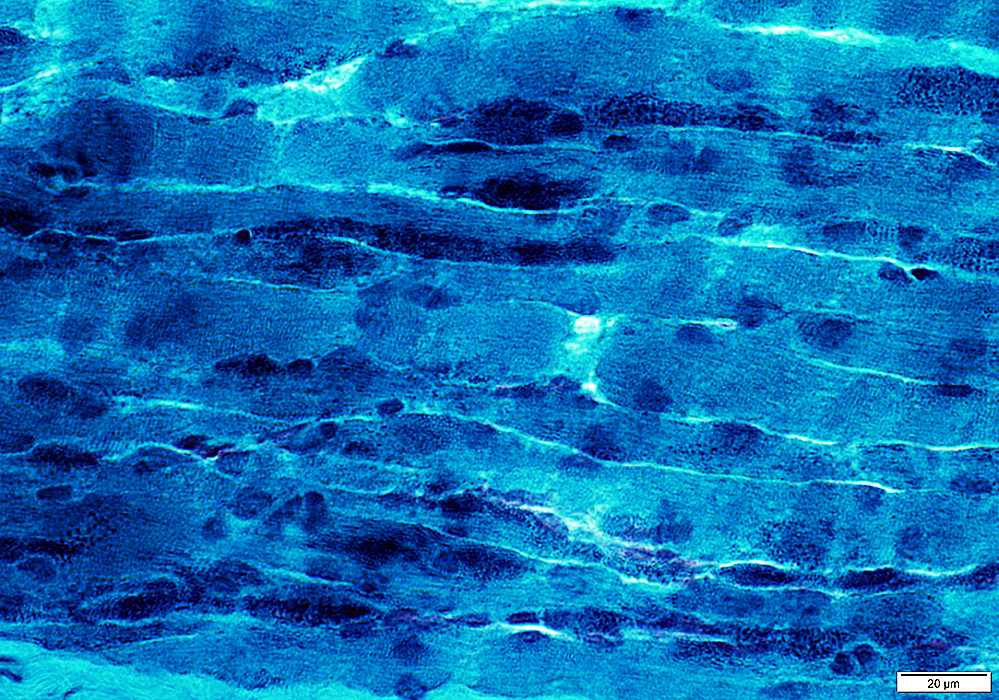
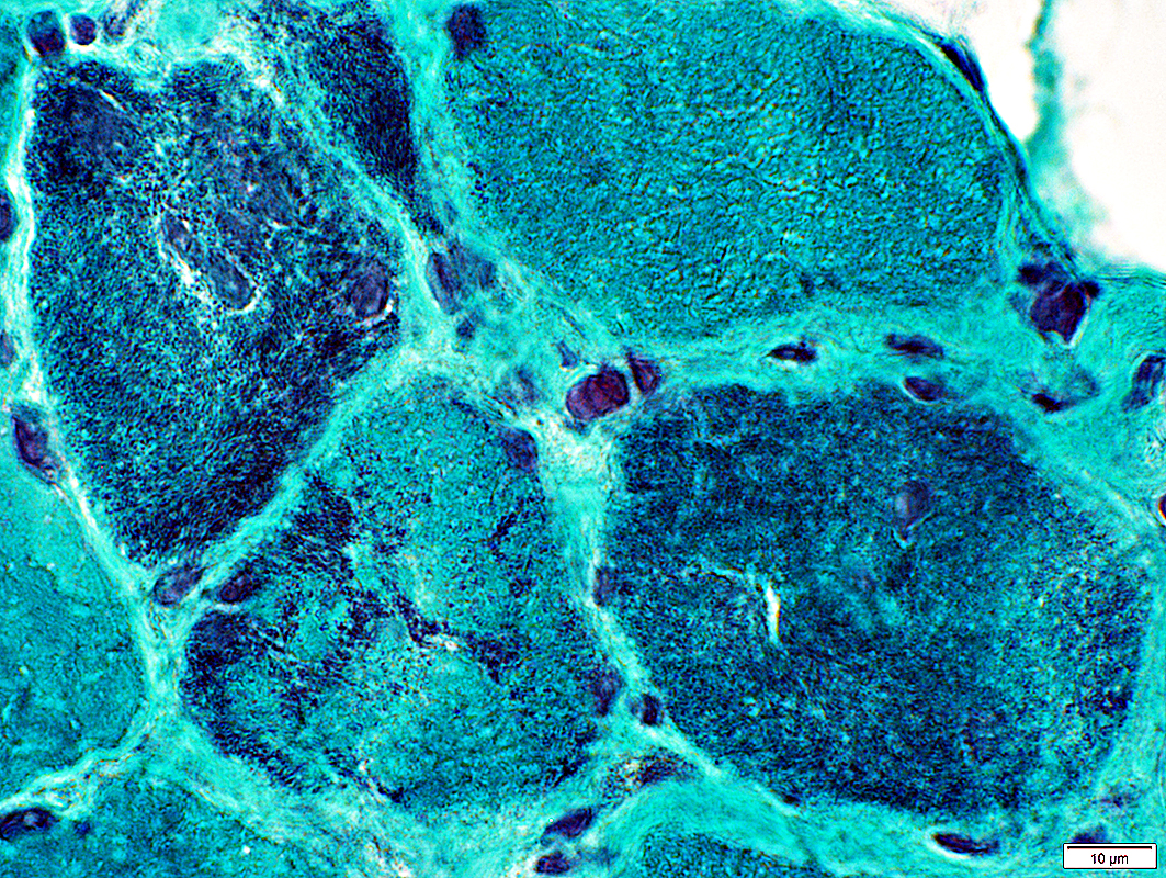
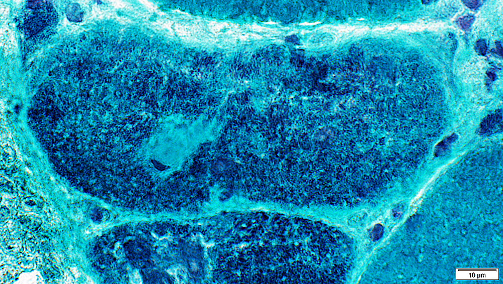
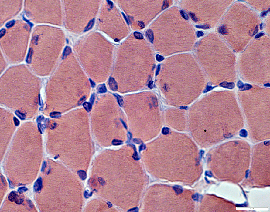
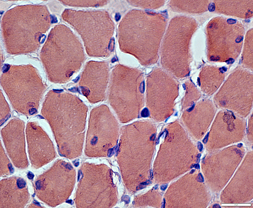
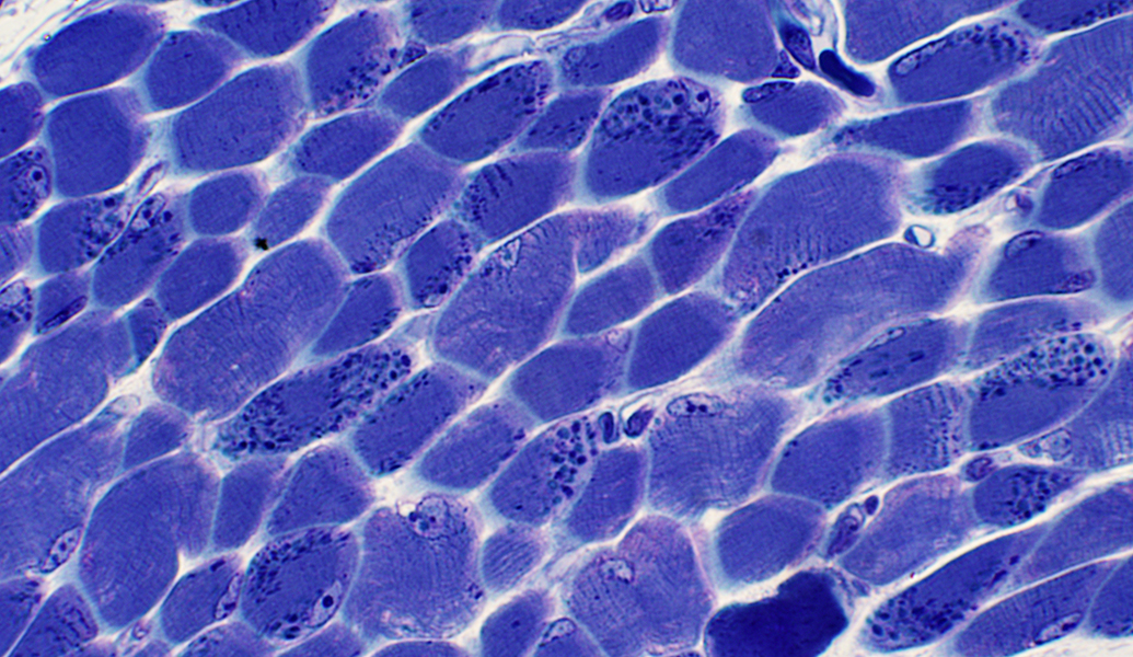 Toluidine blue stain |
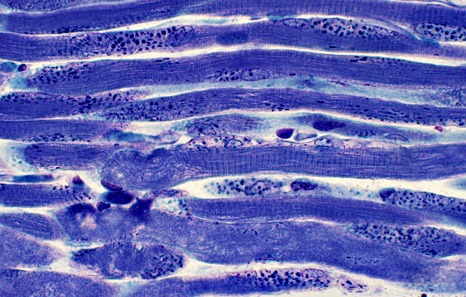 Toluidine blue stain |
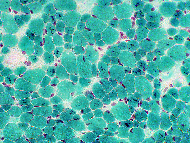
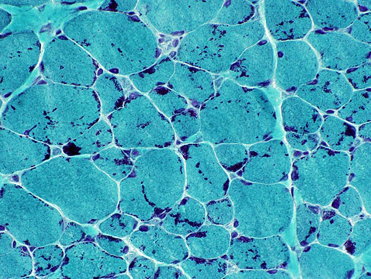
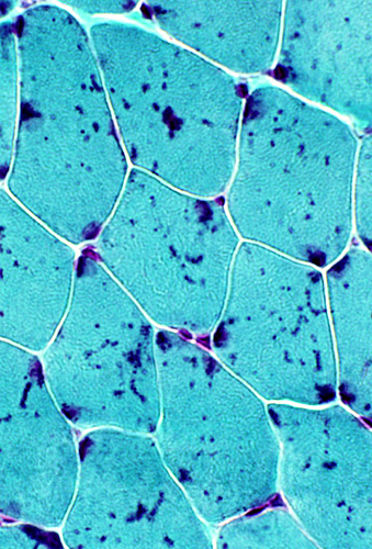
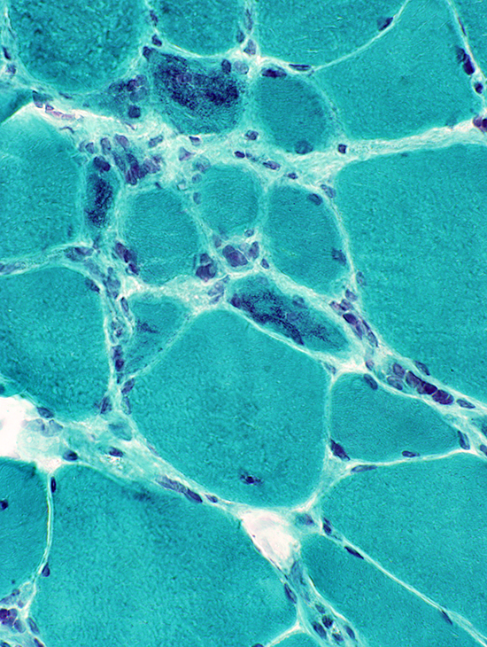
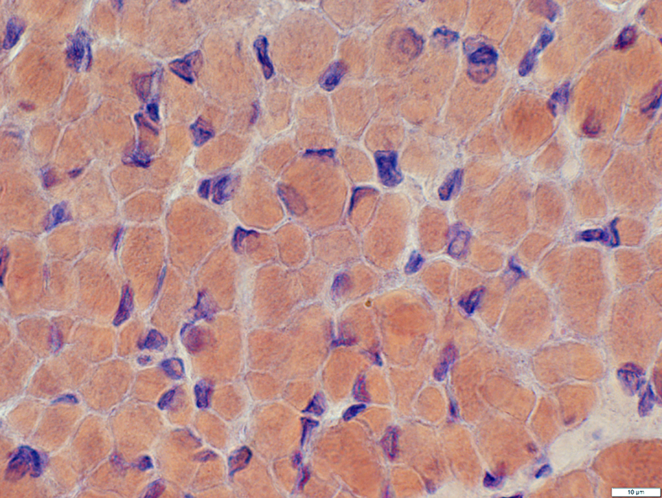
Nemaline rod myopathy, Childhood: 2 Different patterns
Rods: In smaller muscle fibers
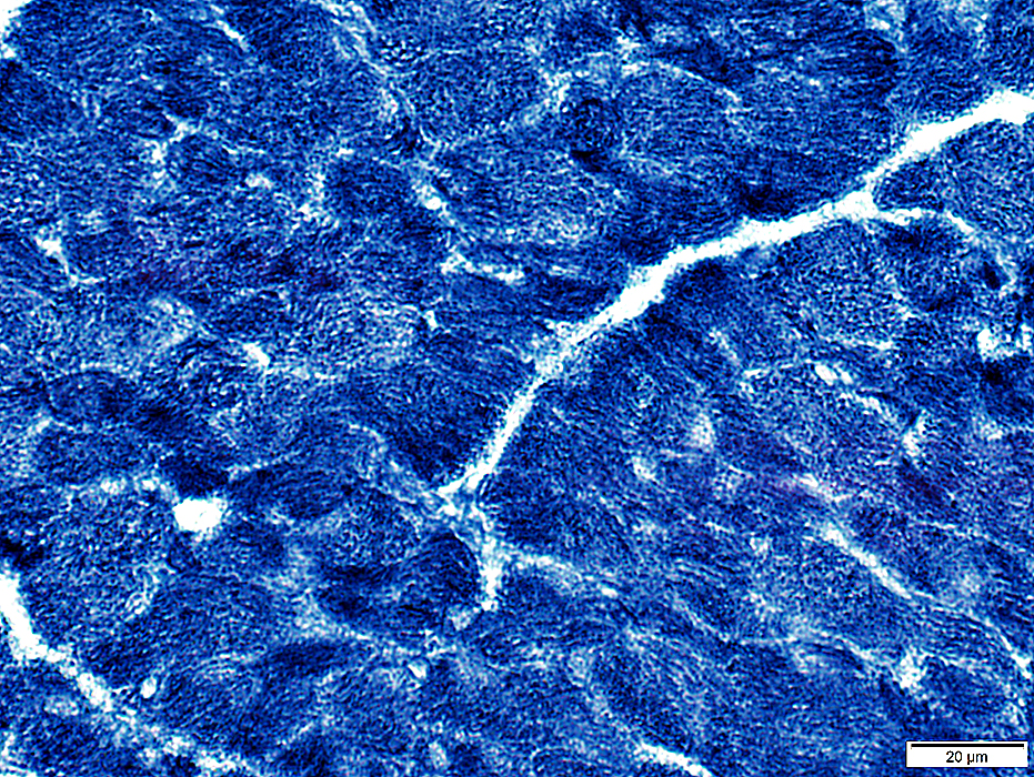
 GT stain |
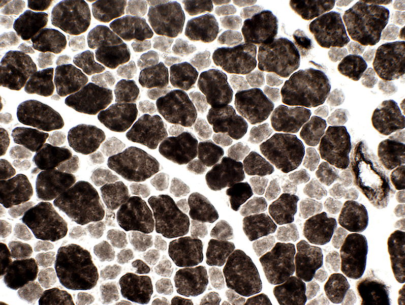
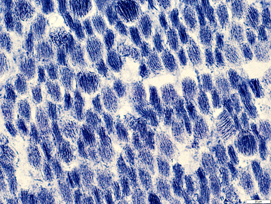
 ATPase stain, pH 9.4 Type I muscle fibers: Smaller than type II |
 GT stain |
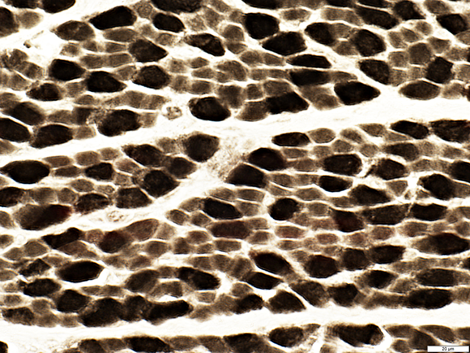
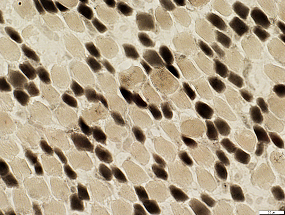
Type I predominance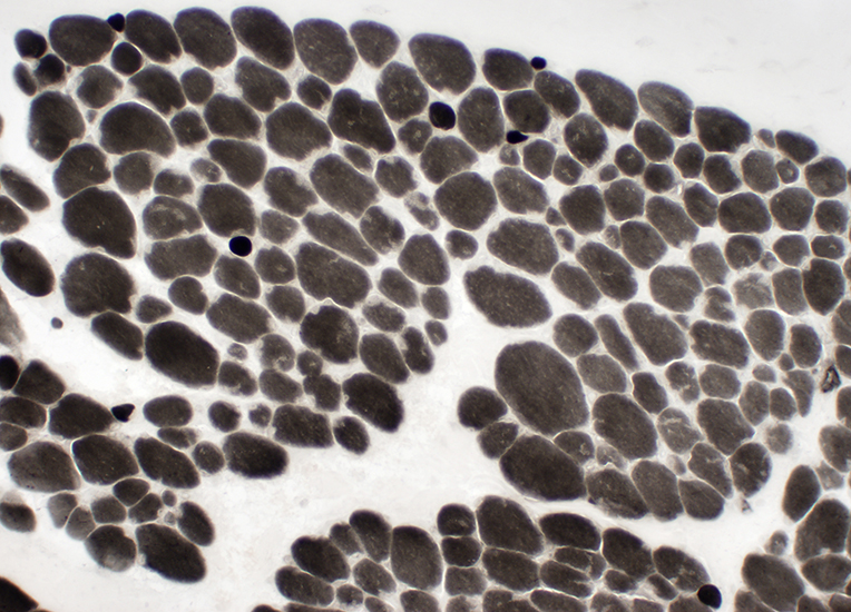 ATPase stain, pH 9.4 |
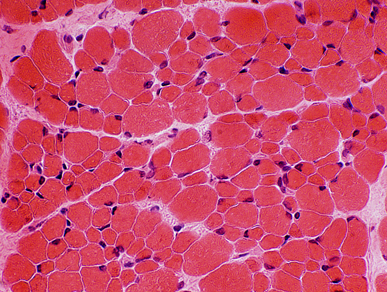
Fiber size variation: Bimodal distribution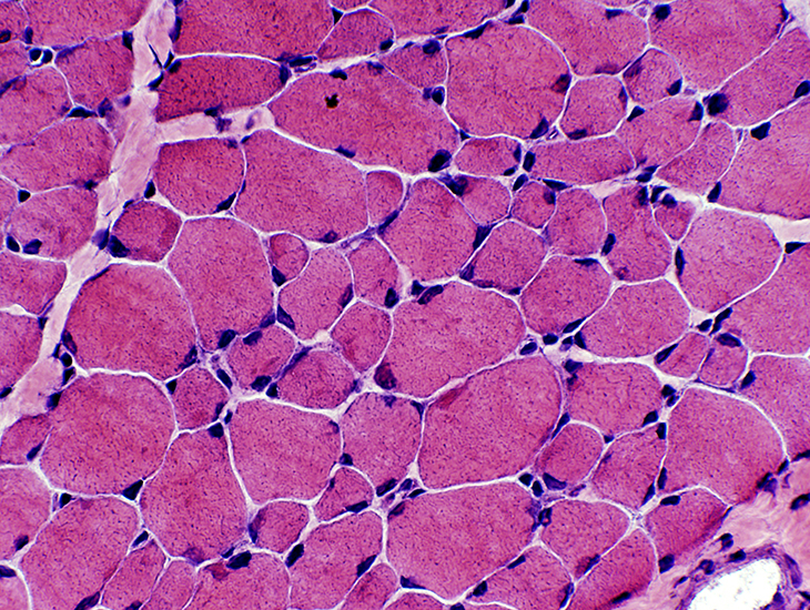 H&E stain |
Rods: Adult nemaline myopathy
Several patterns
Rods: Larger & Aggregated
 Gomori trichrome |
 Gomori trichrome |
Rods: Small & Diffusely distributed in some muscle fibers
Myopathic features: Varied muscle fiber size; Internal nuclei; Endomysial connective tissue increased
 Gomori trichrome |
 Gomori trichrome |
 Gomori trichrome |
 Gomori trichrome |
 H&E stain |
Rods: Atypical
 Gomori trichrome stain |
Stain dark on Gomori trichrome
Near myotendinous junctions
Gomori trichrome stain |
 Toluidine blue stain |
|
|
|
|
Dark stained
Internal structure: Amorphous
Shape: Irregular; Polygonal
Halo: Contains linear structures
|
|
Rods: ACTA1 mutation (M46T)
13 yo male with Rod myopathyNo rods in muscle biopsy of same patient at 1 year old
 H&E stain |
Small
Mild variation
Myonuclei
Mildly large
Shapes: Irregular
 H&E stain |
 Congo red stain |
Abundant
Shapes: Irregular
Sizes: Large
 Congo red stain |
 ATPase pH 4.3 stain |
 Gomori trichrome stain |
Irregular shaped clusters of dark stained material in myofiber cytoplasm
 Gomori trichrome stain |
 Gomori trichrome stain |
 Phalloidin stain for Actin |
Clustered in regions of muscle fiber cytoplasm
 Phalloidin stain for Actin |
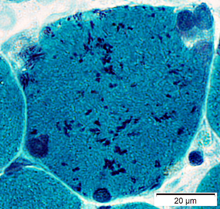
 Toluidine blue stain |
 Toluidine blue stain |
Morphology: Dark-stained rod-shaped inclusions
Distribution: Clustered or scattered in myofiber cytoplasm
 Toluidine blue stain |
 Toluidine blue stain |
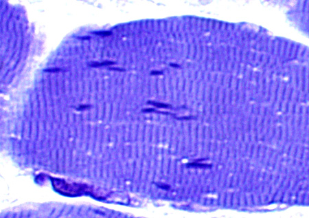
|
Dark-stained rod-shaped inclusions: Often aligned along Z-bands
|

|
|
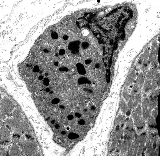
Rods occuring in a subsarcolemmal cluster
|
|
|
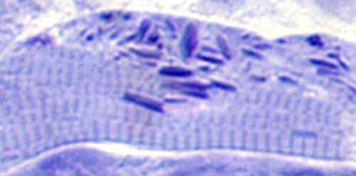
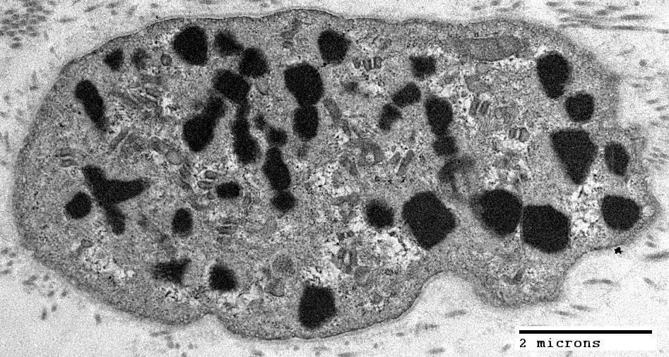
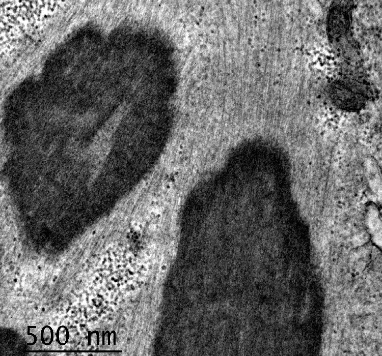
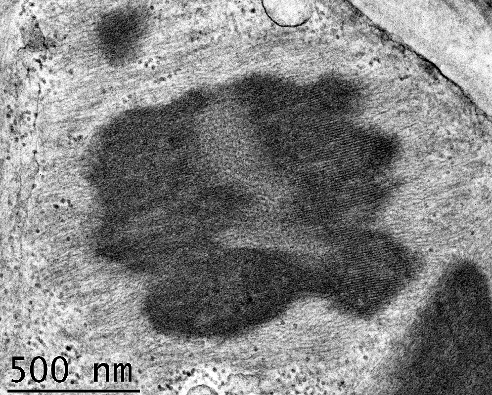
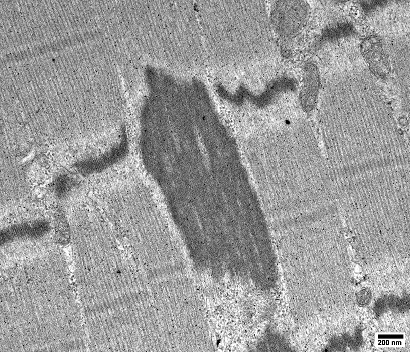
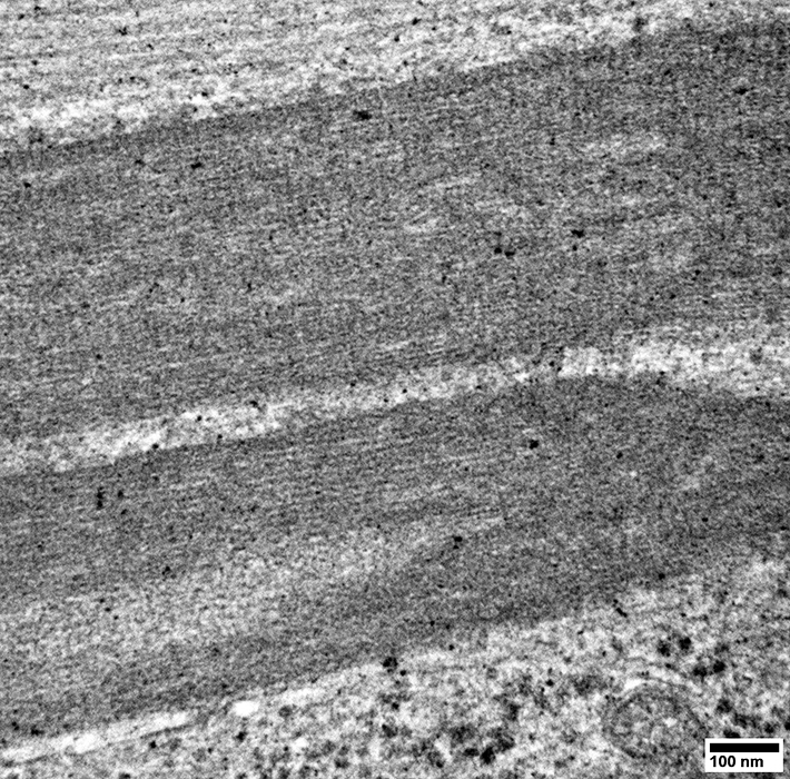
Electron dense
Sizes: Length 1-7 μm; Width 0.3-2 μm
Long axis: Parallel to muscle fiber
Longitudinal section: Striations parallel & perpendicular to long axis
Transverse sections: Lattice structure like Z disk
Thin filaments: May be in continuity with rods
|
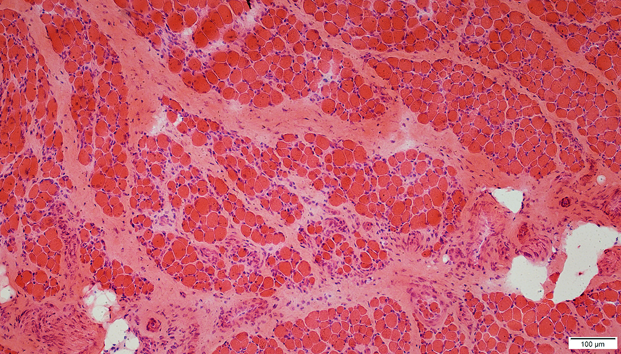
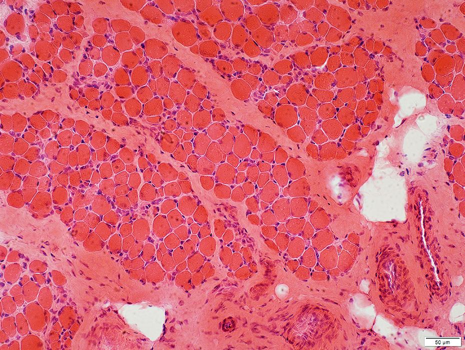
Rod Myopathy: Same patient as above at 1 year old
No rods
Type 1 muscle fibers: Small
 H&E stain |
 Gomori trichrome stain |
 Gomori trichrome stain |
 Congo red stain |
Mucle fiber sizes: Varied
 NADH stain |
 ATPase pH 9.4 stain |
 ATPase pH 4.3 stain |
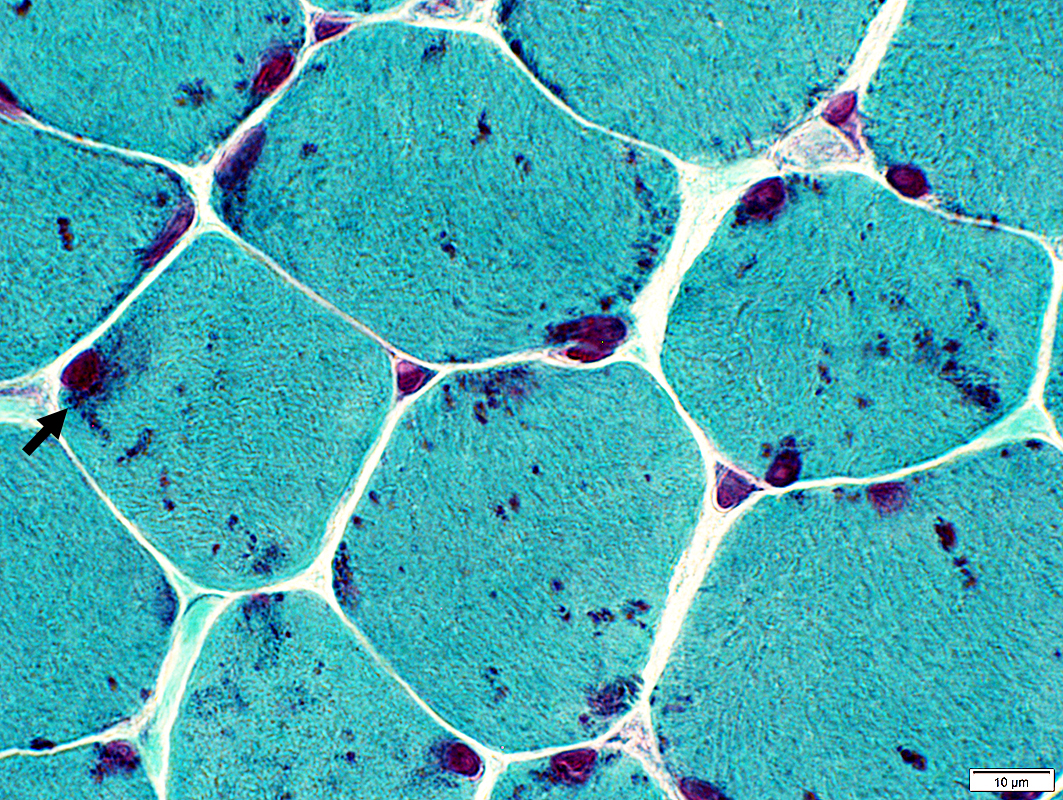
Z-Disk: Streaming & Other disorders
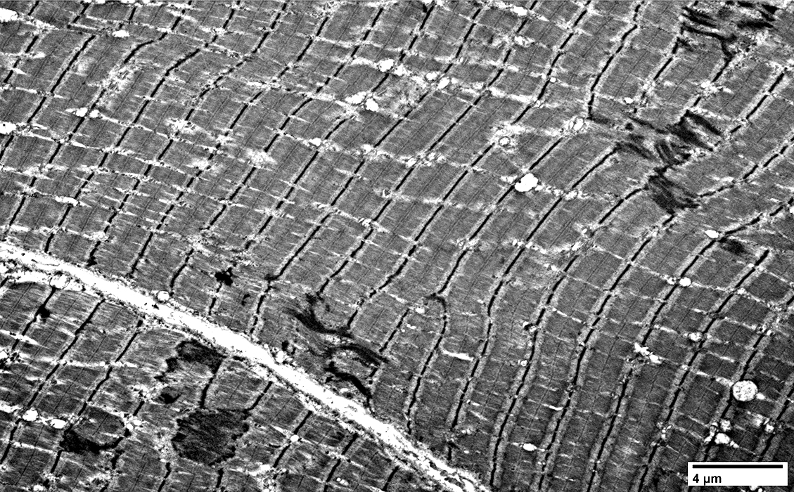 |
- Z-Streaming Definition: Dense Z-band material extends from Z-Disk
- Into sarcomere
- May reach adjacent Z-Disk
- More common: In areas without mitochondria
- Anatomical Association: Loss of thick filaments, or of entire sarcomere
- Z-Streaming: Associations
- Normal: Image
- Pathology: Targets; Cores
- Disorders: Myotonia; Dermatomyositis; Myofibrillar myopathy
- Other: May occur in otherwise normal muscle
- Other Z-line disorders
- Irregularities
- Duplication
- Loss: Muscle fiber necrosis
- Misorientation: Myosin-loss myopathy
- Rods
- Cytoplasmic bodies
- Myofibrillar myopathies
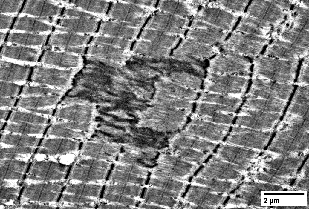 |
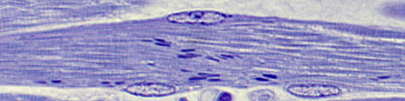
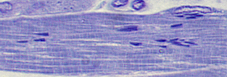
Crosses up to 2 sarcomeres
Z-disk Streaming
Streamed Z-band material extends from Z-band (Arrow)
Other Z-band material is aggregated/streamed at top left of image
 |
Return to Rod myopathies
References
1. J Neuropathol Exp Neurol 2022;81:304-307
10/3/2024