Capillary Pathology: Muscle
|
General Analysis ELS in BIM Regional microvasculopathies Dermatomyopathy syndromes Diffuse microvasculopathy Dermatomyopathies NXP2 SLE Minimal Myopathy Systemic Sclerosis Myopathy Capillary ultrastructure GvHD Also see Pipestem capillaries |
Connective Tissue Disease Endomysial capillary Δ CREST: 1; 2 SLE Systemic Sclerosis Th/To antibody RUV B1/2 antibody + Myopathy Ro-52 antibody Myopathy Systemic Sclerosis Ro-52 antibody |
Capillary Pathology with Minimal Myopathy: Systemic Sclerosis 1 & Other
Clinical associations- Muscle
- Less weakness
- Serum CK: High but lower than myopathy patients
- Systemic: Less frequent
- Pulmonary: Interstitial lung disease; Arterial hypertension
- Renal crises
- Skin: Diffuse involvement
- Histochemistry
- Size: Large
- Stains: NADH; ATPase pH 4.3; Acid phosphatase; PAS
- Neighboring cells: Pericytes & Histiocytes
- Numbers: Reduced
- Ultrastructure
- Basement membrane: Thick & Reduplicated
- Endothelium: Activation
- Pericytes (PDGFR-β stain): Proliferation
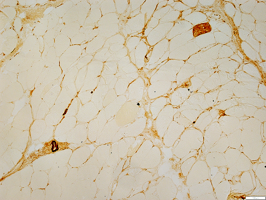
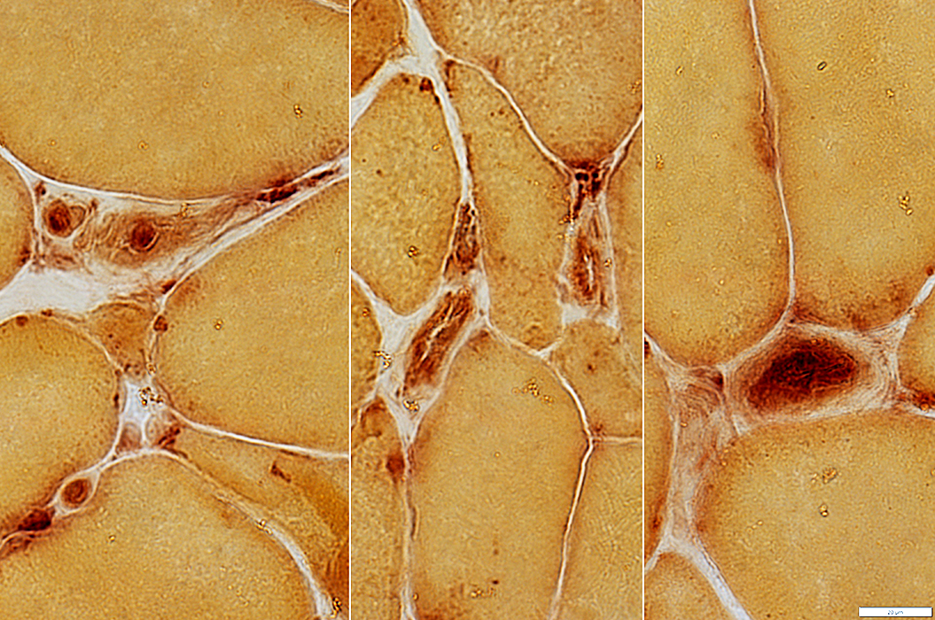
Capillary Features: Stain Analysis
| Basal Lamina | Endothelium | Immune |
|
Morphology H&E GT/VvG Ultrastructure Molecular Decorin/Col IV PAS: + or - Pericytes PDGFRβ |
H&E: Morphology Ulex: Distribution; #; Size; Stain intensity ATPase pH 4.3: #+ Alkaline phosphatase: #+ NADH: + or - |
Humoral C5b-9 Cellular Acid phosphatase HAM56 Cell involvement Muscle: MHC I Capillary: MxA |
Systemic Immune Disorders: Capillary ± Muscle Pathology
SLE
Patient Features
38 yo female
Clinical diagnoses: Systemic Lupus Erythematosis; ILD
Serum Antibodies: Ro52; ANA 1:640, Speckled; ± NT5C1a, PL-12 & MGT-30
Treatment: Prednisone
Capillary pathology
Morphology: Basal lamina mildly to moderately thick
Basal lamina: PAS+; Decorin mildly thick
Endothelium: Ulex large & reduced #; Alkaline phosphatase -; ATPase +; NADH ++
Immune: C5b-9 scattered capillaries; MxA & Acid phosphatase cells
Muscle: MHC1+
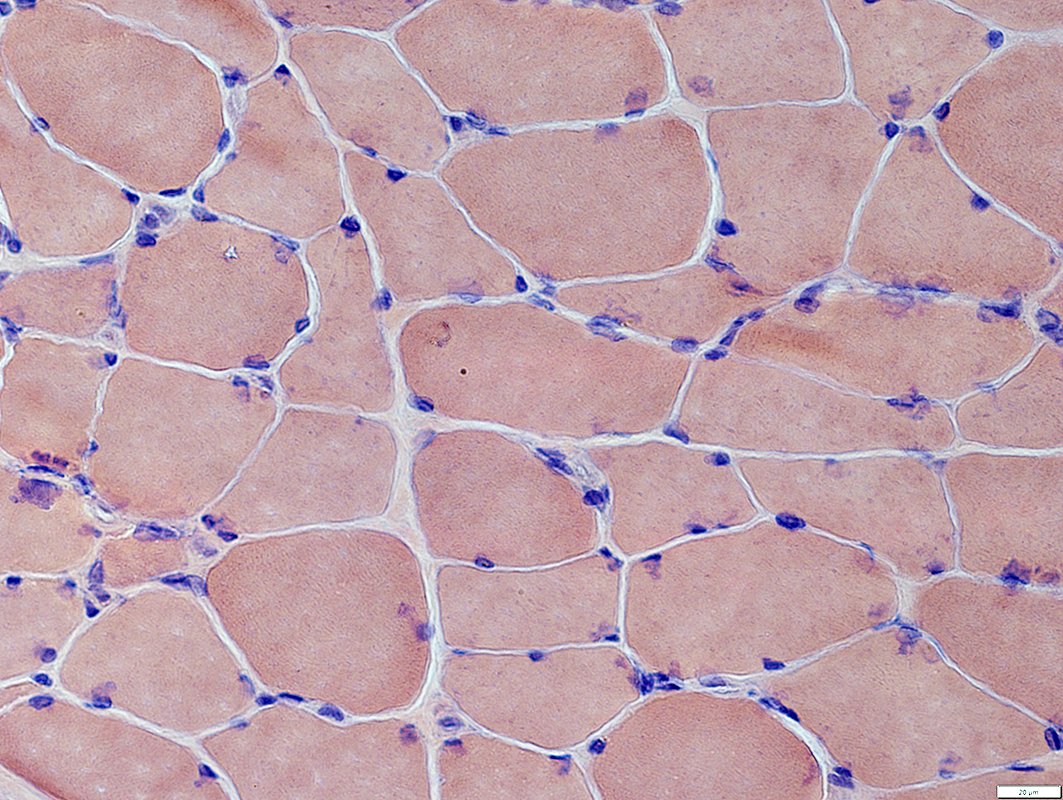
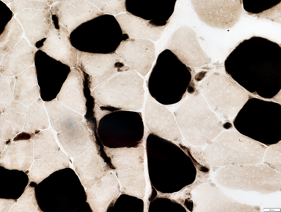
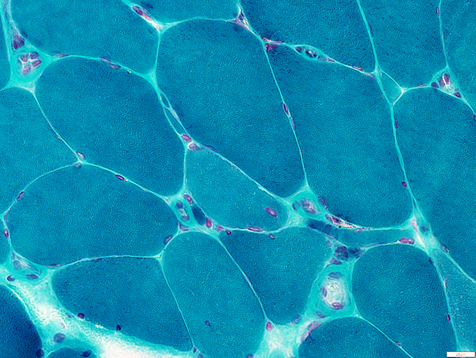
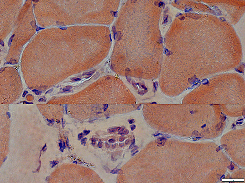
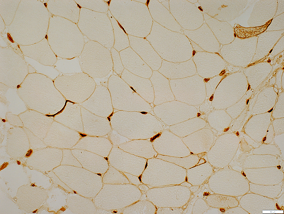
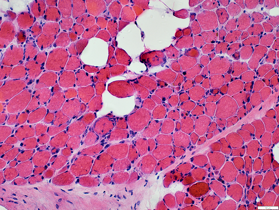
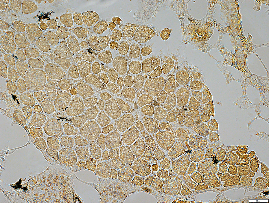
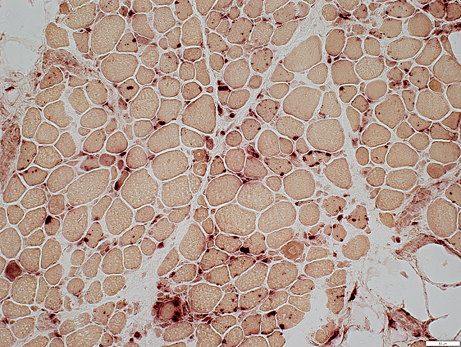
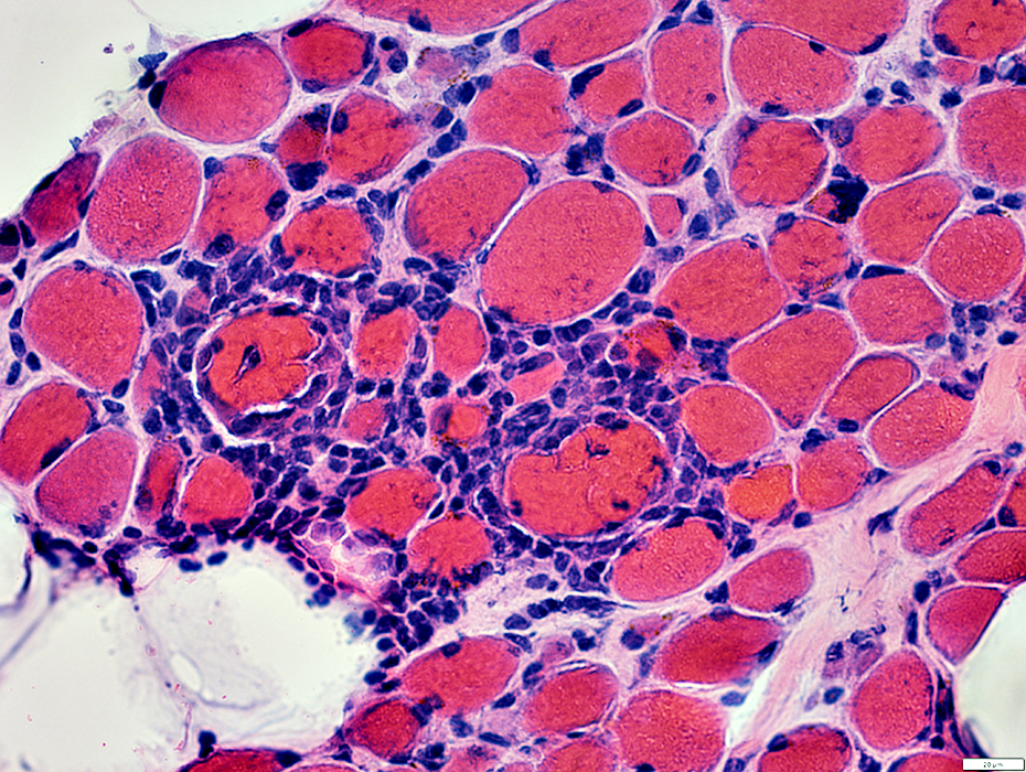
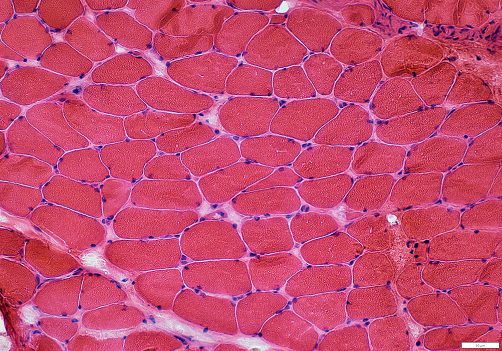
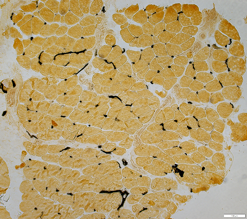
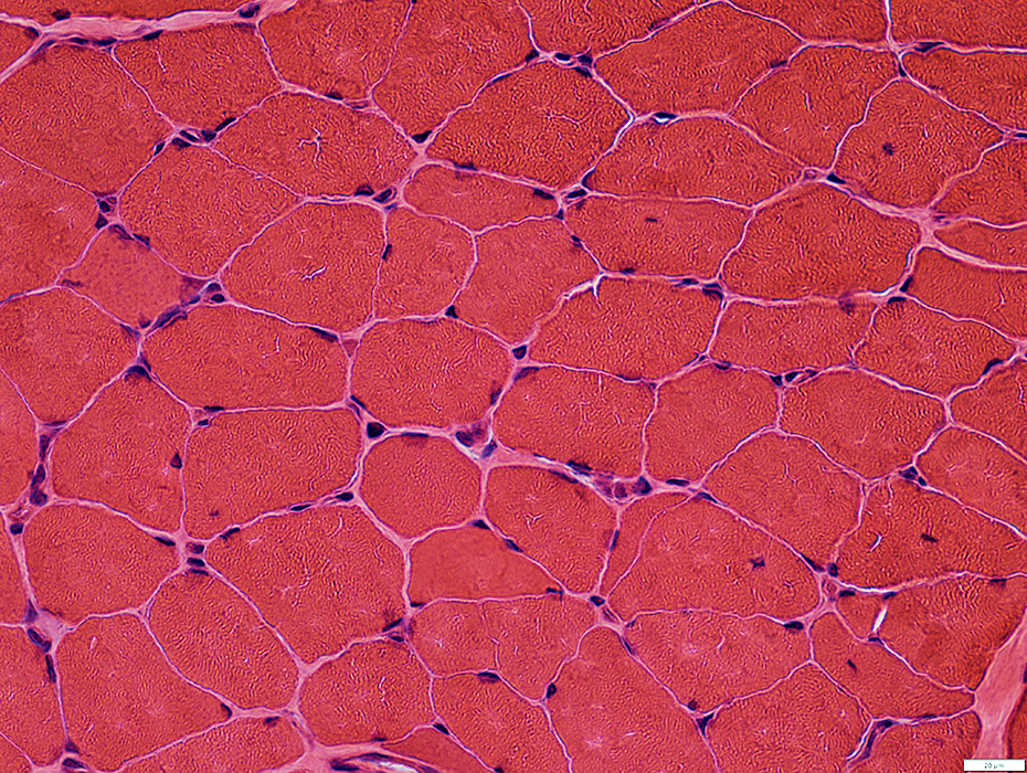
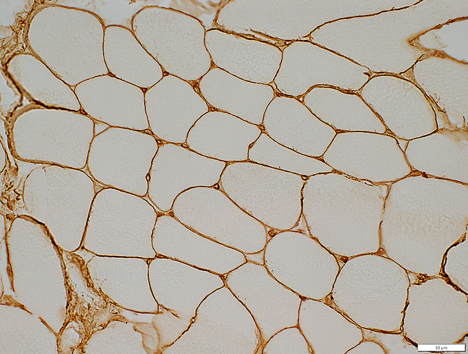
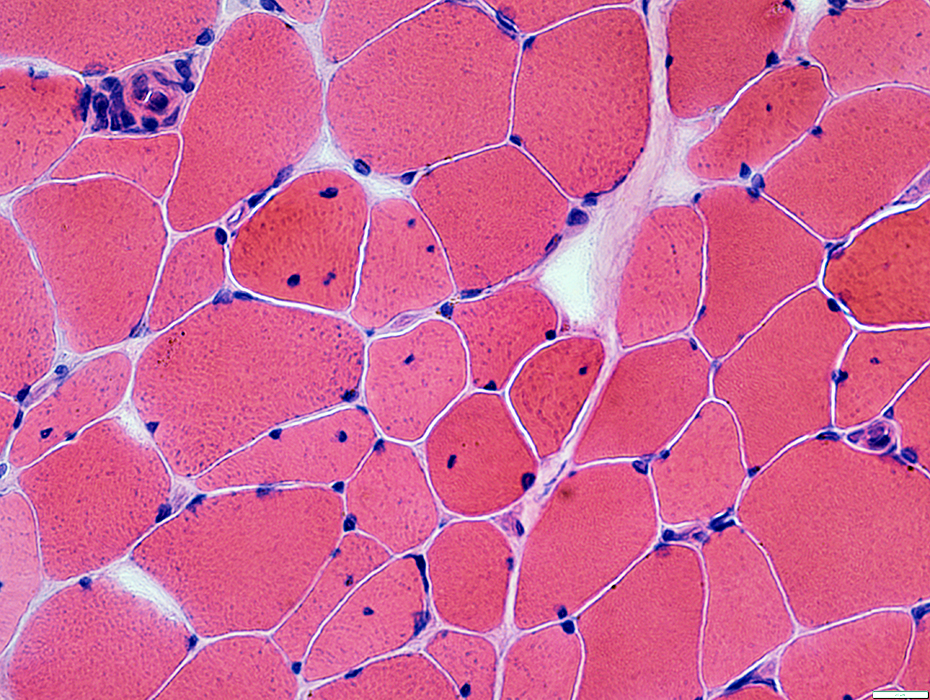
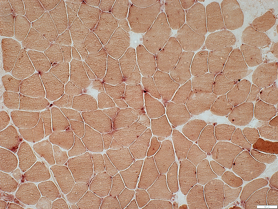
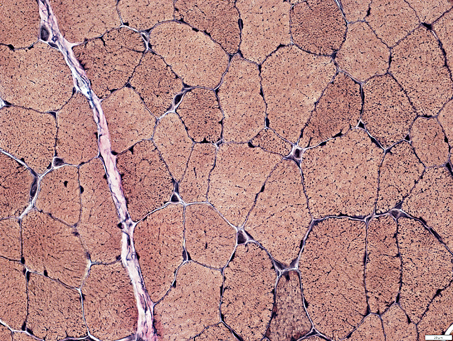
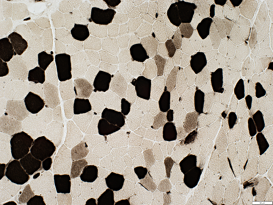
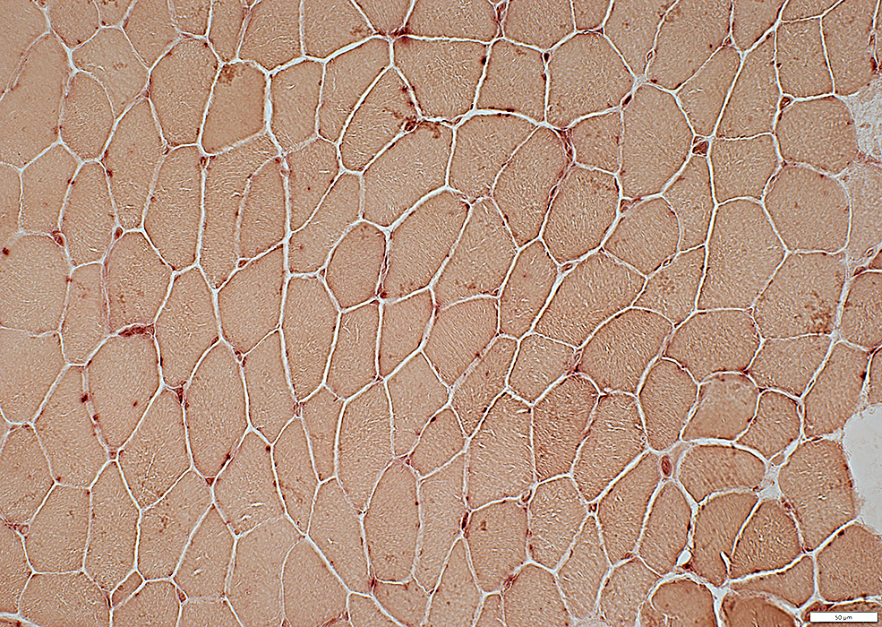
Endomysial Capillaries: Morphology
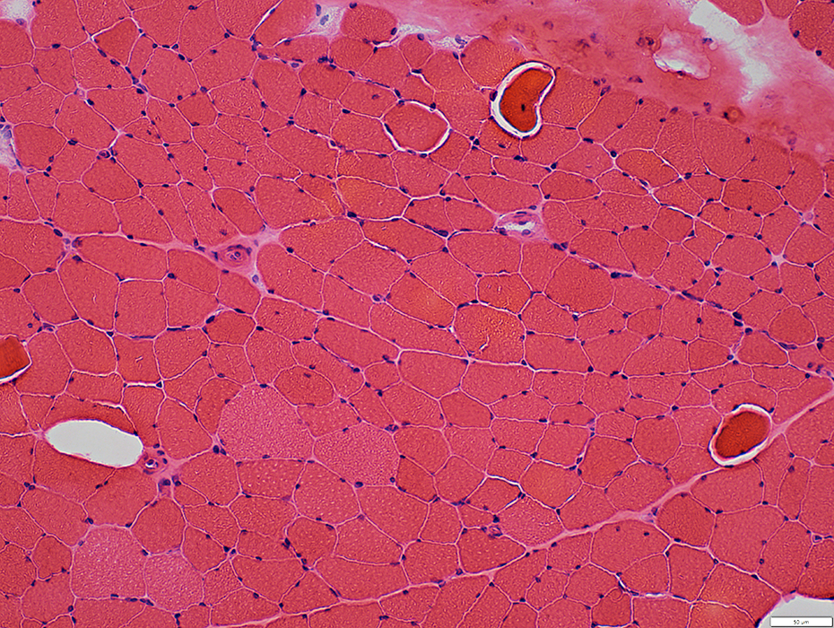
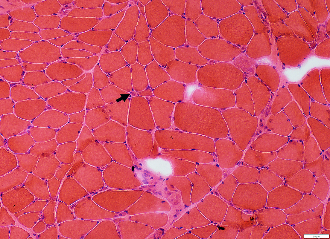 H&E stain |
Moderately enlarged
Muscle fibers
Fiber sizes: Moderately varied
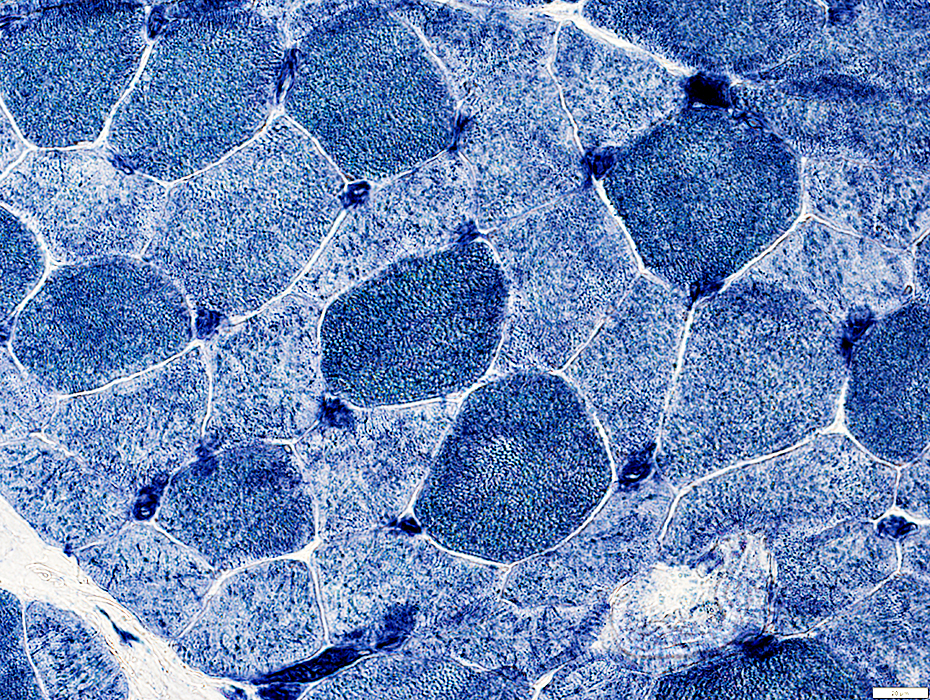
 H&E stain |
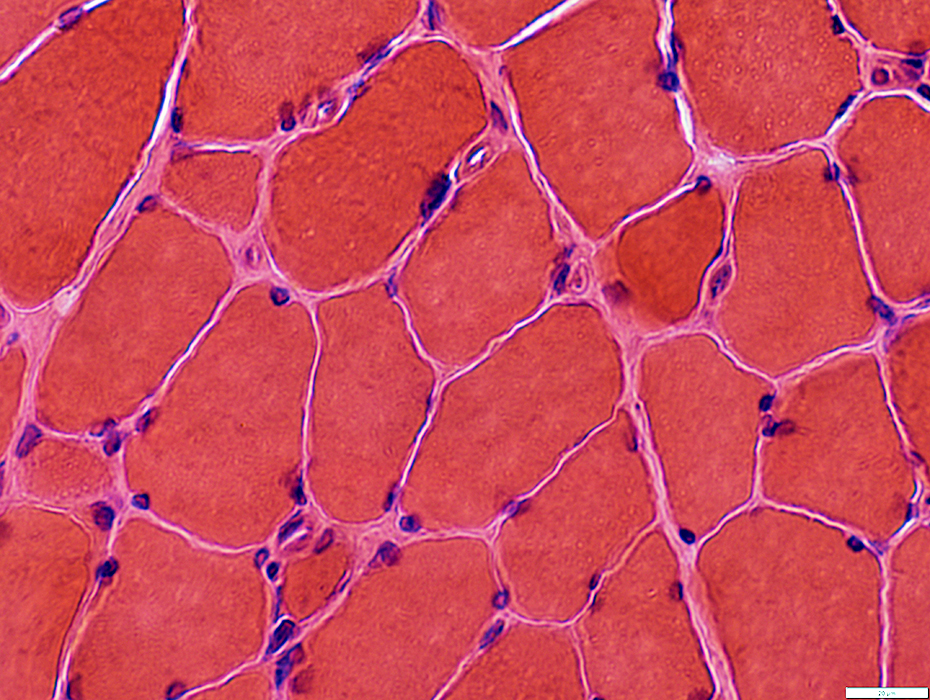
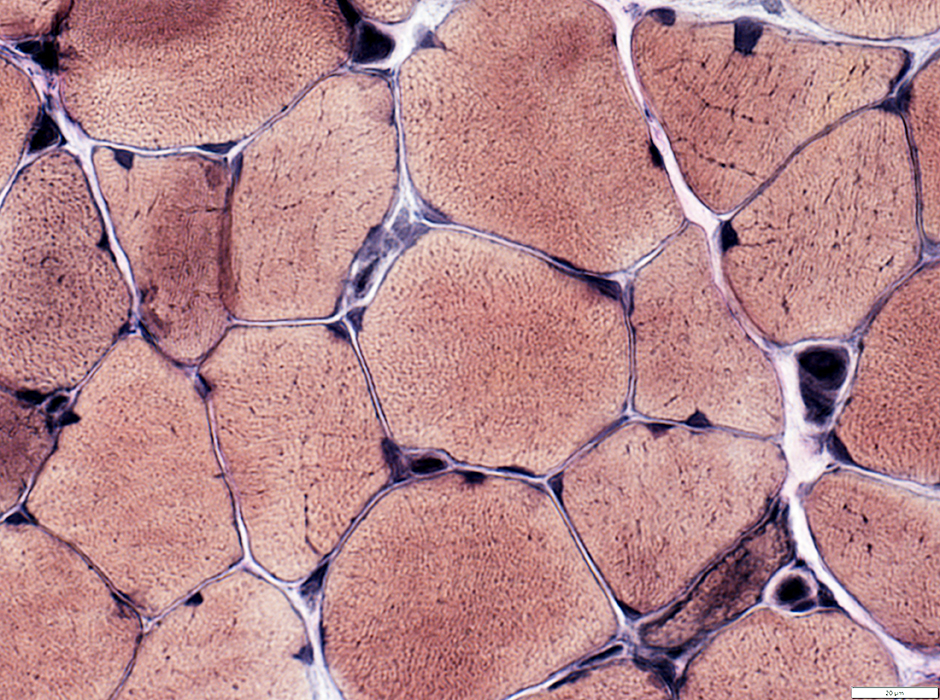
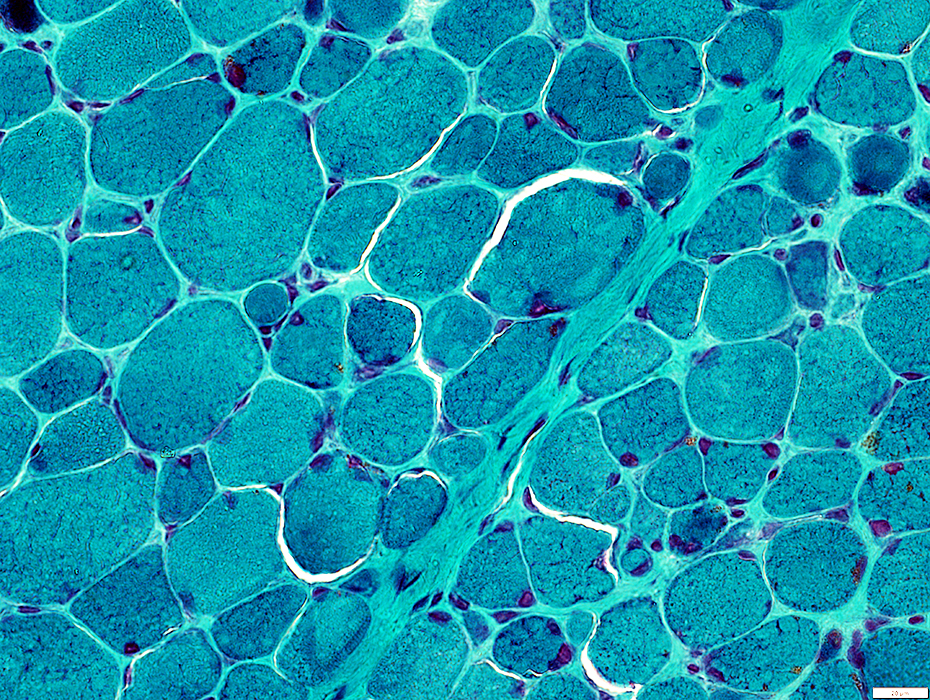
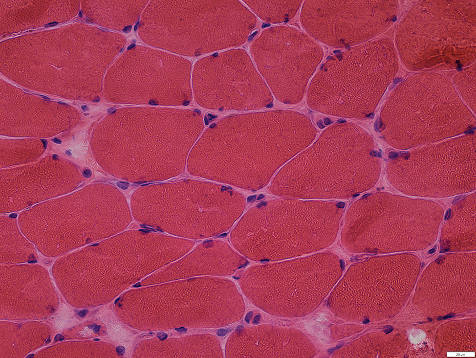
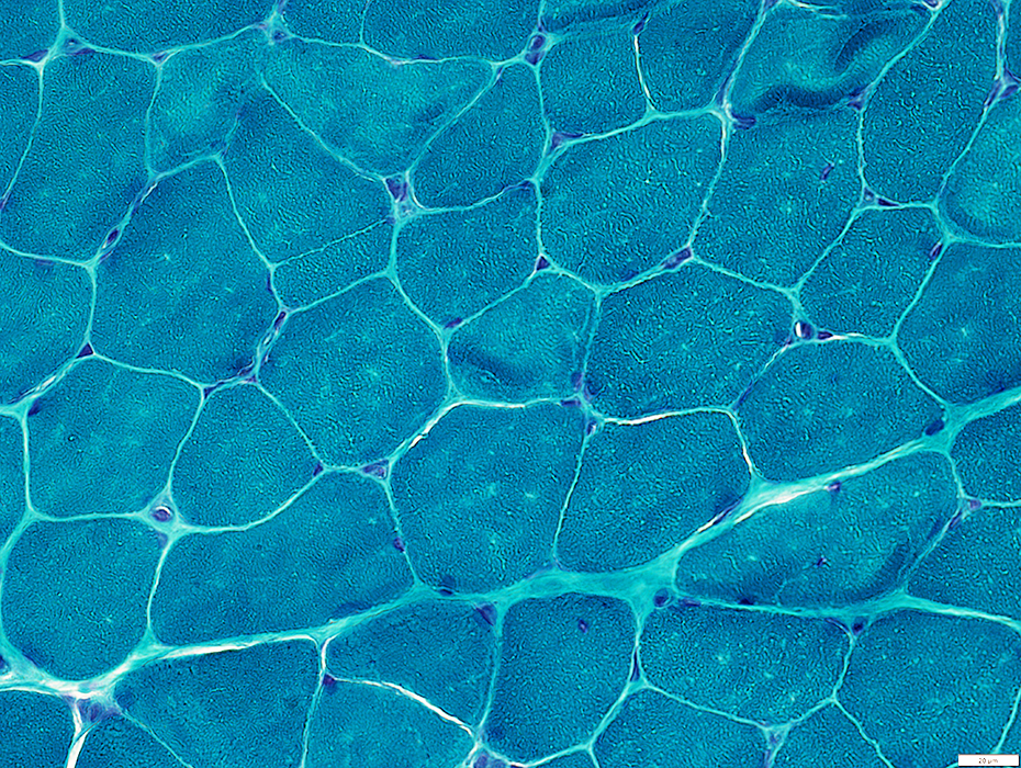
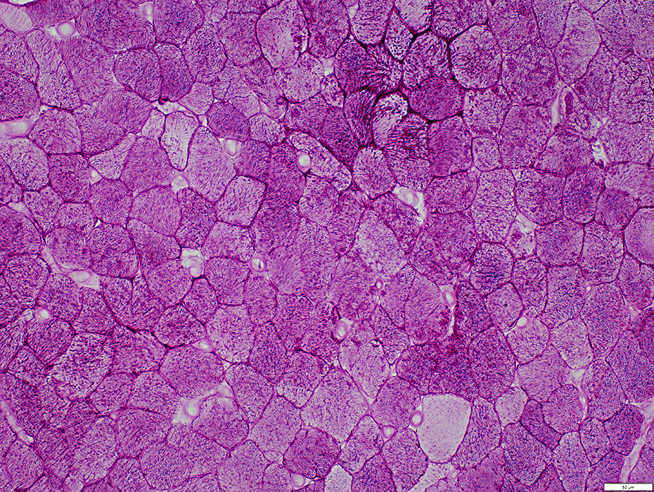
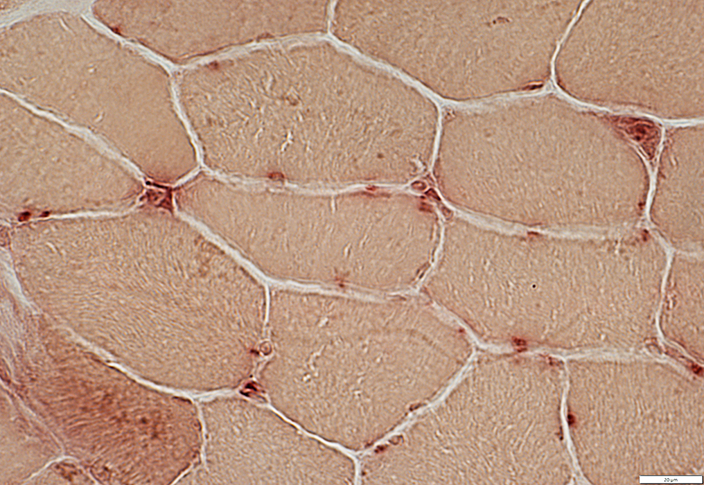
 Gomori trichrome stain |
Moderately enlarged
Some have thick basal lamina
 Gomori trichrome stain |
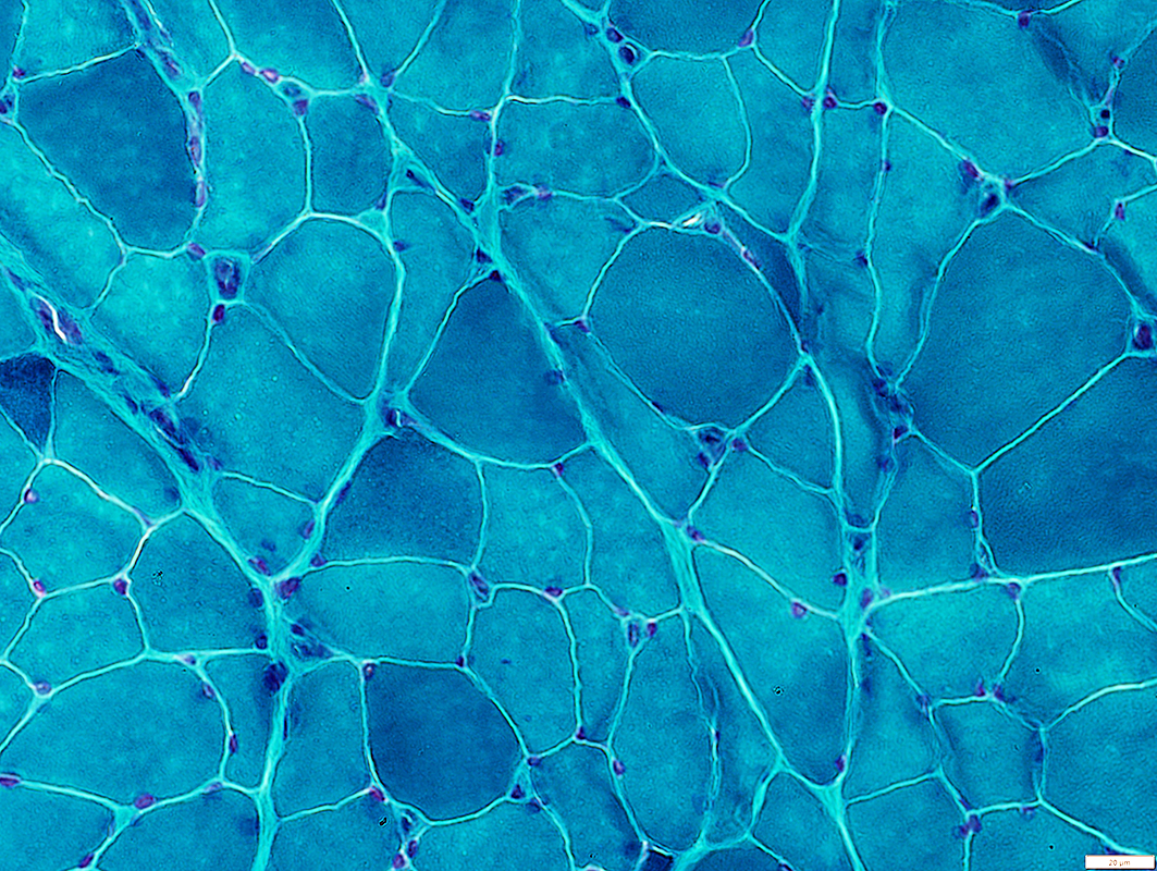
Endomysial Capillaries
Moderately enlarged
 Congo red stain |
 VvG stain |
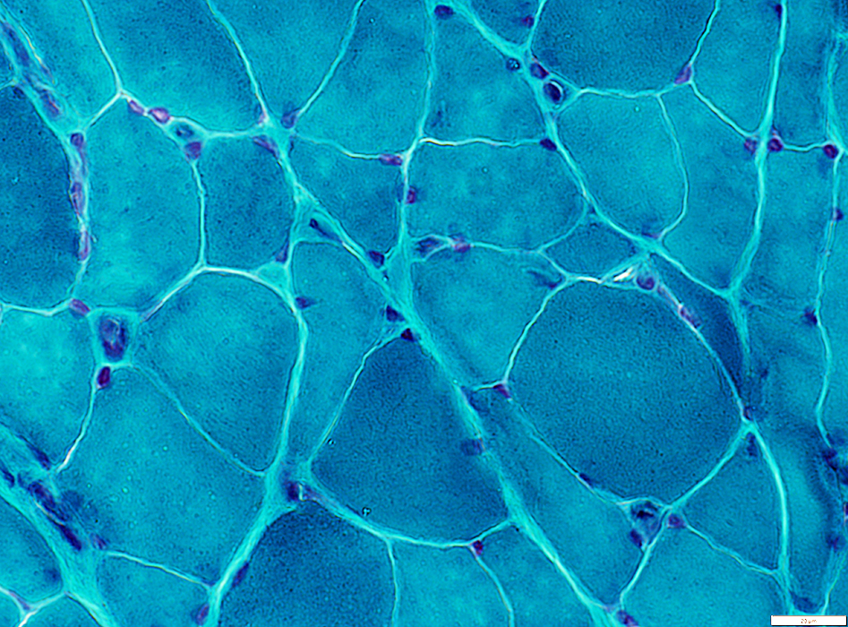
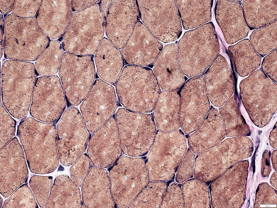
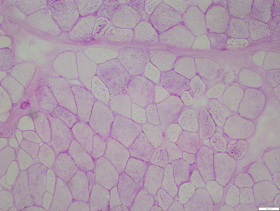
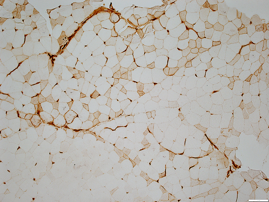
Endomysial Capillaries: Basal lamina
PAS stains capillary basal lamina
 PAS stain |
 Decorin stain |
Moderately enlarged
Basal lamina: Commonly thick
 Decorin stain |
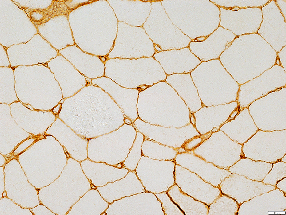
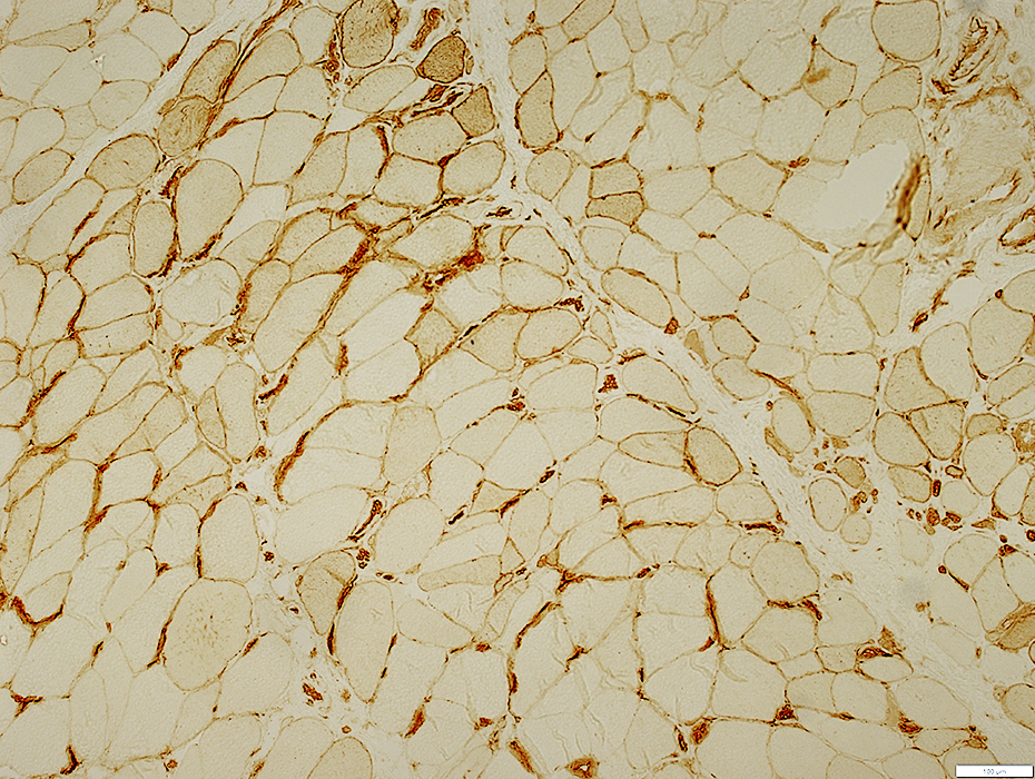
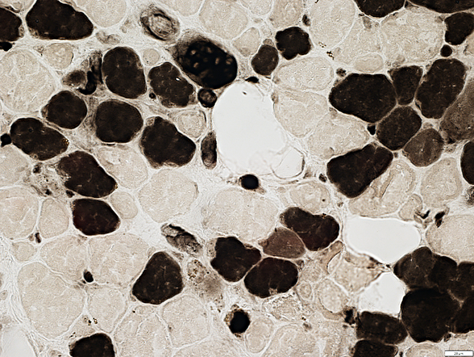
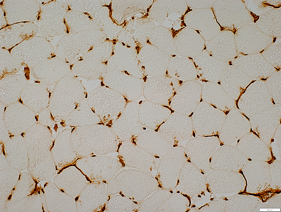
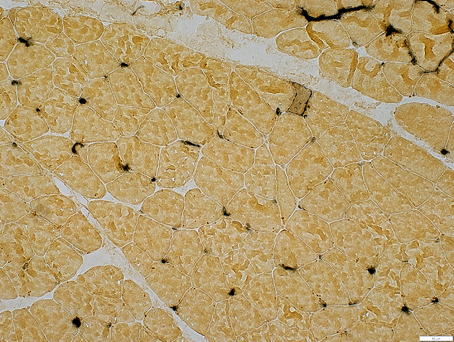
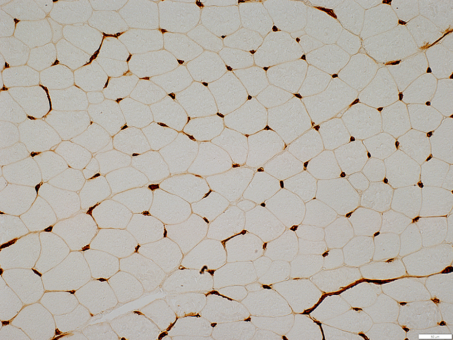
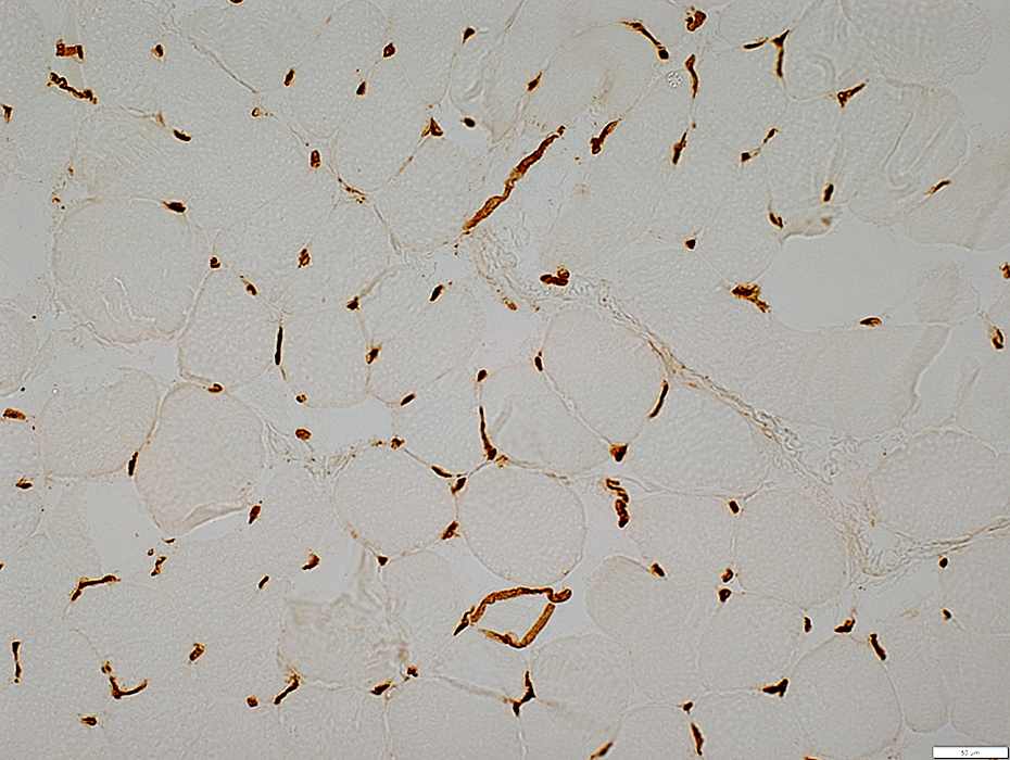
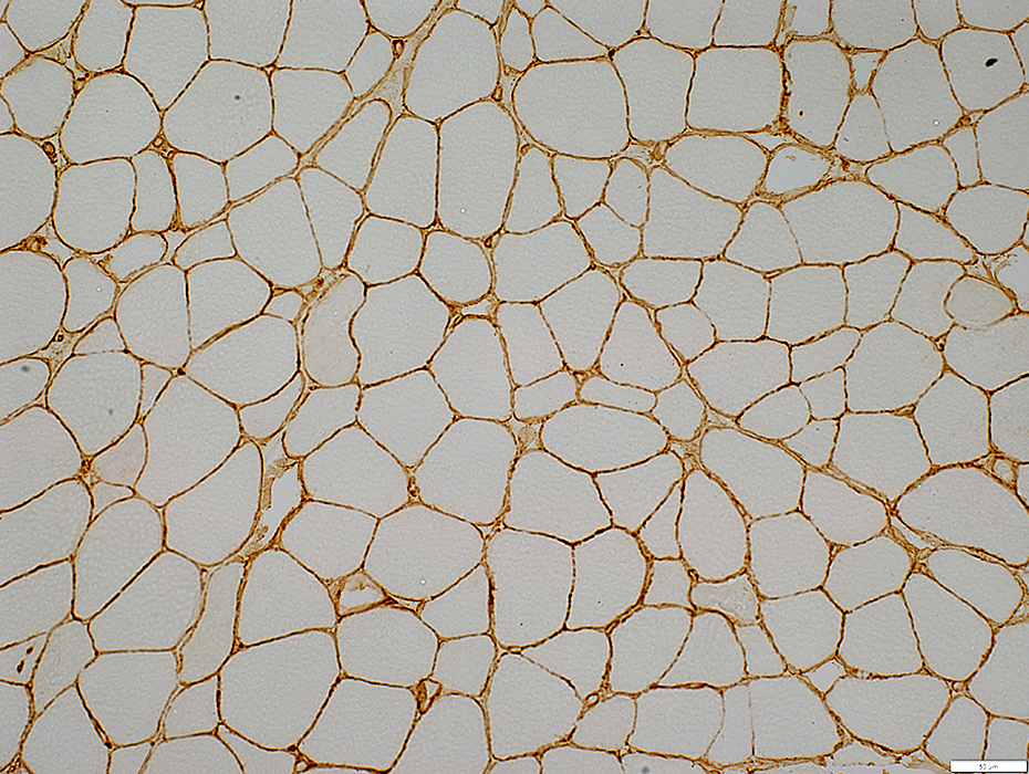
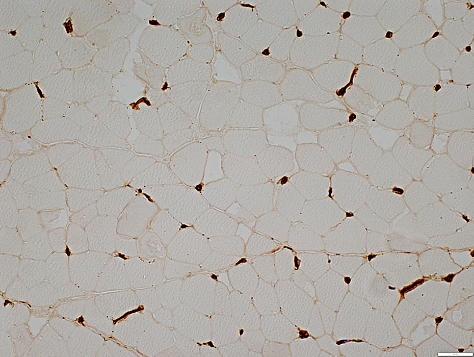
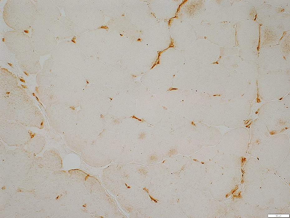
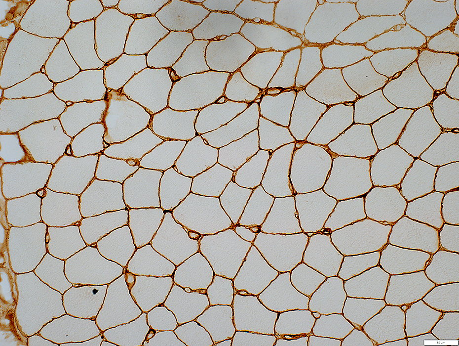
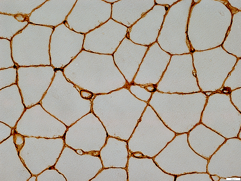
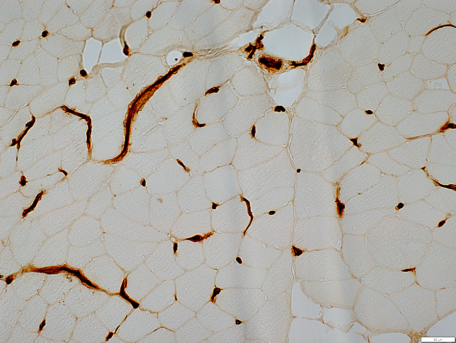
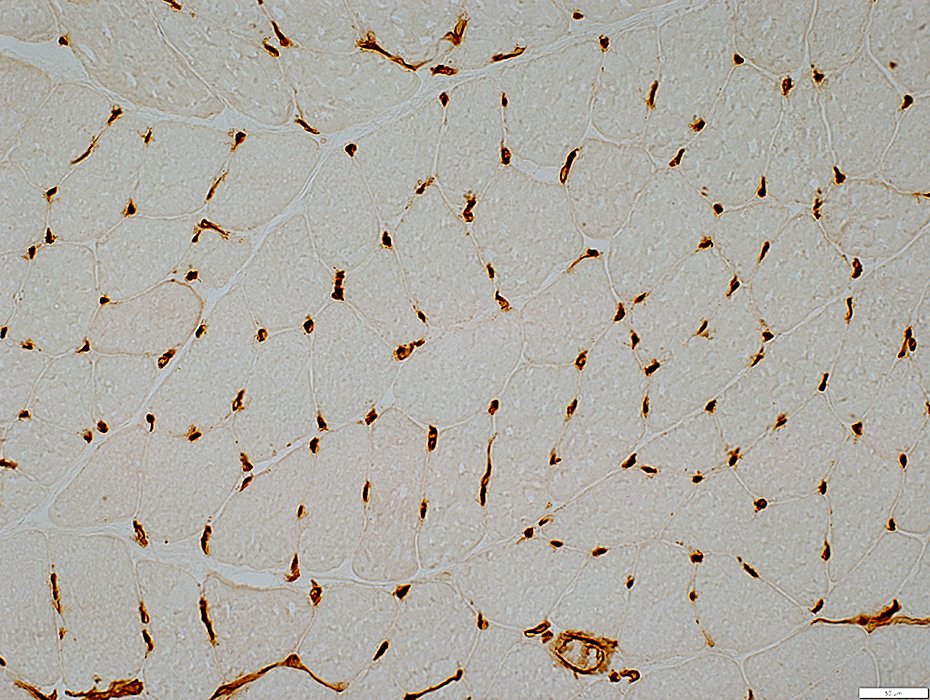
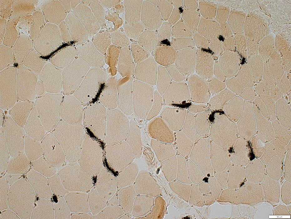
Endomysial capillaries: Endothelium
 UEA I stain |
 Alkaline phosphatase phosphatase stain |
 ATPase pH 4.3 stain |
ATPase positive (Arrow)
 ATPase pH 4.3 stain |
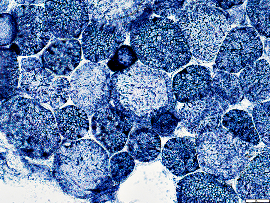
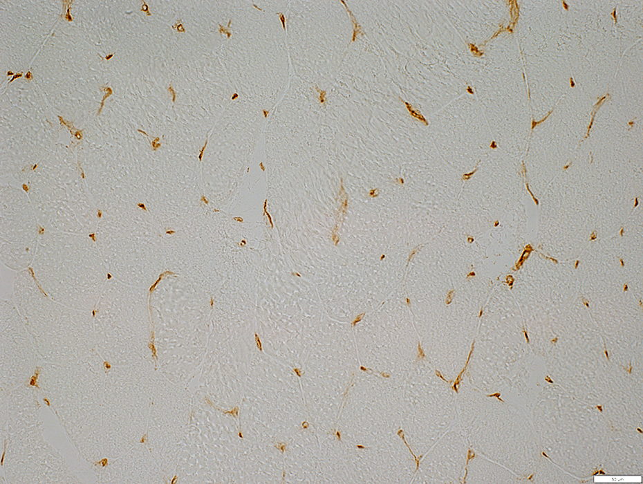
 NADH stain |
NADH positive
 NADH stain |
 NADH stain |
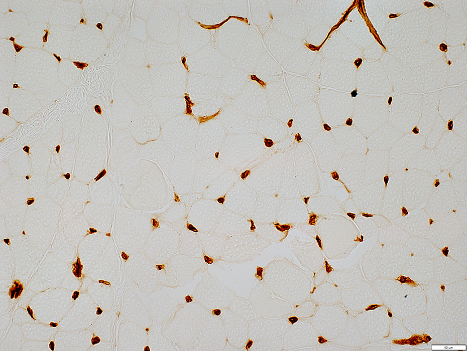
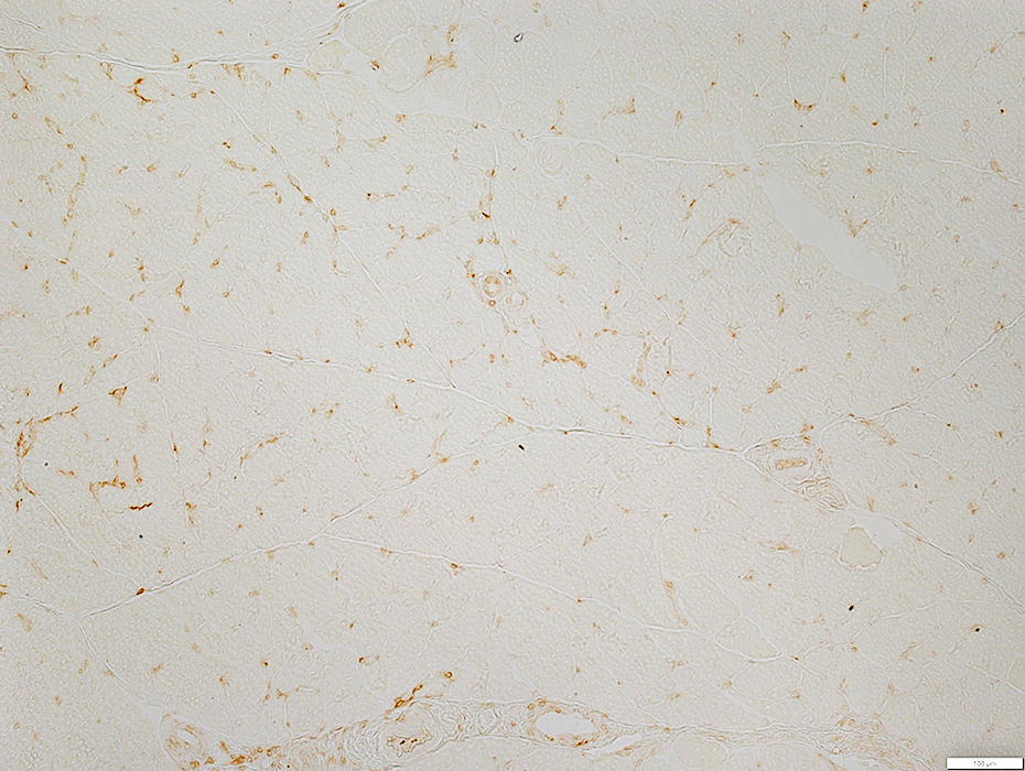
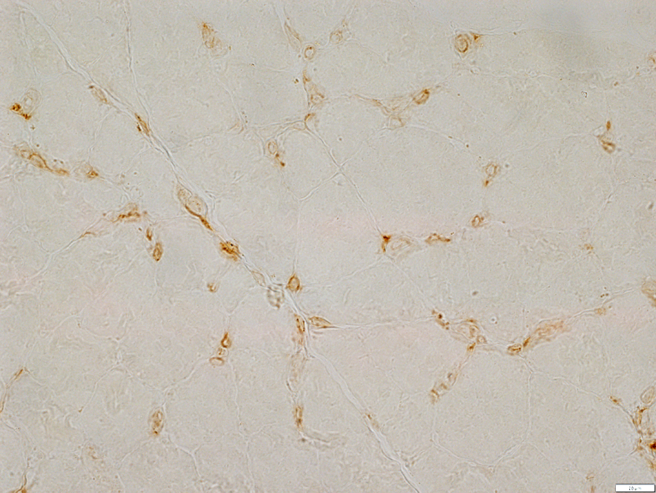
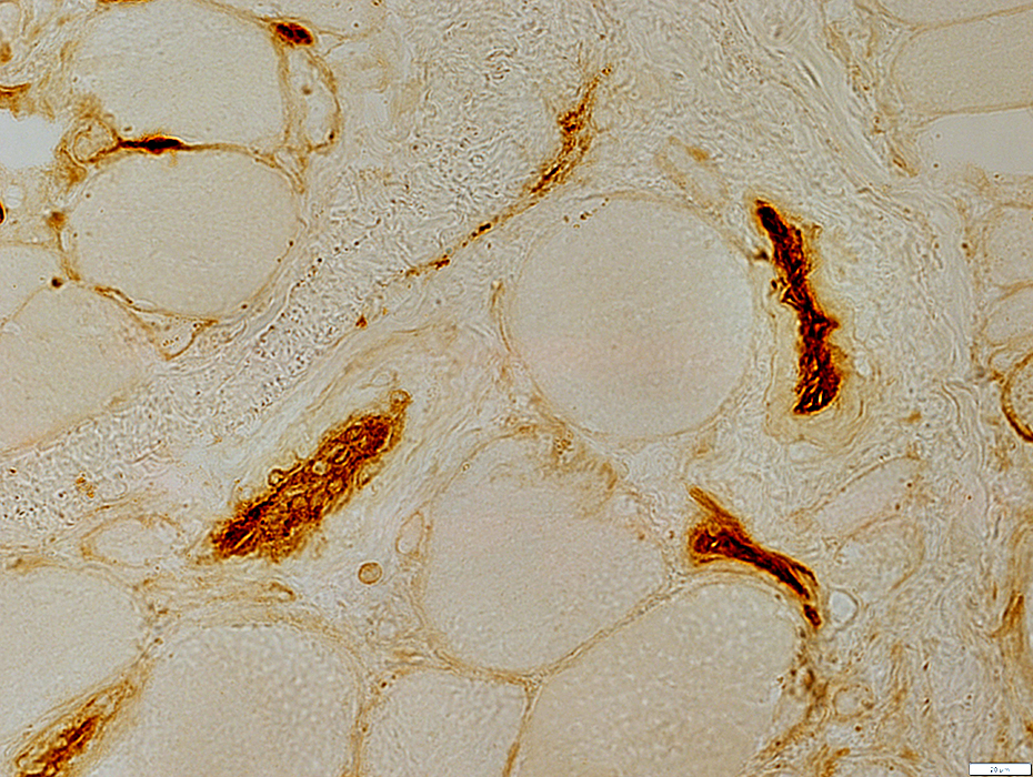
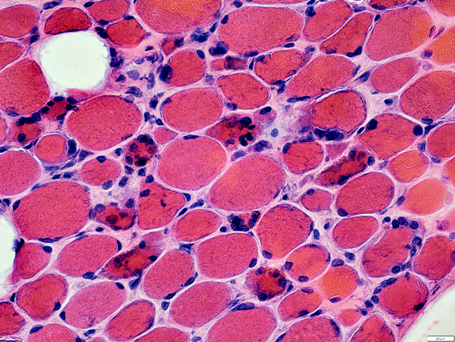
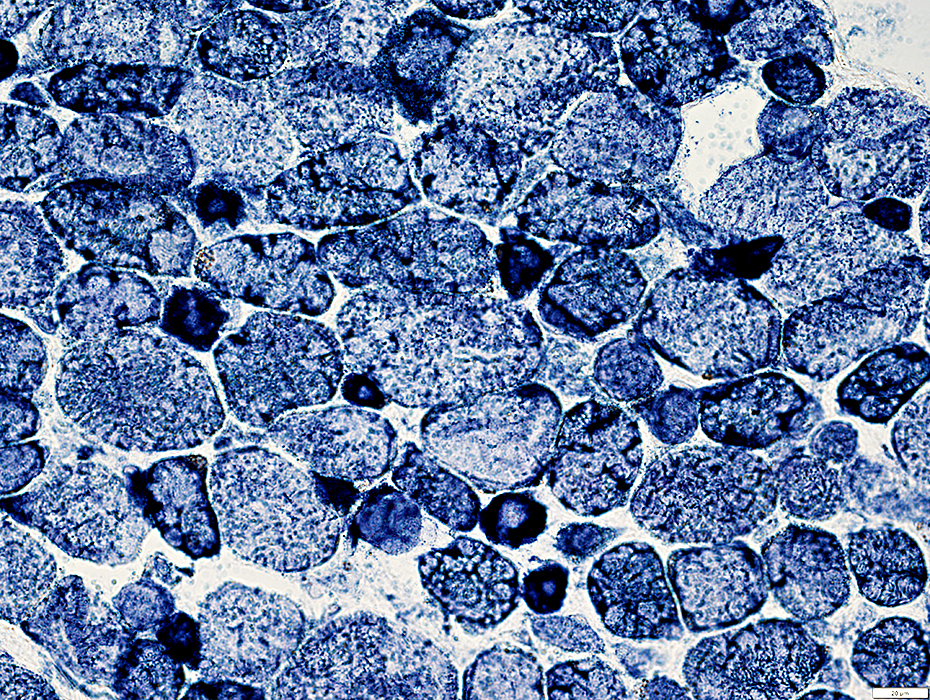
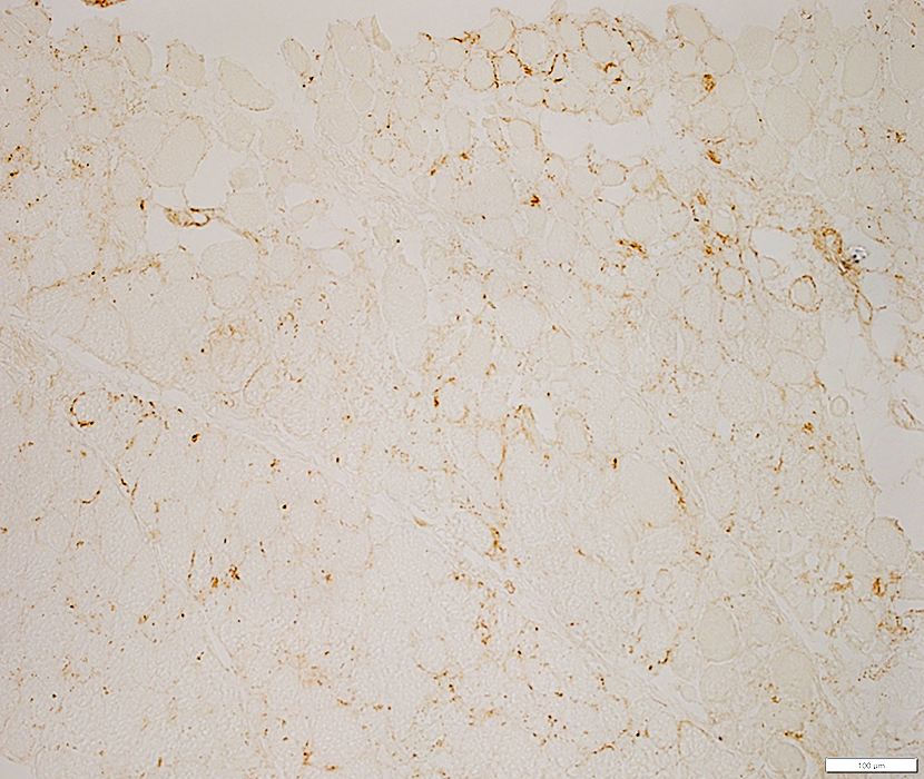
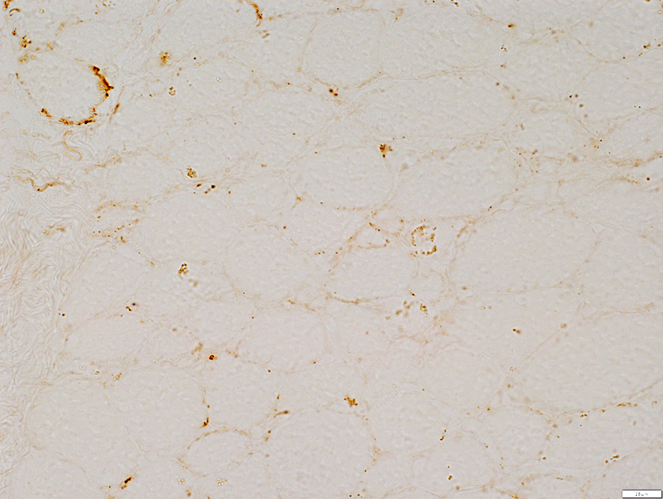
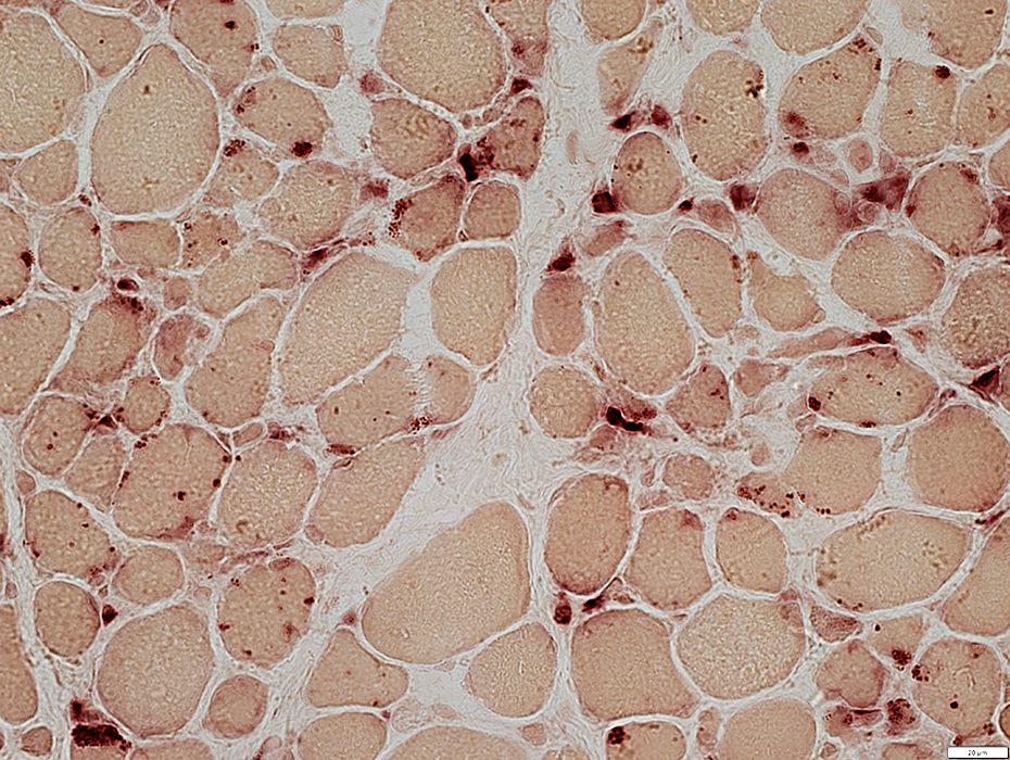
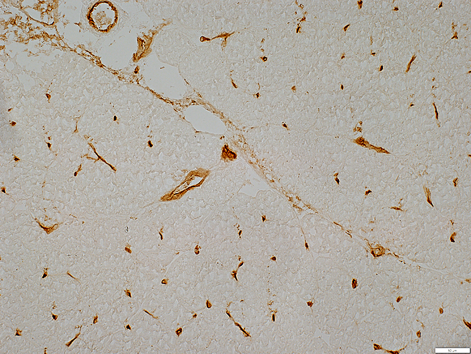
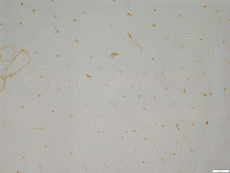
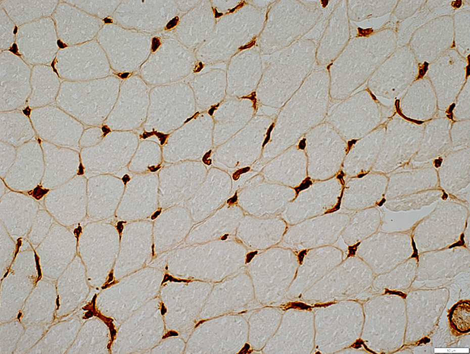
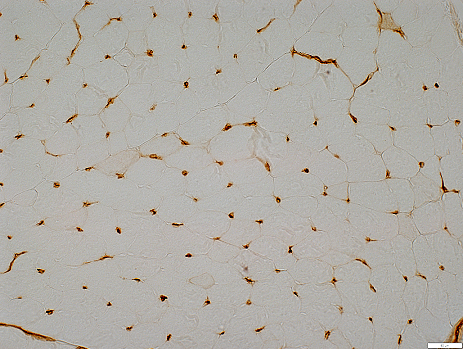
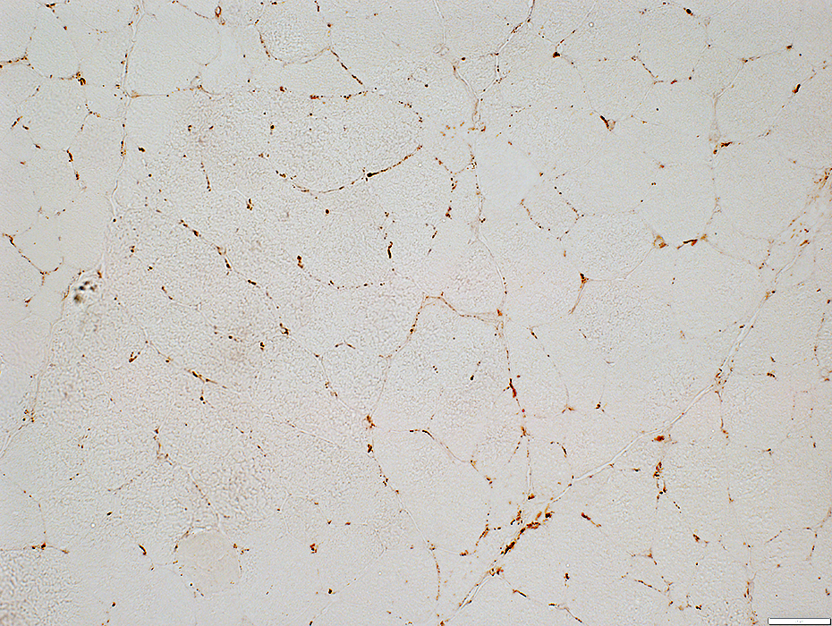
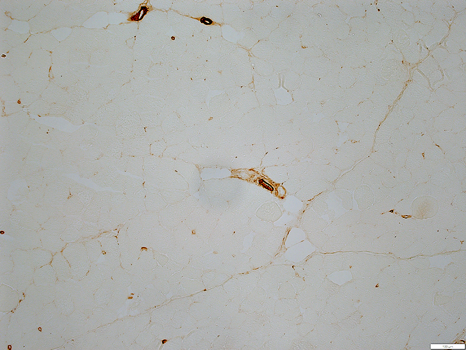
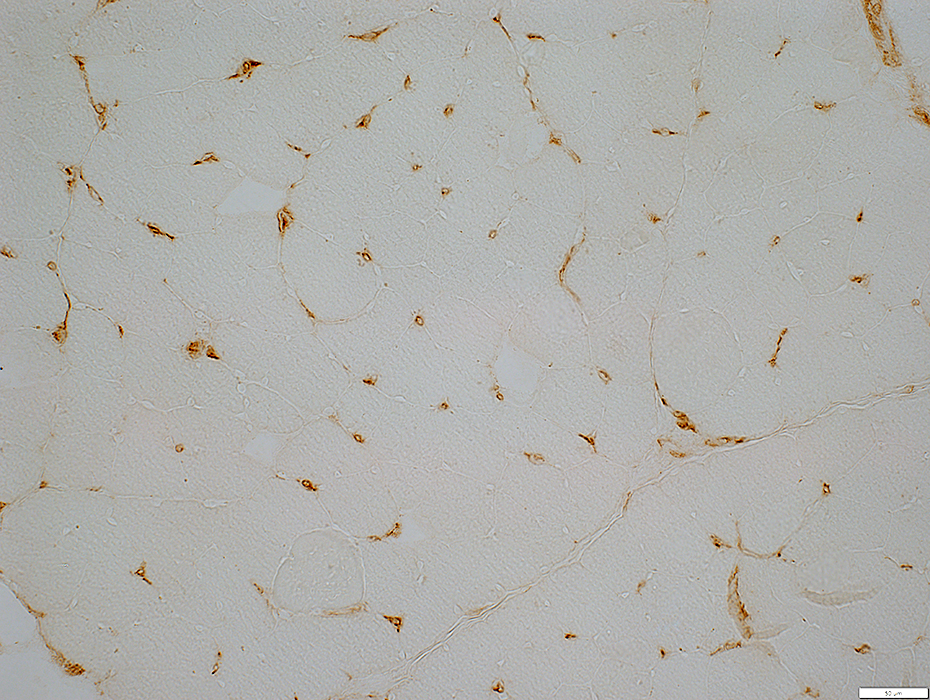
Endomysial Capillaries: Immune
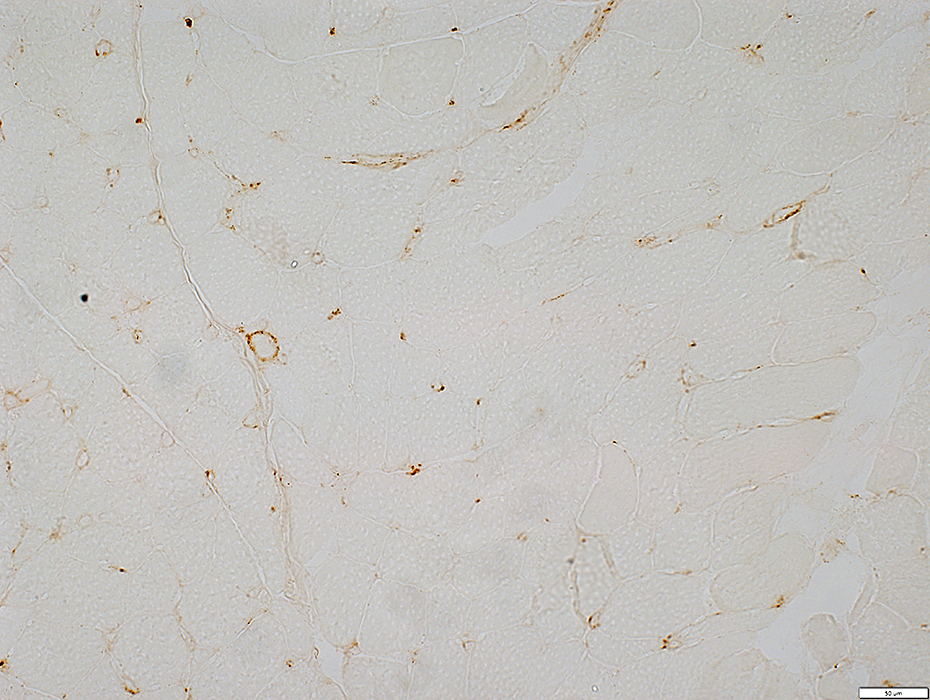 C5b-9 stain |
 MxA stain |
Stains cells in & around endomysial capillary walls
 MxA stain |
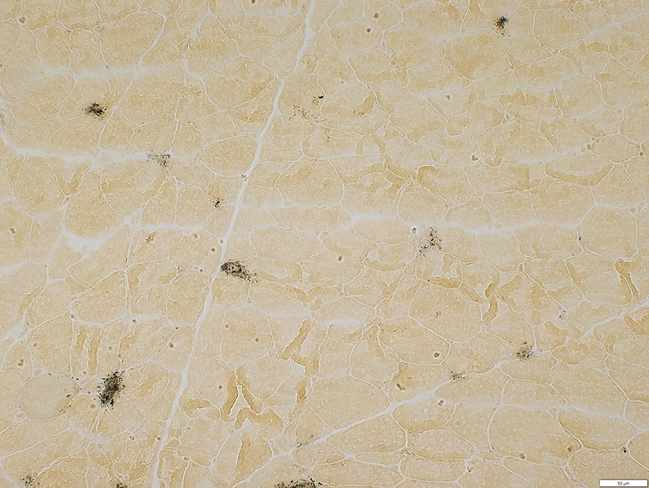
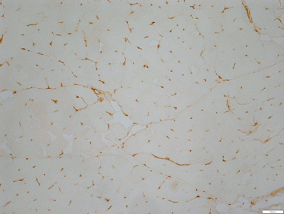
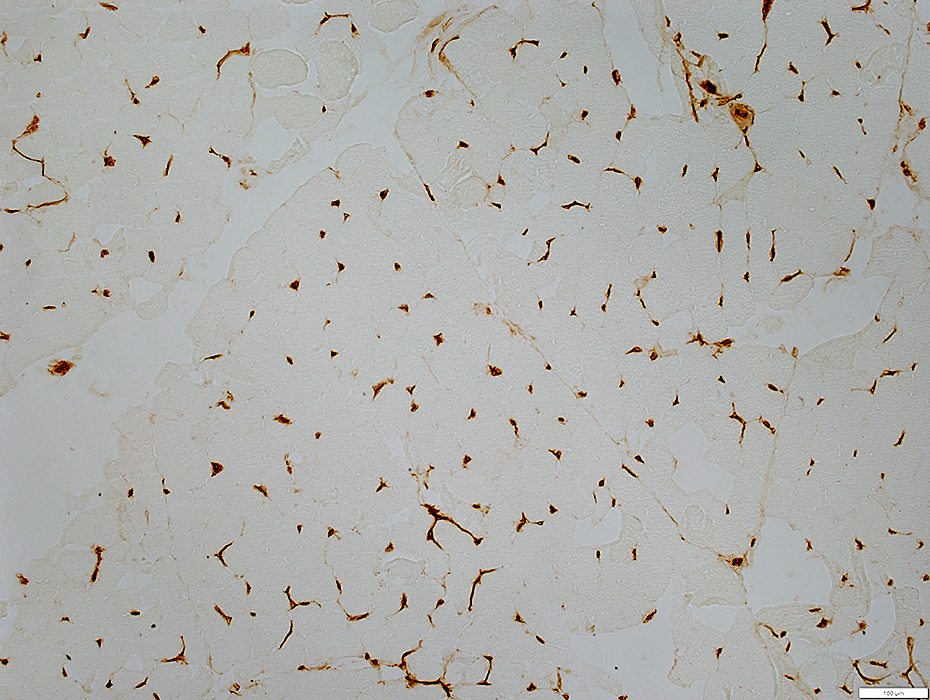
 Acid phosphatase stain |
Acid phosphatase: Stains cells in & around (Arrows) endomysial capillary walls
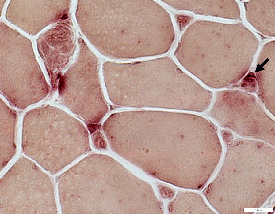 Acid phosphatase stain |
|
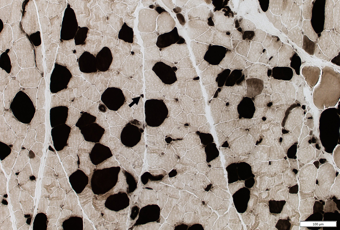
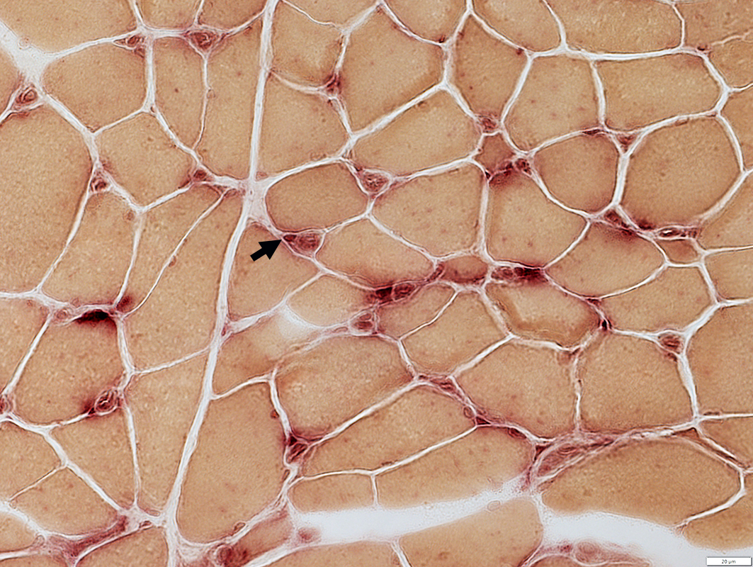
Muscle fiber pathology
MHC I up regulated on muscle fibers
 MHC Class I stain |
Systemic Sclerosis: Capillary Pathology + Myopathy 2
Patient features52 yo female
Clinical diagnosis: Systemic Sclerosis; ILD
Serum Antibodies: ANA 1:1280, Speckled; Ro52; AChR binding
Capillary pathology
Morphology: Basal lamina very thick
Basal lamina: PAS-; Decorin dark & moderately thick
Endothelium: Ulex large & reduced #; Alkaline phosphatase -; ATPase +; NADH ++
Immune: C5b-9 scattered capillaries; Acid phosphatase cells & endothelium
Muscle: MHC1+
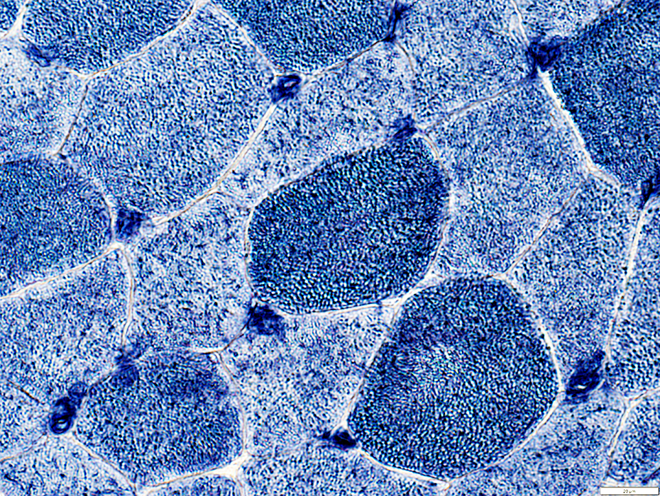
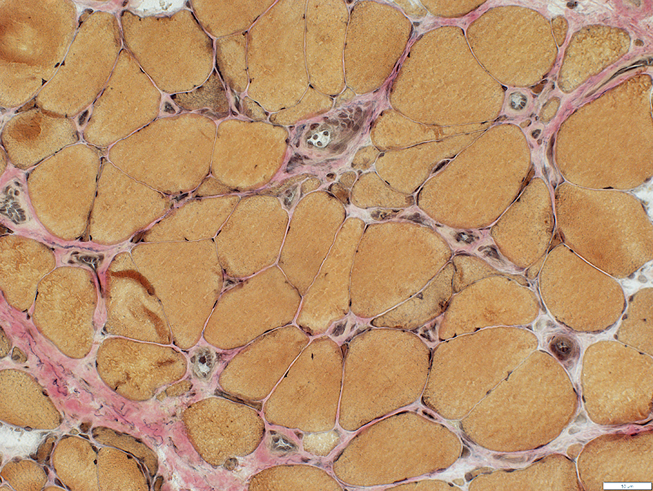
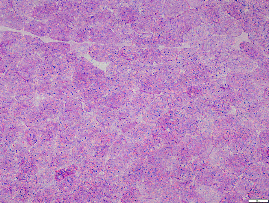
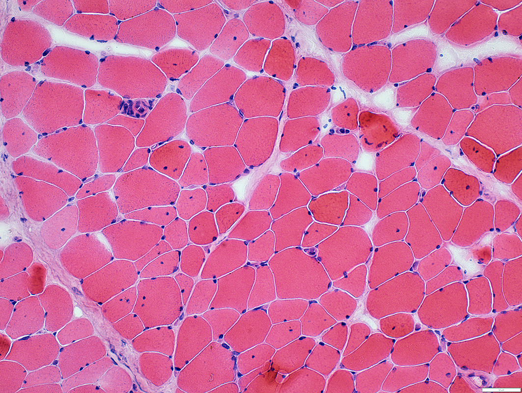
 H&E stain |
Muscle Fiber sizes: Varied
Small, Immature fibers: Scattered
Internal nuclei: Some muscle fibers
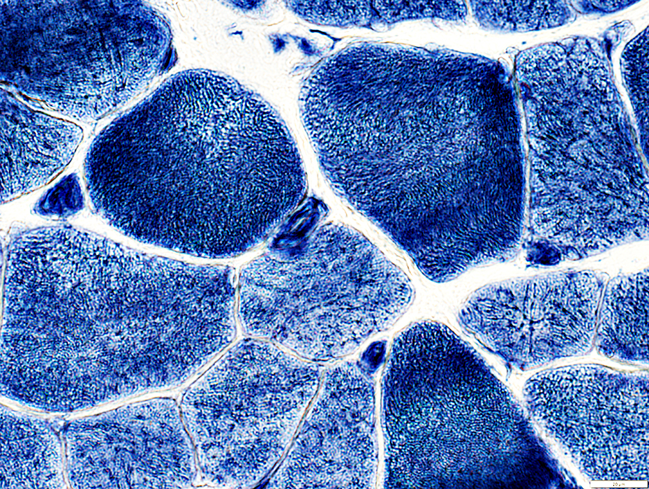
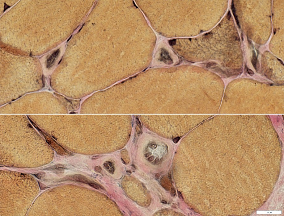
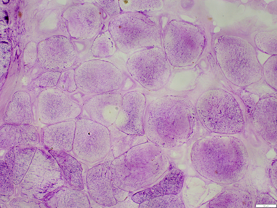
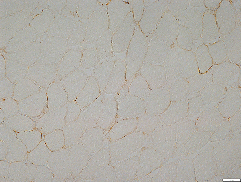
Endomysial Capillary Pathology
Capillary sizes: Large
Capillary basal lamina: Thick
Endothelial cells: Large in enlarged capillaries
 Gomori trichrome stain |
Endomysial capillaries: Large
 VvG stain |
Muscle Fiber sizes: Varied
Small, Immature fibers: Scattered
Endomysial Capillary Pathology
Capillary sizes: Large
Capillary basal lamina: Thick
Endothelial cells: Large in enlarged capillaries
 VvG stain |
 VvG stain |
Capillary sizes: Large
Capillary basal lamina: Thick
Endothelial cells: Large in enlarged capillaries
 Congo red stain |
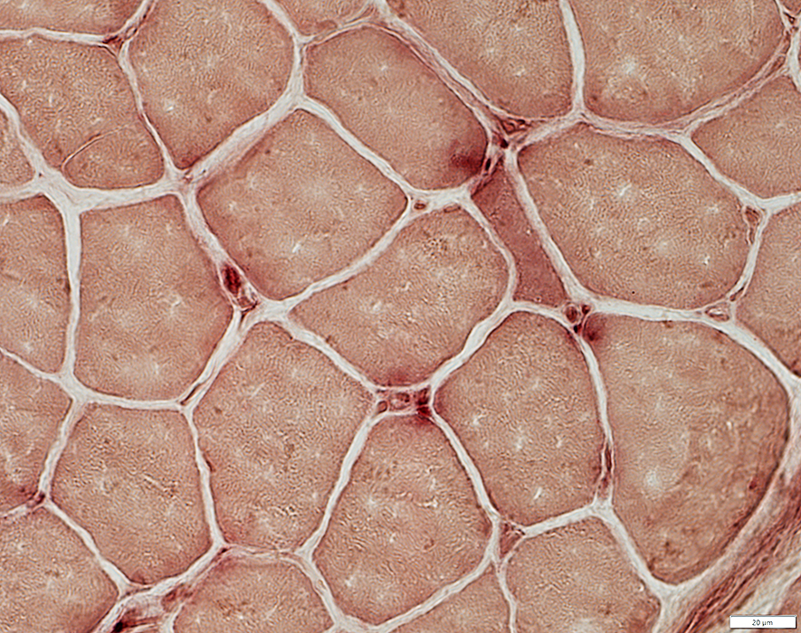
Capillary pathology: Basal lamina
 PAS stain |
 Decorin stain |
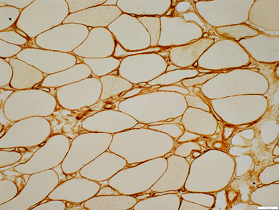
Capillary pathology: Endothelium
 UEA I stain |
Number: Reduced; Many fibers with no adjacent capillary
Endothelial cells
Location: Inside thick basal lamina
Size: Large
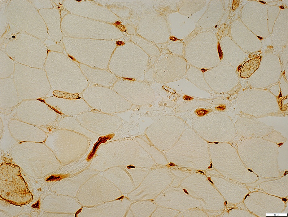
 UEA I stain |
 UEA I stain |
Size: Often large
Endothelial cells
Stain for UEAI & MHC I
Location: Inside thick basal lamina
Size: Large
Basal lamina (Unstained): Thick
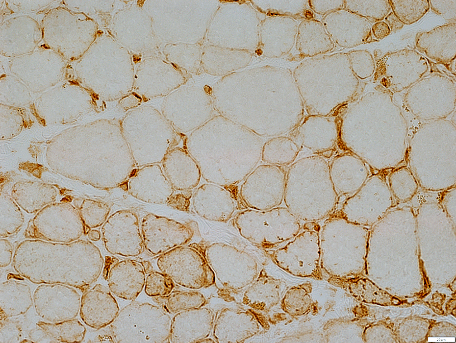
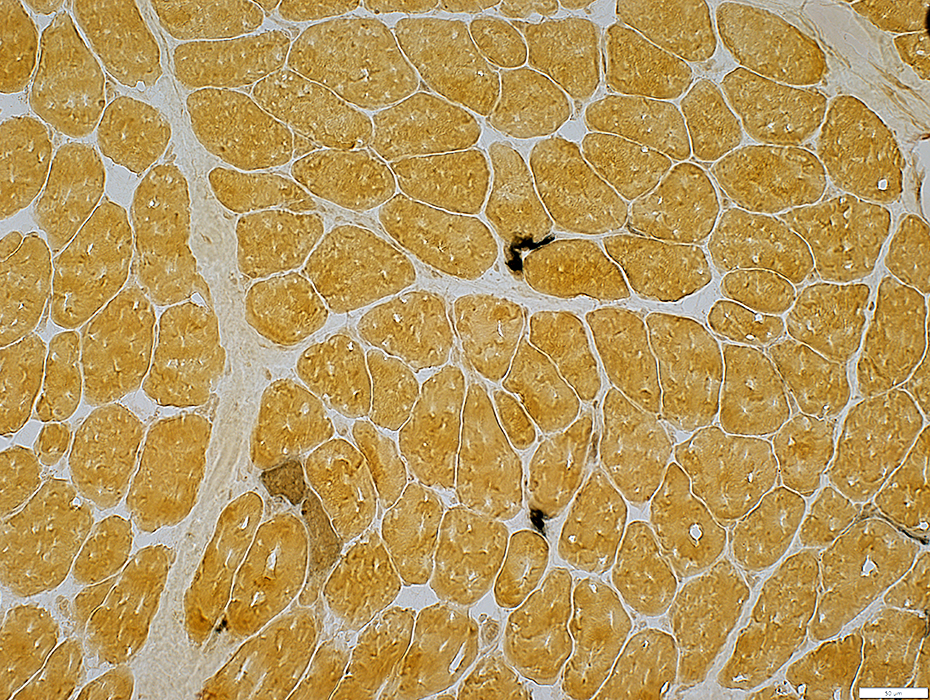
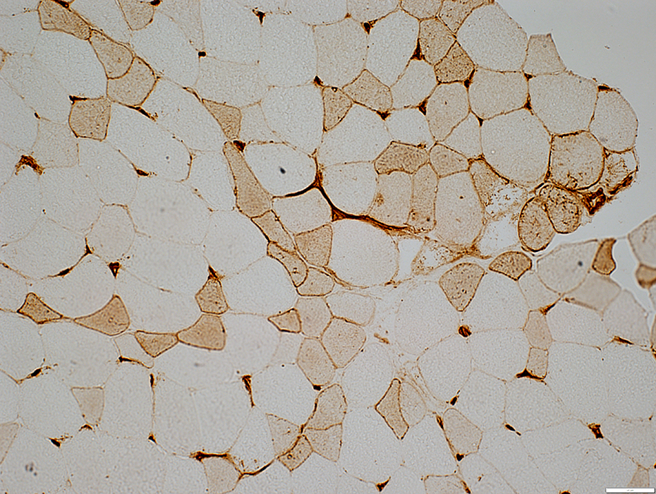
 MHC I stain |
Size: Often large
Endothelial cells
Stain for MHC Class I
Enlarged inside thick basal lamina
Basal lamina (Unstained): Thick
 MHC I stain |
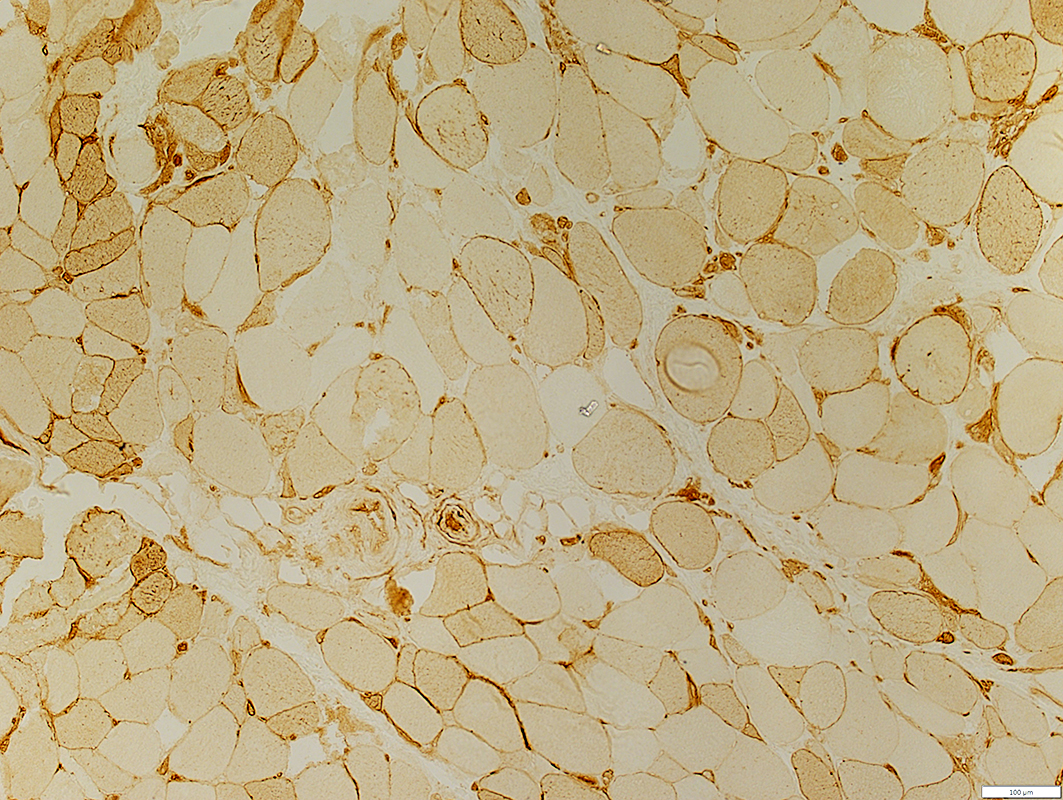
 Alkaline phosphatase stain |
 NADH stain |
Capillary sizes: Large
Capillary basal lamina: Thick
Endothelial cells: Large; NADH positive
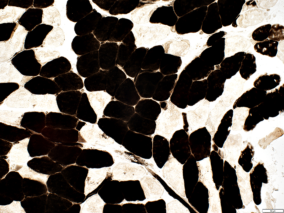
 ATPase pH 4.3 stain |
Endomysial Capillaries: Large (Arrows)
 ATPase pH 4.3 stain |
Endothelial cells: ATPase positive
Capillary basal lamina: Often thick around endothelial cells
 ATPase pH 4.3 stain |
 ATPase pH 4.3 stain |
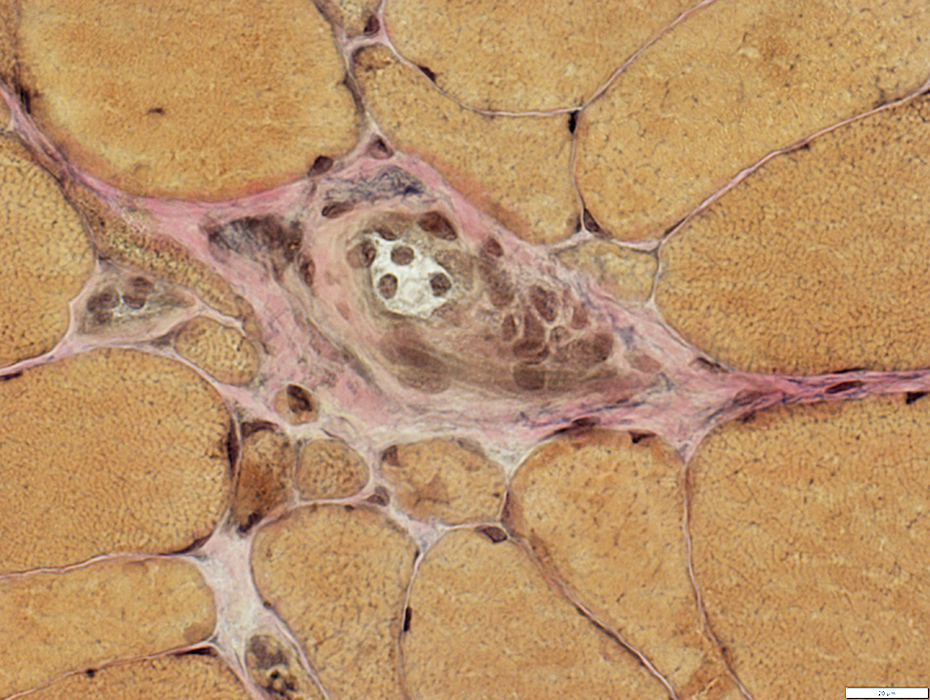
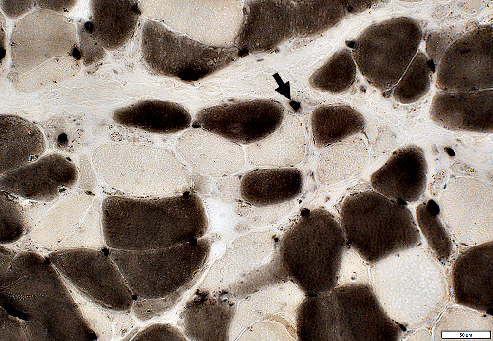
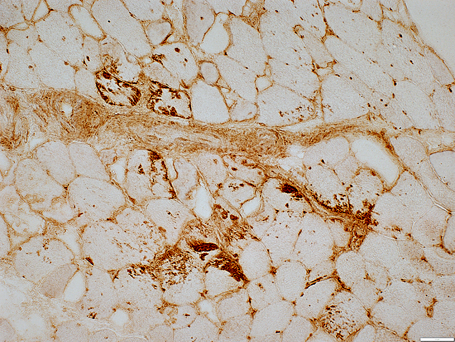
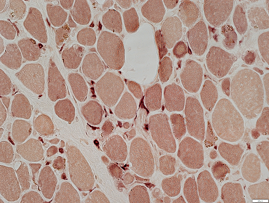
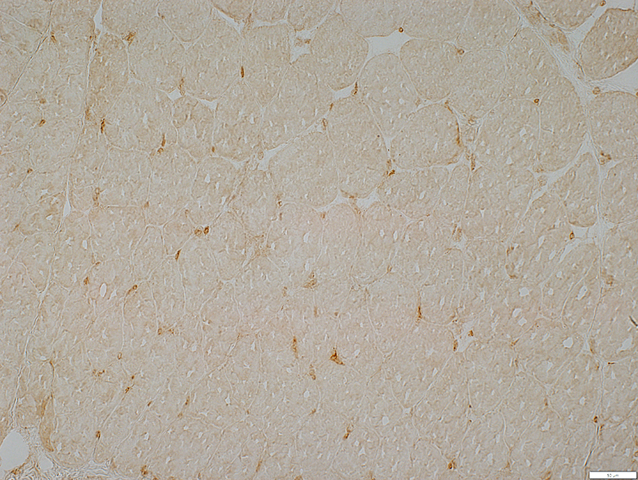
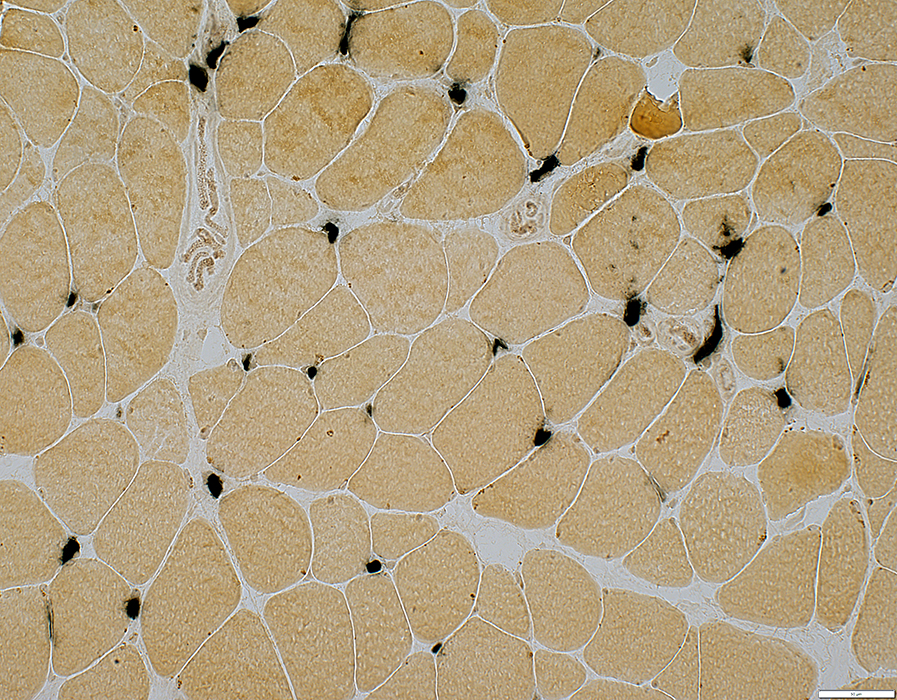
C5b-9 stain: Endomysial Capillaries
 C5b-9 stain |
Endomysial Capillaries
Endothelial cells: Acid phosphatase positive
 Acid phosphatase stain |
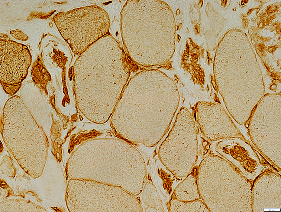
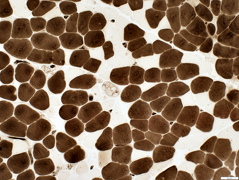
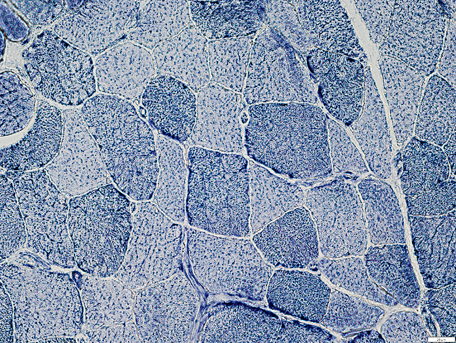
Systemic Sclerosis: Myopathy
 MHC Class I stain |
Muscle fibers
Varied sizes
MHC1: Patchy upregulation
 MHC Class I stain |
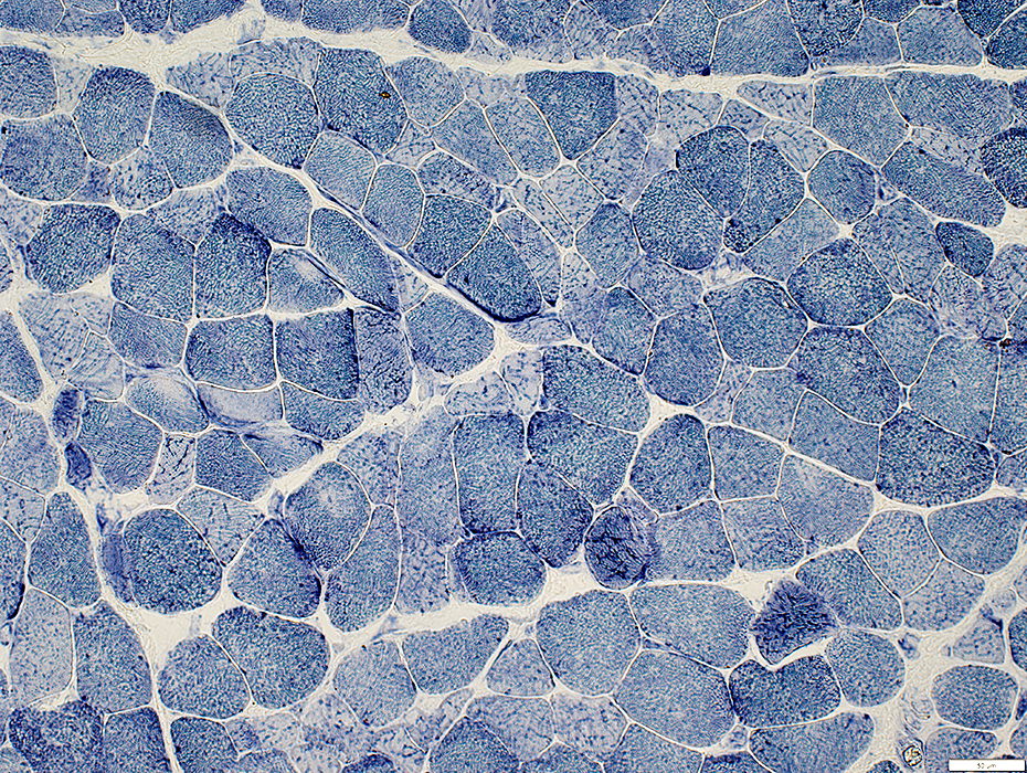
 LC3 stain |
Clusters of muscle fibers have multiple, irregular collectons of LC3 aggregates
 LC3 stain |
Systemic Sclerosis: Myopathy ± Capillary Pathology
Patient featuresClinical diagnosis: Systemic Sclerosis
Serum Antibodies: ANA high; Ro; NT5C1a
Capillary pathology
Morphology: Some capillaries large
Basal lamina: PAS-; Decorin mildly thick walls
Endothelium: Ulex large; Alkaline phosphatase -; ATPase +; NADH -
Immune: C5b-9 scattered capillaries; Acid phosphatase cells & endothelium
Muscle: MHC1+; Endomysial inflammation; Sarcoplasmic pads; Atrophy; LC3 -
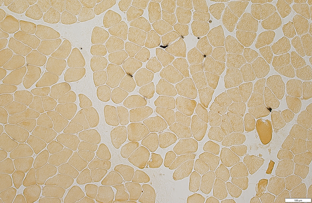
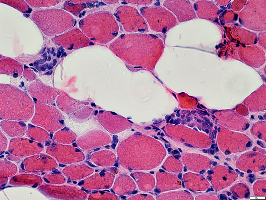
Systemic Sclerosis Myopathy: Morphology
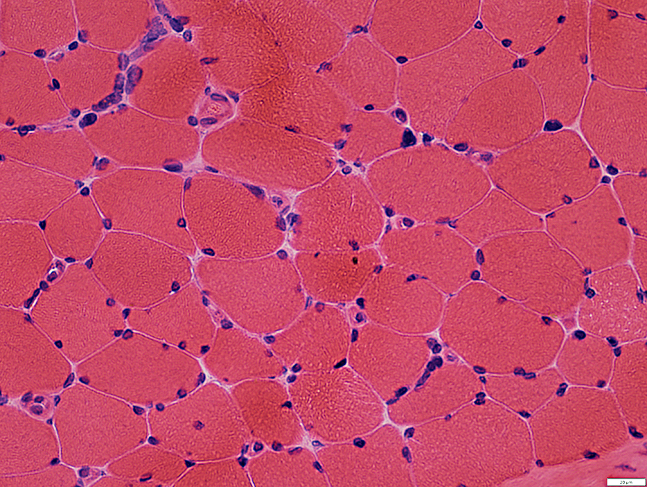
 H&E stain |
Mildly large
Often diffficult to visualize
Muscle fibers
Sizes: Bimodal distribution
All fibers are small
Some fibers: Very small, Round, Large nuclei
 H&E stain |
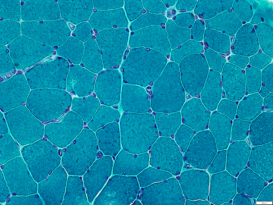
 GT stain |
Mildly large
Often diffficult to visualize
Muscle fibers
Sizes: Bimodal distribution
All fibers are small
Some fibers: Very small, Round, Large nuclei
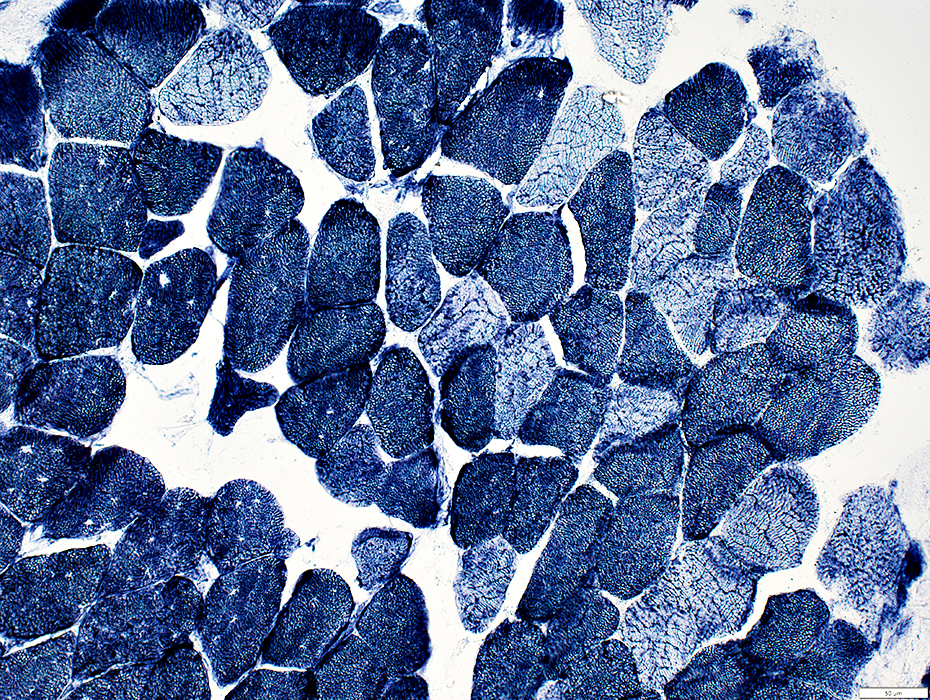
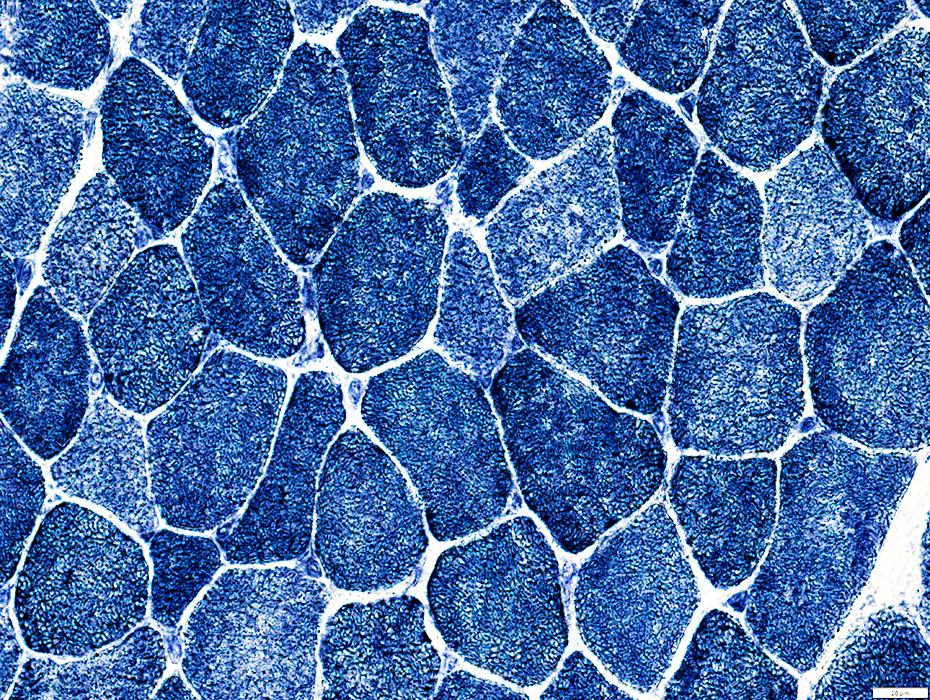
 VvG stain |
 PAS stain |
Decorin: Endomysial capillaries have thick walls
 Decorin stain |
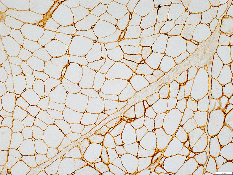
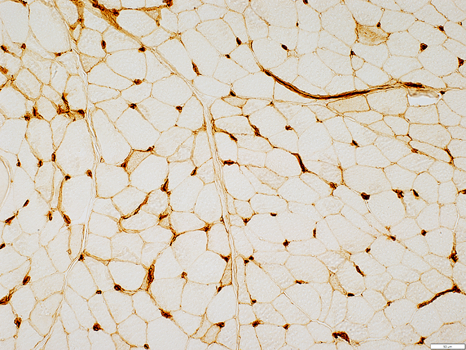
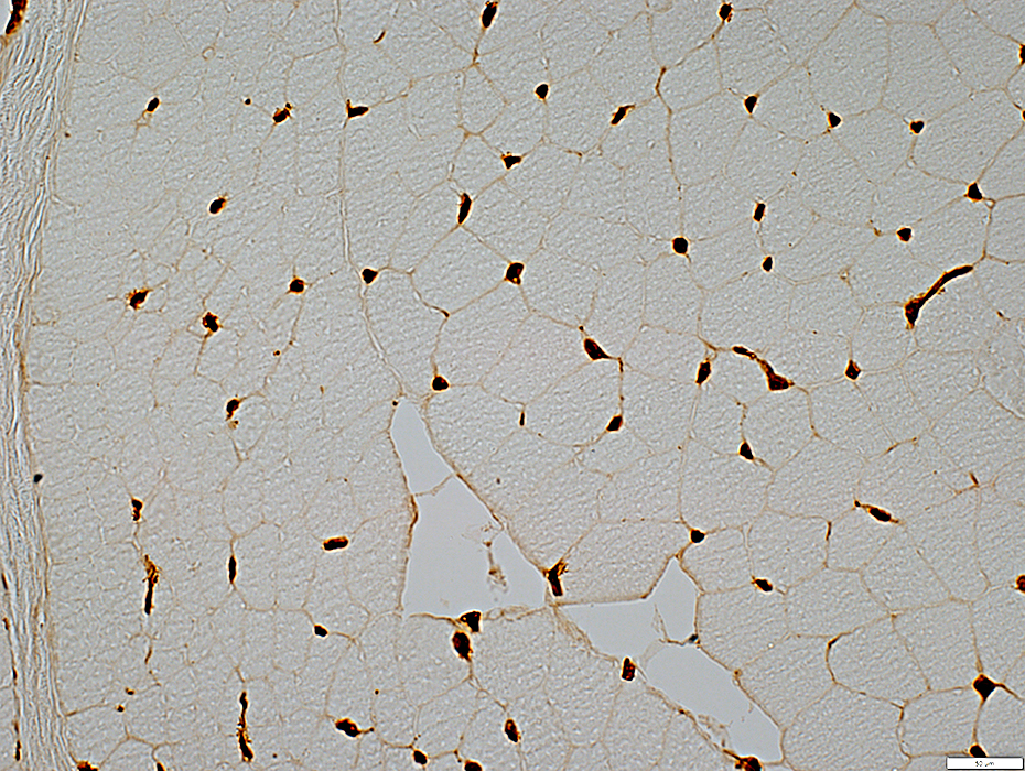
 UEA I stain |
Control
 UEA I stain |
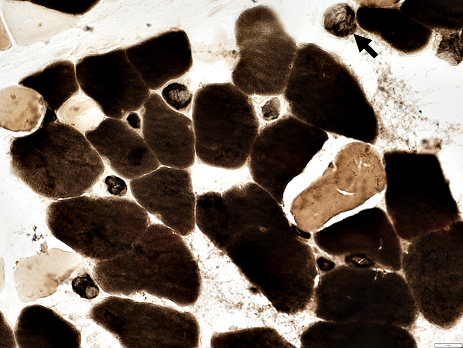
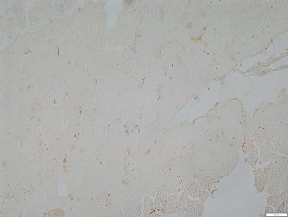
 Alkaline Phosphatase stain |
ATPase pH 4.3: Stains larger endomysial capillaries
 ATPase pH 4.3 stain |
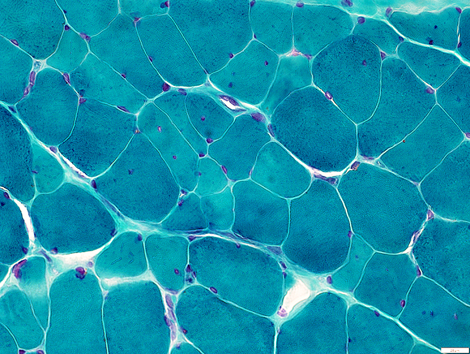
 NADH stain |
Capillaries: No prominent staining
Muscle fibers
Internal architecture: Coarse
Scattered fibers: Rings or Sarcoplasmic pads
 NADH stain |
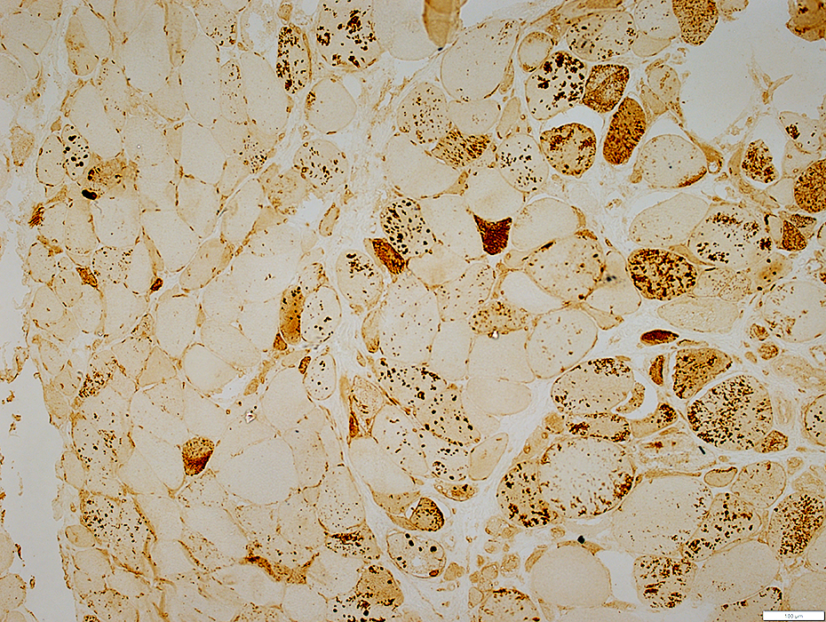
 C5b-9 stain |
 C5b-9 stain |
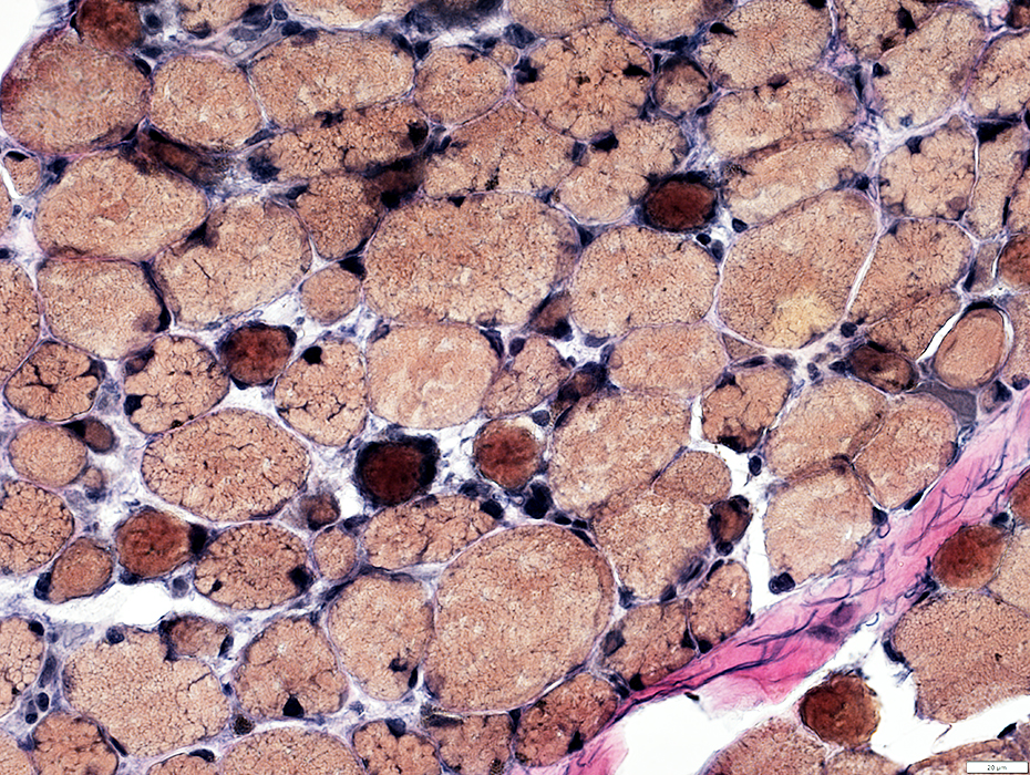
 Acid phosphatase stain |
 Acid phosphatase stain |
 Acid phosphatase stain |
 Esterase stain |
 MxA stain |
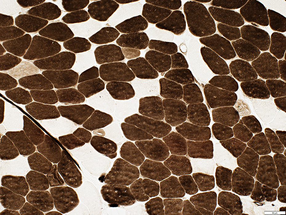
 MHC I stain |
 MHC I stain |
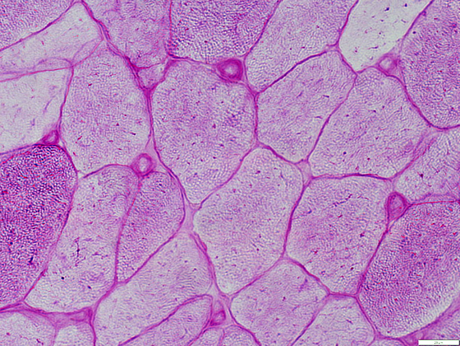
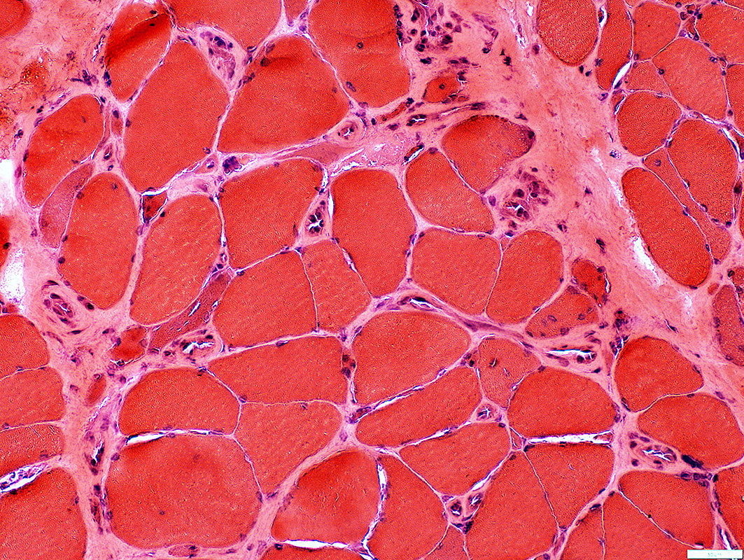
 H&E stain |
 H&E stain |
 H&E stain |
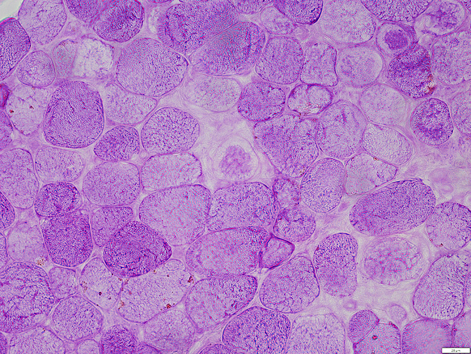
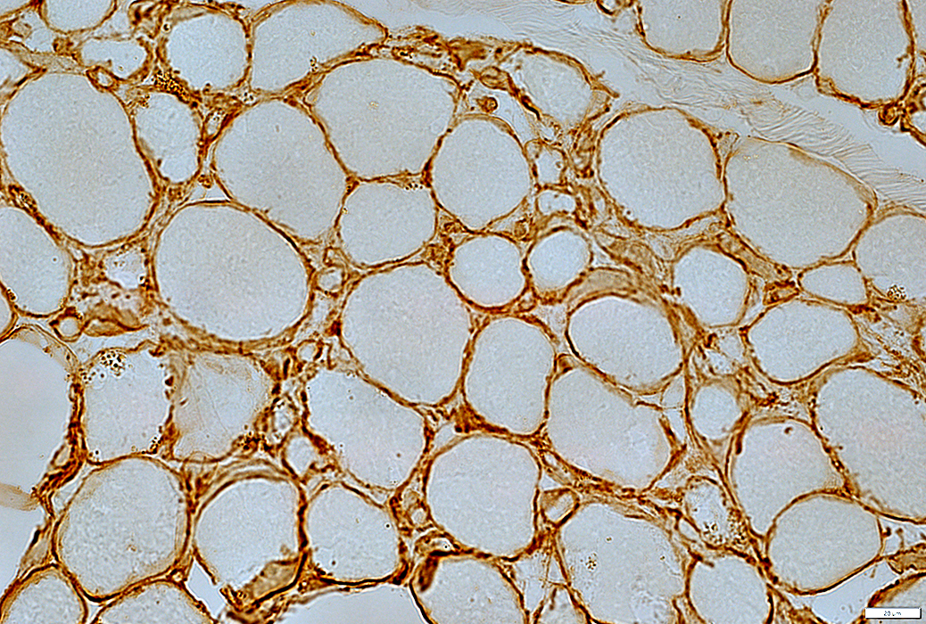
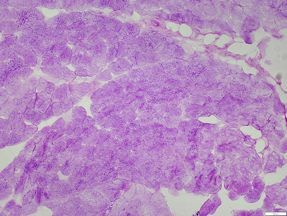
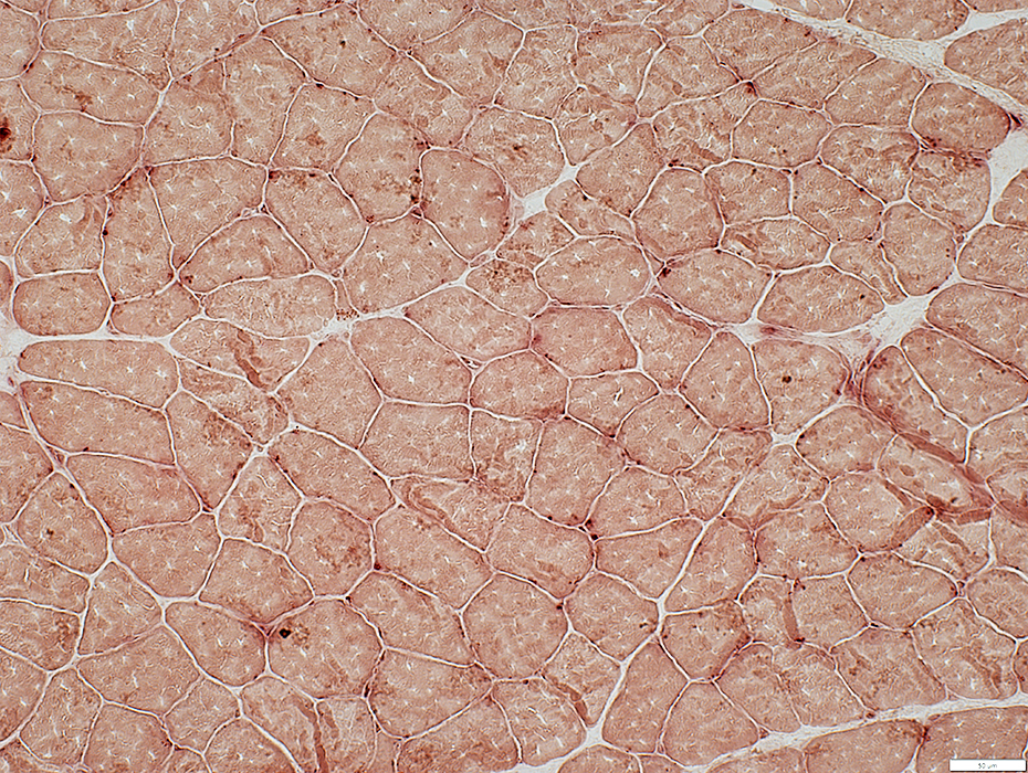
Systemic Sclerosis
Serum Antibody: Th/ToCapillary pathology
Morphology: Capillary sizes normal to slightly large
Basal lamina: PAS-; Decorin small size, dark staining & moderately thick
Endothelium: Ulex & MHC I reduced intensity of staining; Alkaline phosphatase +; ATPase +; NADH -
Immune: C5b-9 -; Acid phosphatase minor staining of few endomysial capillaries; MxA -
Muscle: MHC1-
 H&E stain |
Muscle fibers: Scattered intermediate sized fibers
Capillaries: Normal to Mildly large sizes
 H&E stain |
Systemic sclerosis + Th/To: Basal Lamina
 PAS stain |
 Decorin stain Capillary Basal lamina: Small size endomysial capillaries with mildly thick, dark-stained walls |
Systemic sclerosis + Th/To: Endomysial Capillary Endothelium
 UEA I stain |
 UEA I stain |
 MHC Class I stain |
Muscle: No upregulation of MHC class I
 MHC Class I stain |
 Alkaline phosphatase stain |
 >Alkaline phosphatase stain |
 ATPase pH 4.3 stain |
 NADH stain |
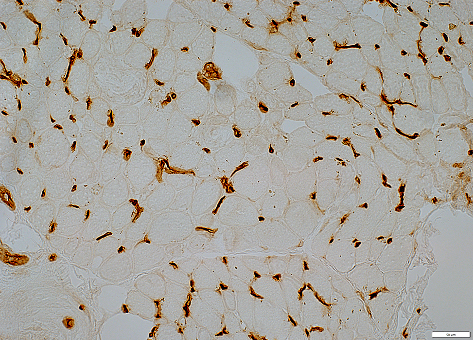
Systemic sclerosis + Th/To: Immune
 Acid phosphatase stain |
 Acid phosphatase stain |
 MxA stain |
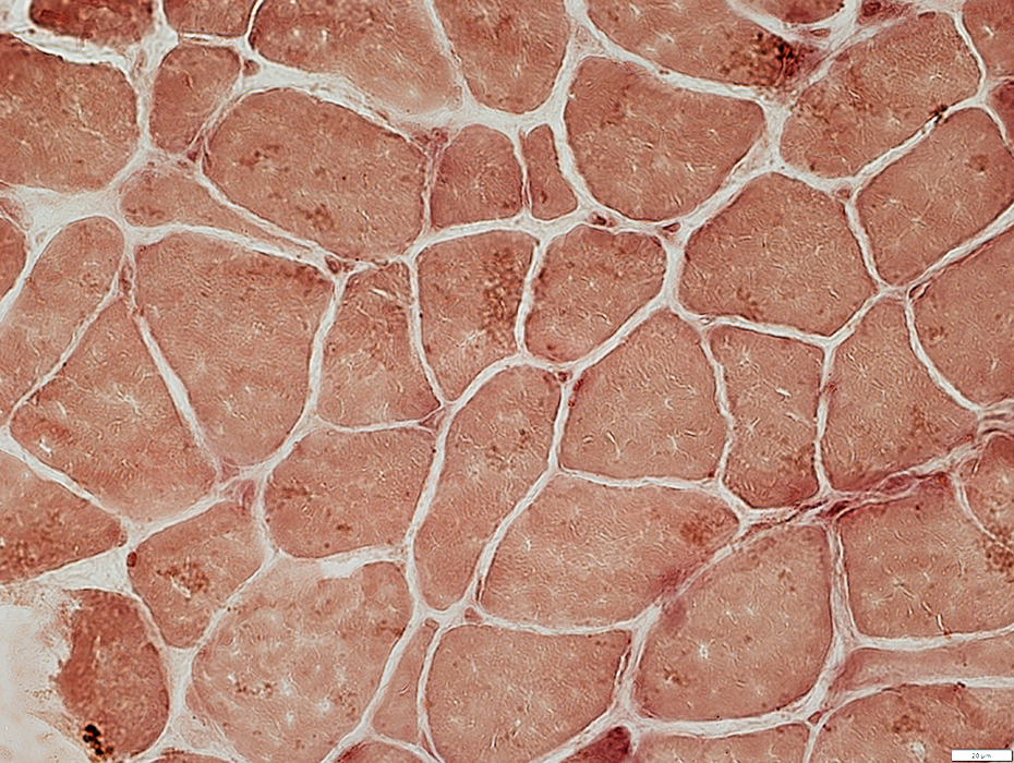
Systemic Sclerosis
Serum Antibody: RUV B1/2Capillary pathology
Morphology: Capillary sizes mildly large
Basal lamina: PAS-; Decorin capillaries moderately thick wall, Normal size to slightly large
Endothelium: Ulex mildly large capillaries; Alkaline phosphatase -; ATPase +- few capillaries; NADH mild +
Immune: C5b-9 no capillary staining; Acid phosphatase +; MxA +-
Muscle: MHC1-; C5b-9 on endomysium; UEA I on muscle fiber surfaces
Systemic Sclerosis + RUV B1/2: Morphology
 H&E stain |
 H&E stain |
 Gomori trichrome stain |
 VvG stain |
Systemic Sclerosis + RUV B1/2: Endomysial Capillary Basal Lamina
 PAS stain |
 Decorin stain |
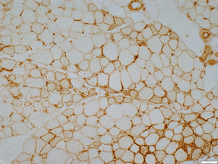
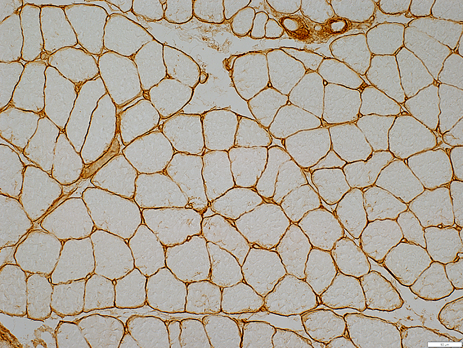
Systemic Sclerosis + RUV B1/2: Capillary endothelium
 UEA I stain |
Moderately large sizes
Borderline reduced #: Scattered small muscle fibers with no associated capillary
Muscle fibers: UEA I stain on fiber surfaces
 UEA I stain |
 MHC class I stain |
Pale (Above) compared to
Control (Below)
Muscle fibers: Mostly Normal
 MHC class I stain |
 Alkaline phosphatase stain |
 ATPase pH 4.3 stain |
 NADH stain |
Systemic Sclerosis + RUV B1/2: Immune features
 C5b-9 stain |
Endomysial capillaries: No staining
Muscle fiber surface membranes: Patchy beaded staining
 Acid phosphatase stain |
Stains capillary endothelial cells & few neighboring histiocytes
 MxA stain |
Stains capillary endothelial cells & few neighboring histiocytes
CREST
Patient 1 features75 yo female
Clinical Diagnosis: CREST
Serum antibodies: Ro-52; Tif1γ; NT5C1a
Capillary pathology
Morphology: Basal lamina normal; Lymphocytes surround small vessels
Basal lamina: PAS+; Decorin small size, dark staining & moderately thick
Endothelium: Ulex large & reduced #; Alkaline phosphatase +; ATPase +; NADH -
Immune: C5b-9 most capillaries; Acid phosphatase cells & endothelium, scattered; MxA +
Muscle: MHC1+
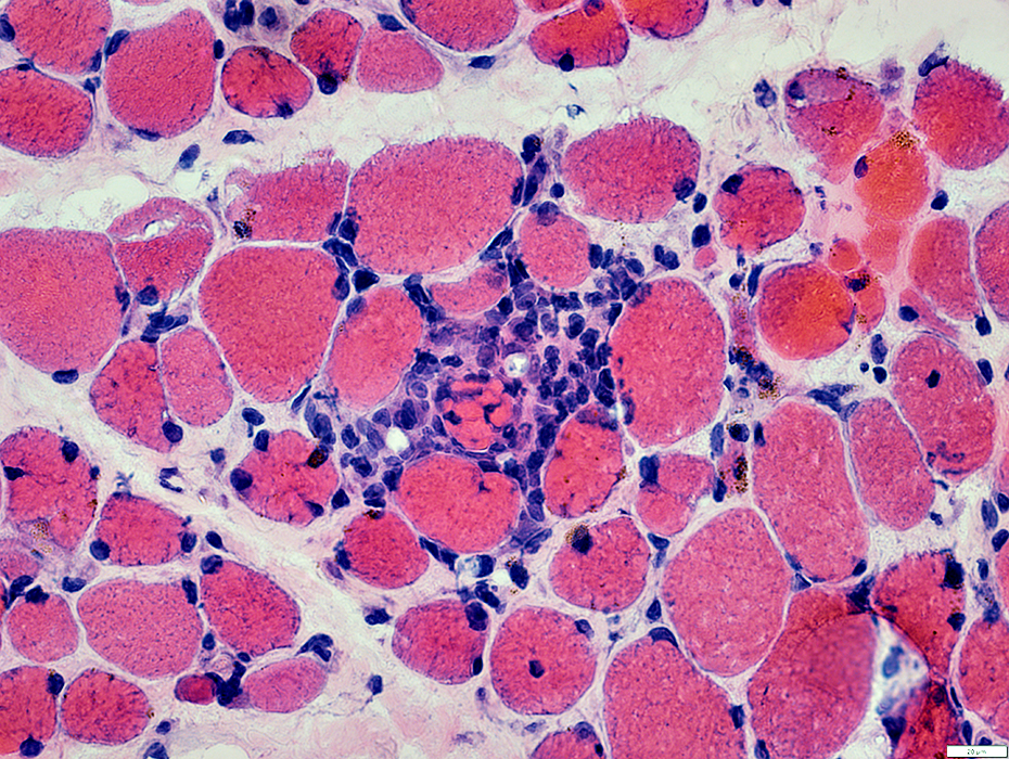
CREST: Endomysial Capillary Morphology
 H&E stain |
Morphology: Normal to Slightly large size
Lymphocytes: Surround a small vessel
 H&E stain |
 Gomori trichrome stain |
CREST: Endomysial Capillary Basal Lamina
 PAS stain |
PAS: Mild staining
 Decorin stain |
Decorin: Dark stained walls
CREST: Endomysial Capillary Endothelium
 UEA I (Ulex) stain |
Reduced numbers: Many muscle fibers have no adjacent capillary
 Alkaline phosphatase stain |
Alkaline phosphatase stain: Increased numbers, especially large capillaries, are positive (Above)
ATPase pH 4.3 stain: Larger capillaries are positive (Below)
 ATPase pH 4.3 stain |
Endomysial Capillaries: No NADH stain
 NADH stain |
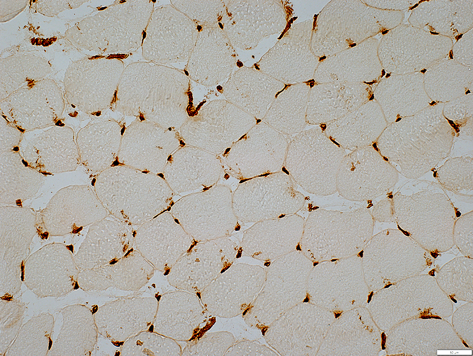
CREST capillaries: Immune
C5b-9 staining
Capillaries: Present on many endomysial capillaries
Muscle fibers: Punctate on some fiber surfaces
 C5b-9 stain |
Endomysial Capillaries
MxA stained cells are present near endomysial capillaries & in perimysium
 MxA stain |
Endomysial Capillaries
Endothelial cells & some surrounding cells: Acid phosphatase positive
 Acid phosphatase stain |
CREST: Muscle fibers
 MHC Class I stain |
Muscle fibers
Varied sizes
MHC1: Patchy upregulation, especially by fibers with
Small size
Location near perimysium
Capillaries: Reduced numbers in some regions
 MHC Class I stain |
CREST
Patient 2 features
30 yo Male
Clinical Diagnosis: CREST
Serum antibodies: Sm; ANA 1:2560
Capillary pathology
Morphology: Basal lamina normal to mildly thick
Basal lamina: PAS +-; Decorin large, dark staining & moderately thick
Endothelium: Ulex large & reduced #; Alkaline phosphatase +; ATPase +; NADH -
Immune: C5b-9 few capillaries; Acid phosphatase cells & endothelium, scattered; MxA ++
Muscle: MHC1 -
 H&E stain |
Morphology
Size: Large
Basal lamina: Mildly thick
 H&E stain |
 Gomori trichrome stain |
Morphology
Size: Large
Basal lamina: Mildly thick
 VvG stain |
CREST: Endomysial Capillary Basal Lamina
 PAS stain |
PAS: Mild staining of larger vessels
 Decorin stain |
Decorin: Dark stained walls; Large size
 Decorin stain |
 UEA1 stain |
Reduced numbers
Many muscle fibers have no adjacent capillary
 UEA1 stain |
Capillaries (Control)
All muscle fibers have adjacent capillary
Endomysial capillary size: Mildly large
 UEA1 stain |
 Alkaline phosphatase stain |
Alkaline phosphatase stain: Increased numbers, especially large capillaries, are positive (Above)
ATPase pH 4.3 stain: Larger capillaries are positive (Below)
 ATPase pH 4.3 stain |
Endomysial Capillaries: NADH stains endothelium in larger capillaries
 NADH stain |
 C5b-9 stain |
C5b-9 staining
Capillaries: Present on a few, scattered endomysial capillaries
 MxA stain |
MxA stains endothelial cells & cells around endomysial capillaries
 MxA stain |
 Acid phosphatase stain |
Endothelial cells & some surrounding cells: Acid phosphatase positive
 Acid phosphatase stain |
CREST: Muscle fibers
 MHC Class I stain |
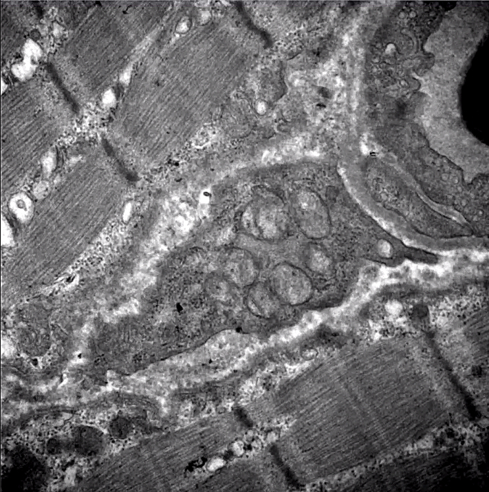
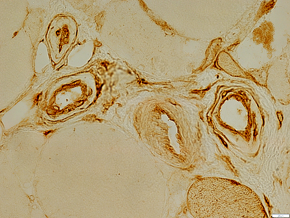
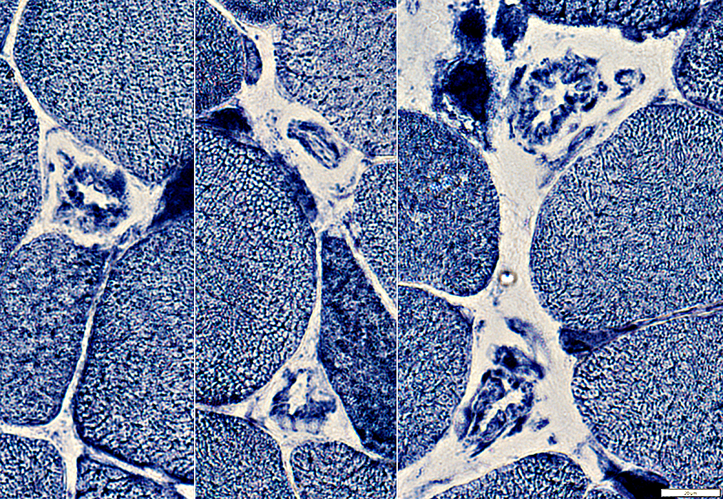
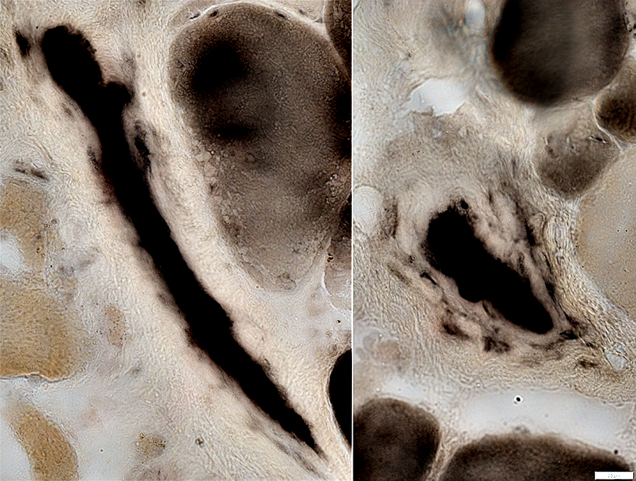
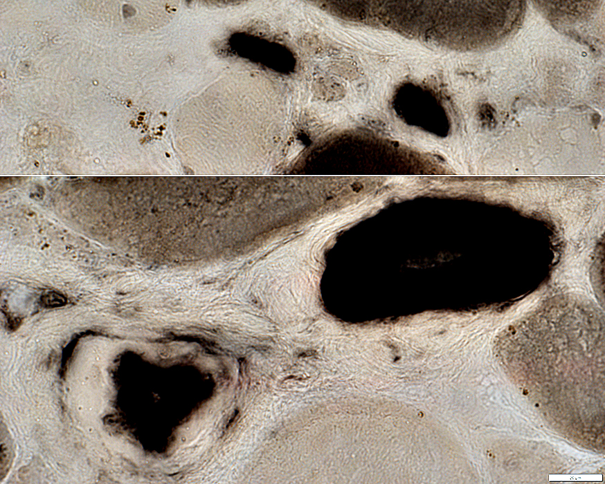
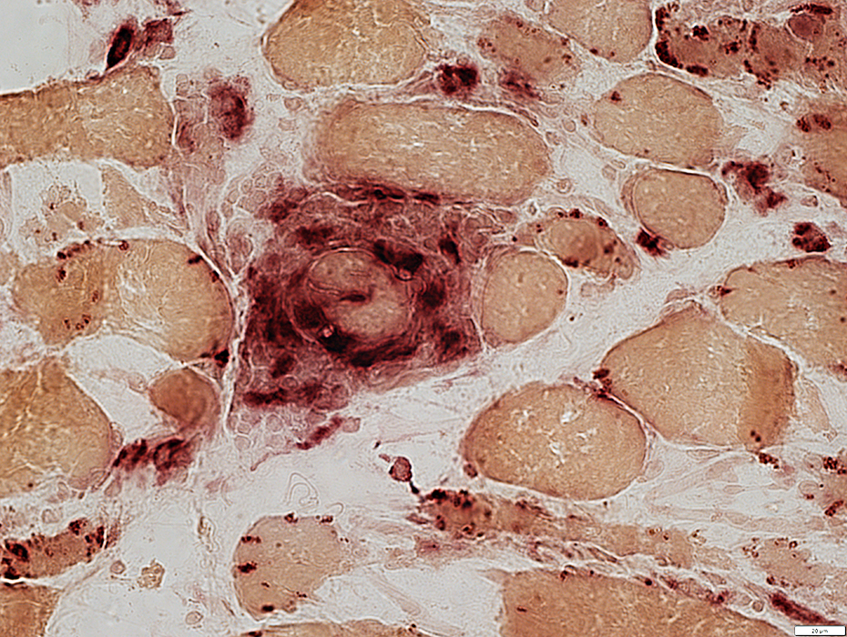
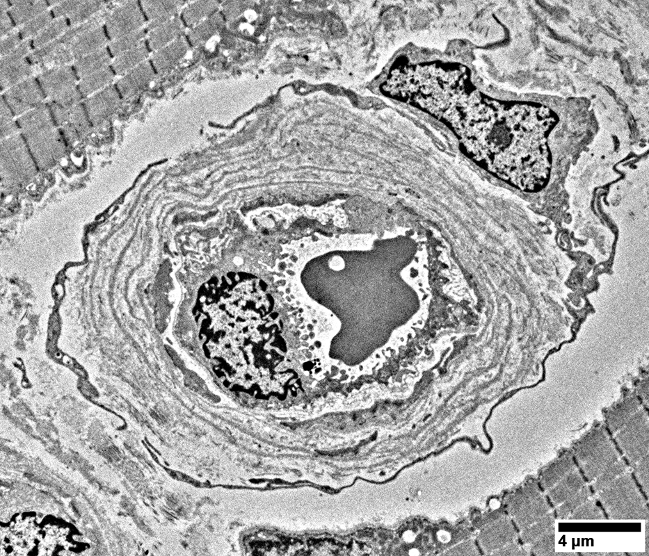
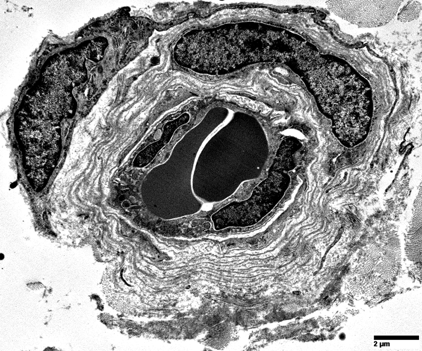
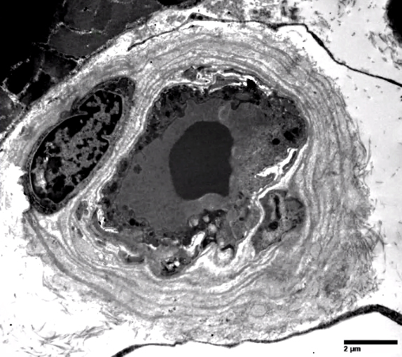
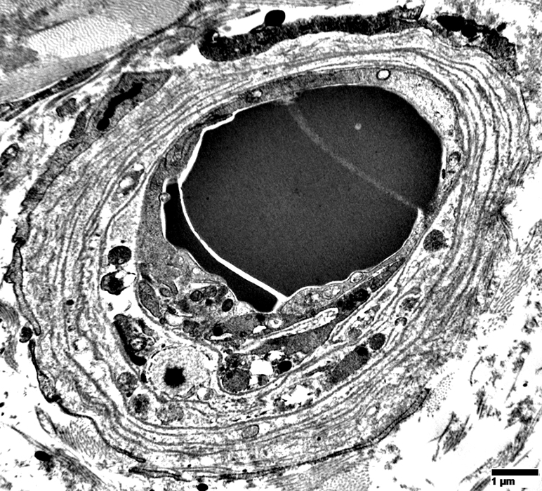
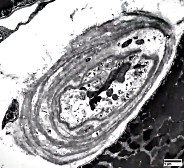
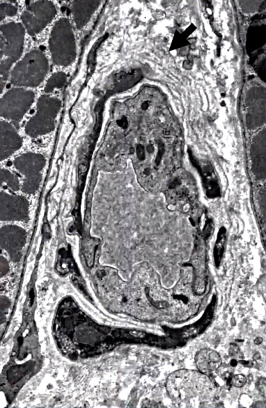
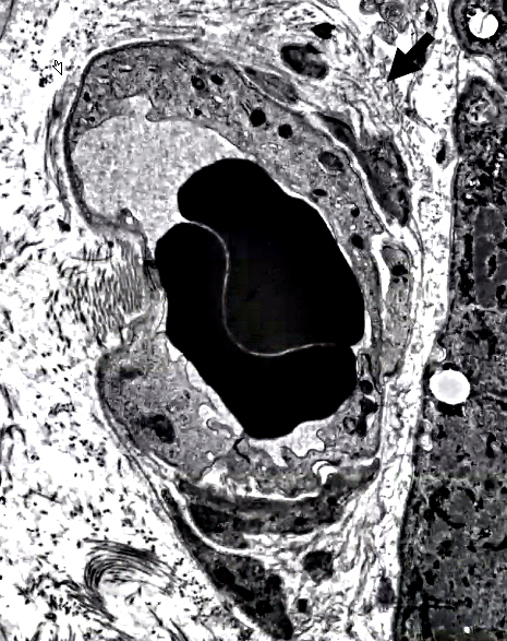
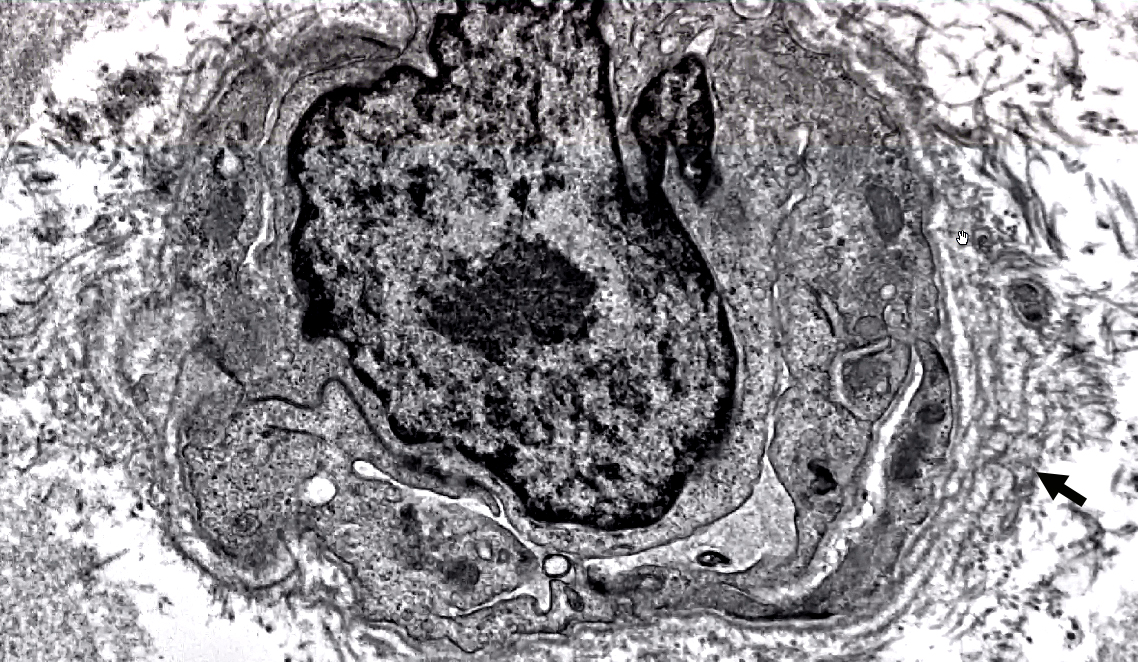
Endomysial Capillary Walls: Multiple circumferential layers of Basal Lamina
|
|
|
|
|
|
|
Irregular increase in basal lamina layers around capillaries (Arrows)
|
Return to Neuromuscular Home Page
Return to Pathology Index
Return to Capillary pathology
References
1. Acta Neuropathol 2021;141:917-927
2. Rheumatology (Oxford) 2023;62(SI):SI82-SI90, Curr Opin Rheumatol. 2023 Aug 23
7/14/2026