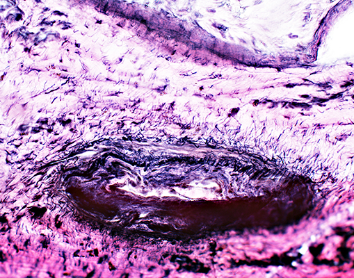
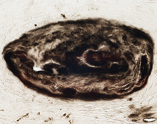
Histiocytic Vasculopathy: Rheumatoid Disorder/IgMκ M-protein/Cryoglobulinemia
Histiocytic Arteriopathy
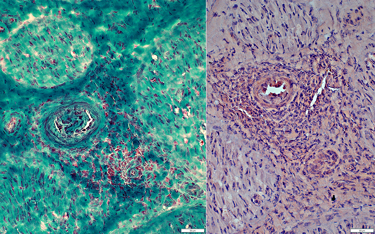
82 year old female with rapidly progressive mononeuritis multiplex & Multi-organ failureRheumatoid factor = 616; ANCA-; NfL 777; C4<4; IgMκ M-protein; Cryoglobulins, borderline
|
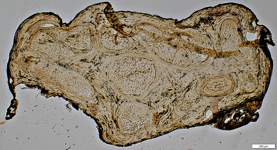
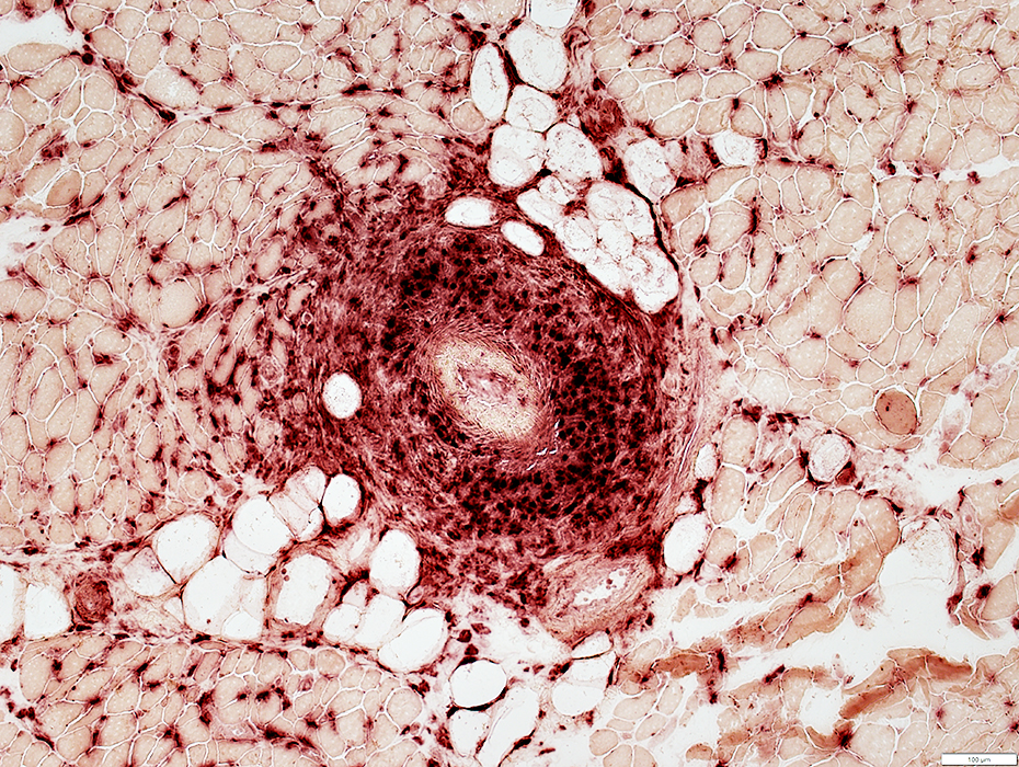
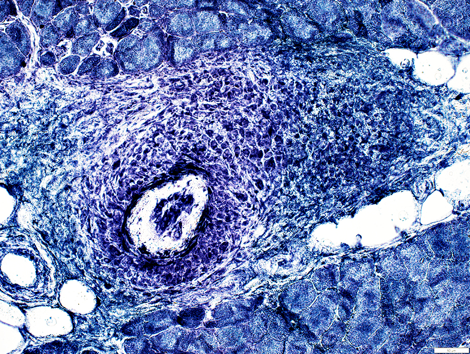
Vessel Pathology Epineurium Artery Occlusion & Damage Also see: pANCA Perimysium: Histiocytic Vasculitis Focal Infarction: Nerve Axon loss: Severe; Regional |
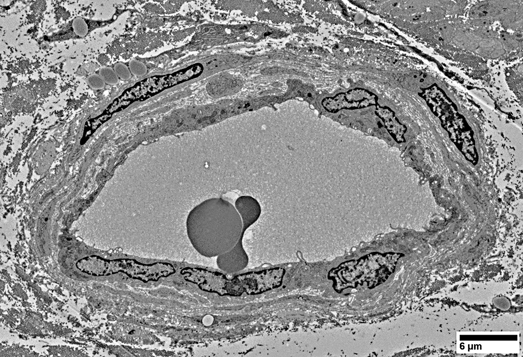
Nerve: Occluded Artery; Focal infarction; Histiocytic/Few lymphocytes
Focal nerve infarction: Also see Behçet
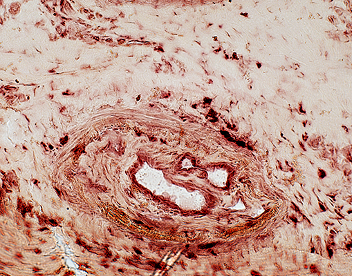
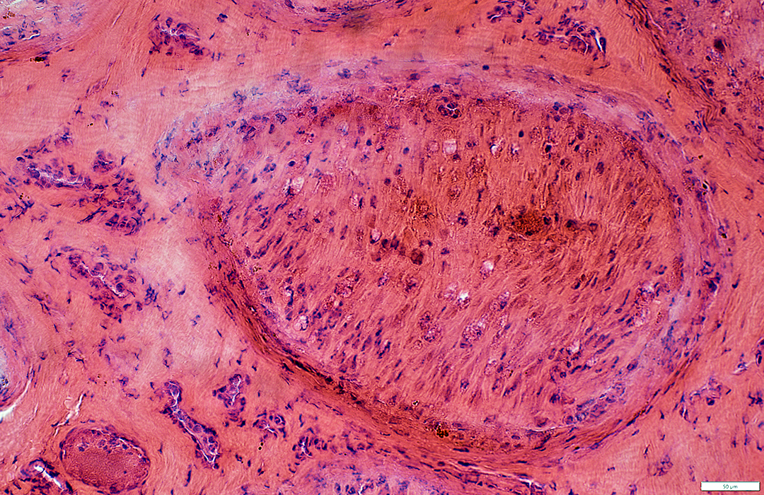
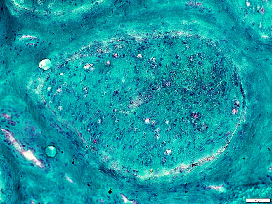
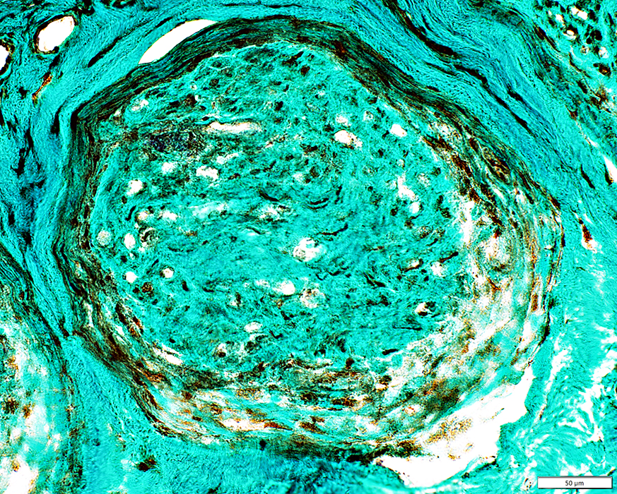
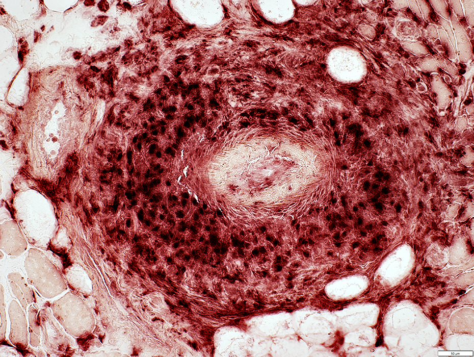
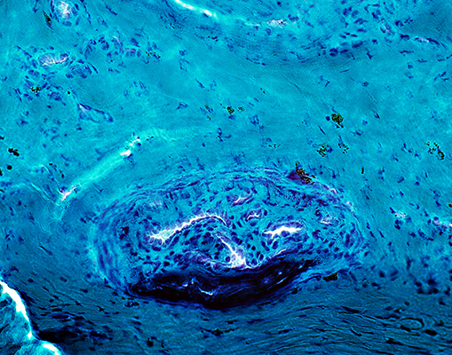 Gomori trichrome stain |
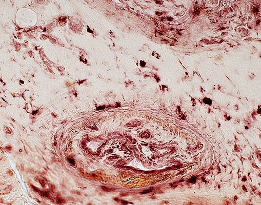
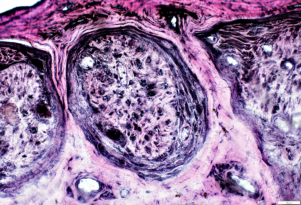
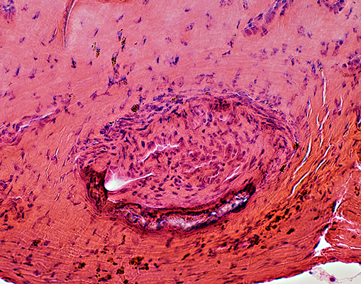 H&E stain |
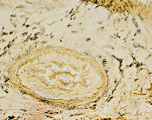
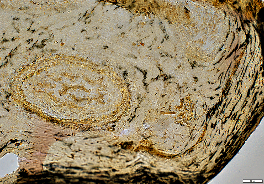
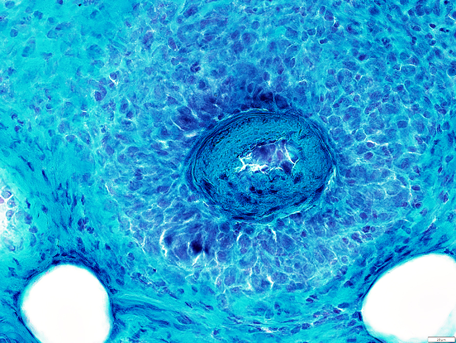
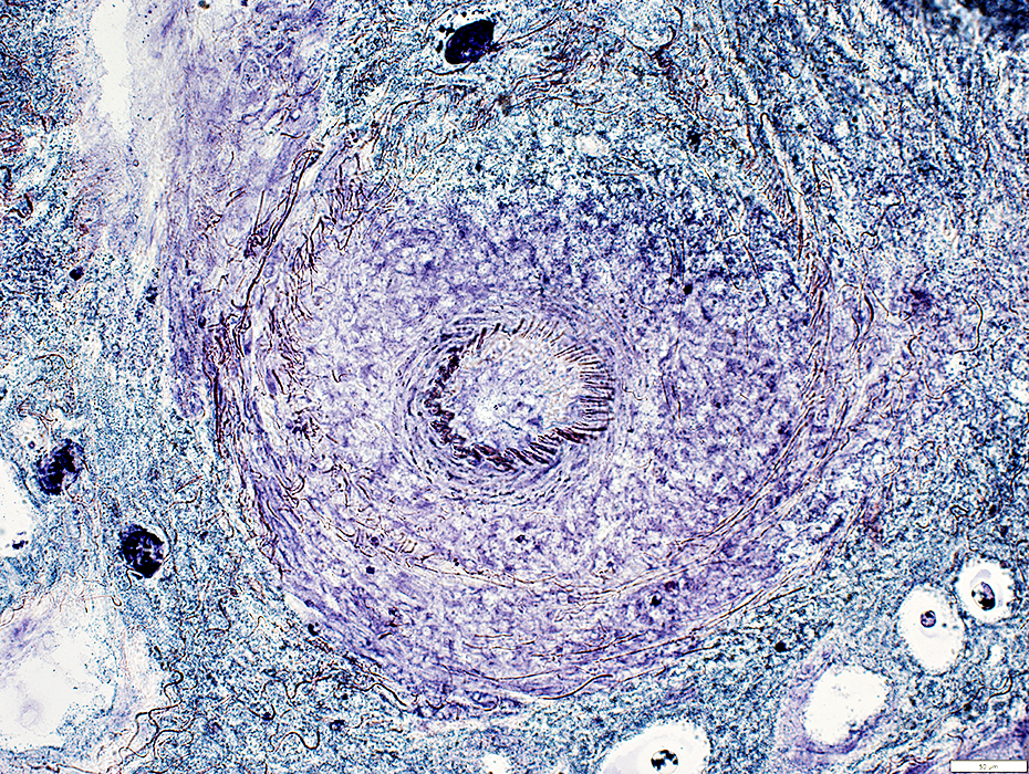
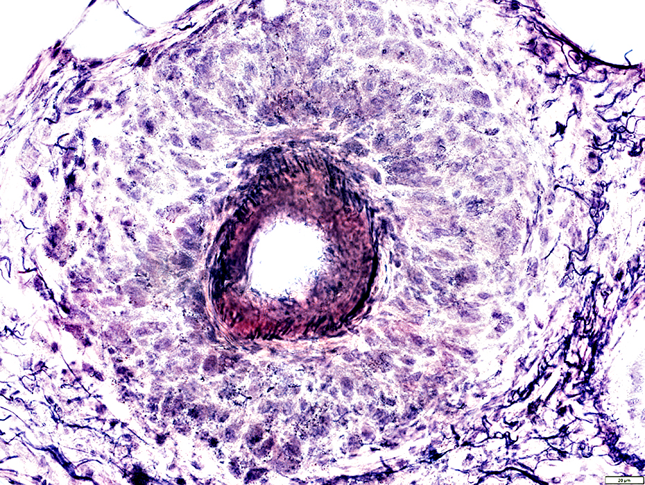
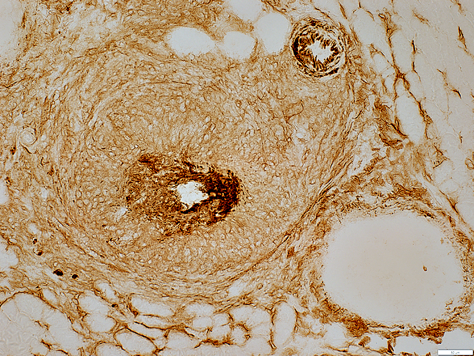
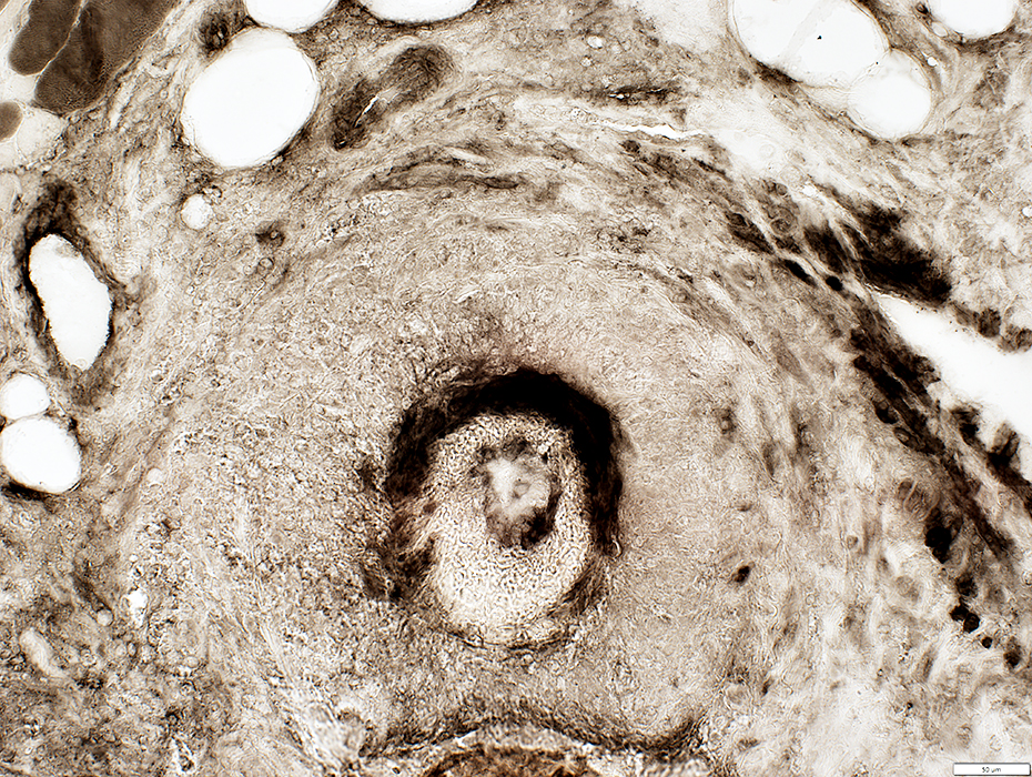
Epineurial Vessel (Artery): Chronic Pathology, Occlusion
Vessel StructureDamaged wall
Elastin layer interrupted & irregular
Irregular ATPase stained smooth muscle
Endothelial cells: Acid phosphatase positive
Lumen: Absent or Contains large cells, possibly endothelium
Neovascularization
Abnormal staining for NCAM
Size: Large at some levels
Calcification: In some regions of vessels wall
Hemosiderin: Present in Epineurium
Epineurial connective tissue Surrounding vessel
Scattered histiocytes
Alkaline phosphatase stained
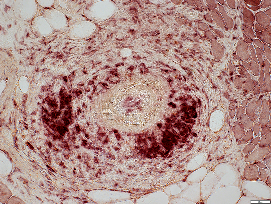
See: Epineurial vessels, control
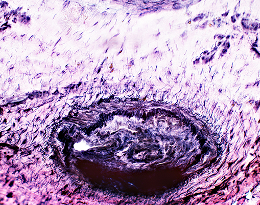 VvG stain |
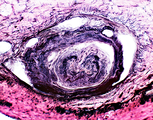 VvG stain |
|
 Acid phosphatase stain |
 Acid phosphatase stain |
 Alkaline phosphatase stain |
 ATPase pH 4.3 stain |
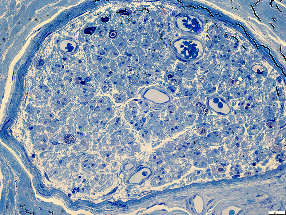
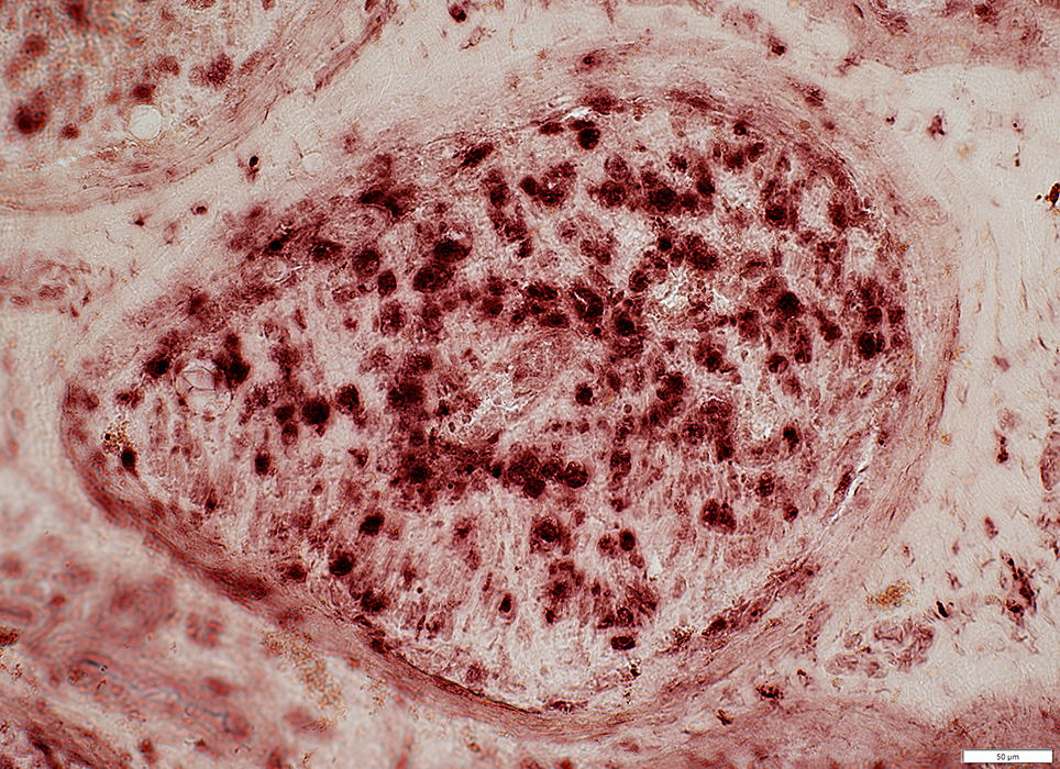
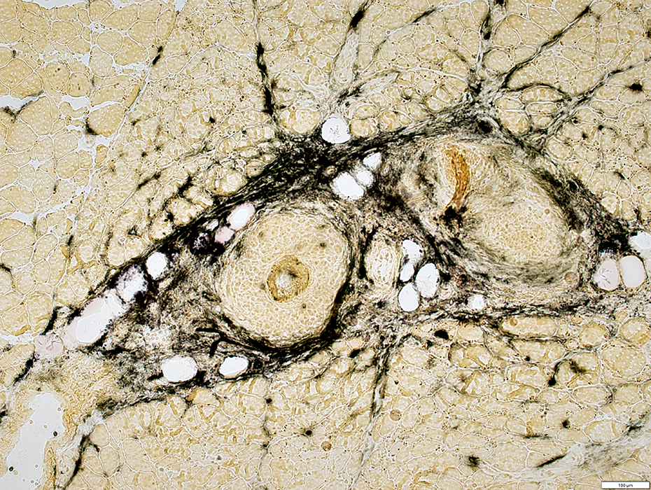
Cryoglobulin/Rheumatoid Vasculitis: Nerve Infarction
 H&E stain |
Endoneurial vessels: Large size & lumen
Epineurium: Increased numbers of small vessels (Above)
Wallerian degeneration: Many Myelin sheaths are abnormally pale-stained (Below)
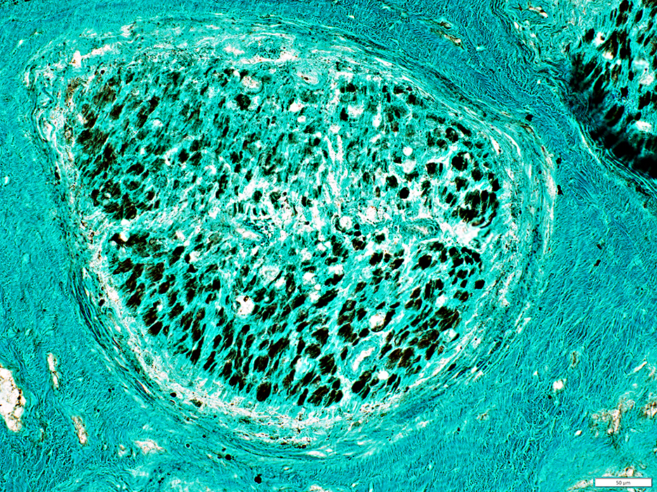
 Gomori trichrome stain |
Endoneurial Vessels: Enlarged with large lumen
 Toluidine blue stain |
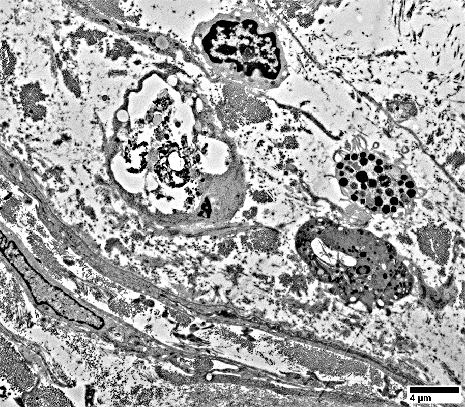
Histiocytes with Lipid droplets: Scattered in endoneurium
Myelin remnants (Ovoids): Scattered in endoneurium
|
Axon Loss
Myelinated Axons: Marked lossEndoneurial Microvessels: Often large; Some dark stained
 VvG stain |
Axons, Large & Small: Nearly complete loss
Perineurium: Thick & Irregular structure
 Neurofilament stain |
Axons, Large & Small: Nearly complete loss in some regions
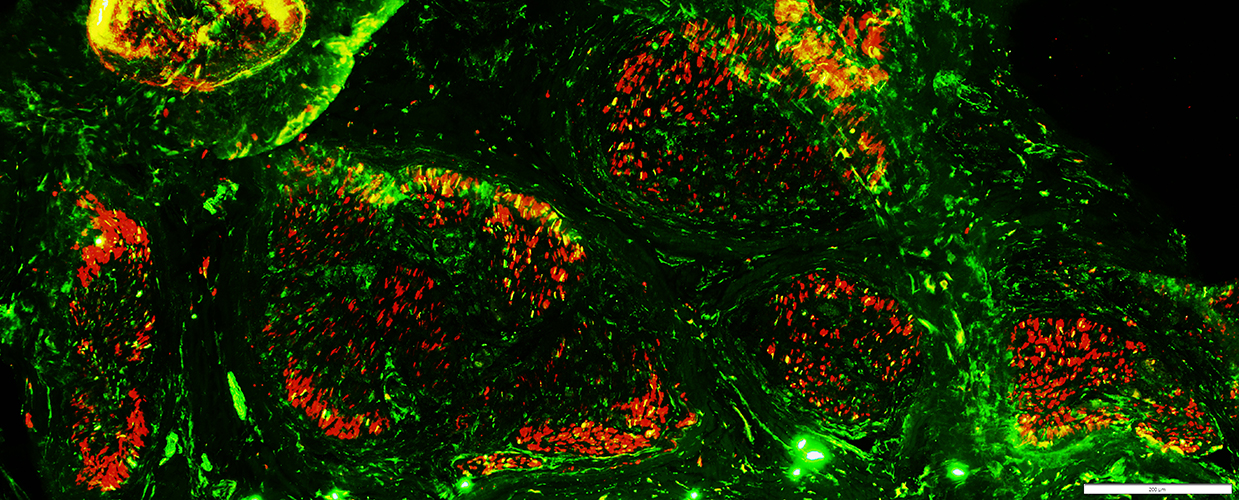 Neurofilament (Green) + NCAM (Red) |
Axons: Complete loss in fascicles with loss of Schwann cells
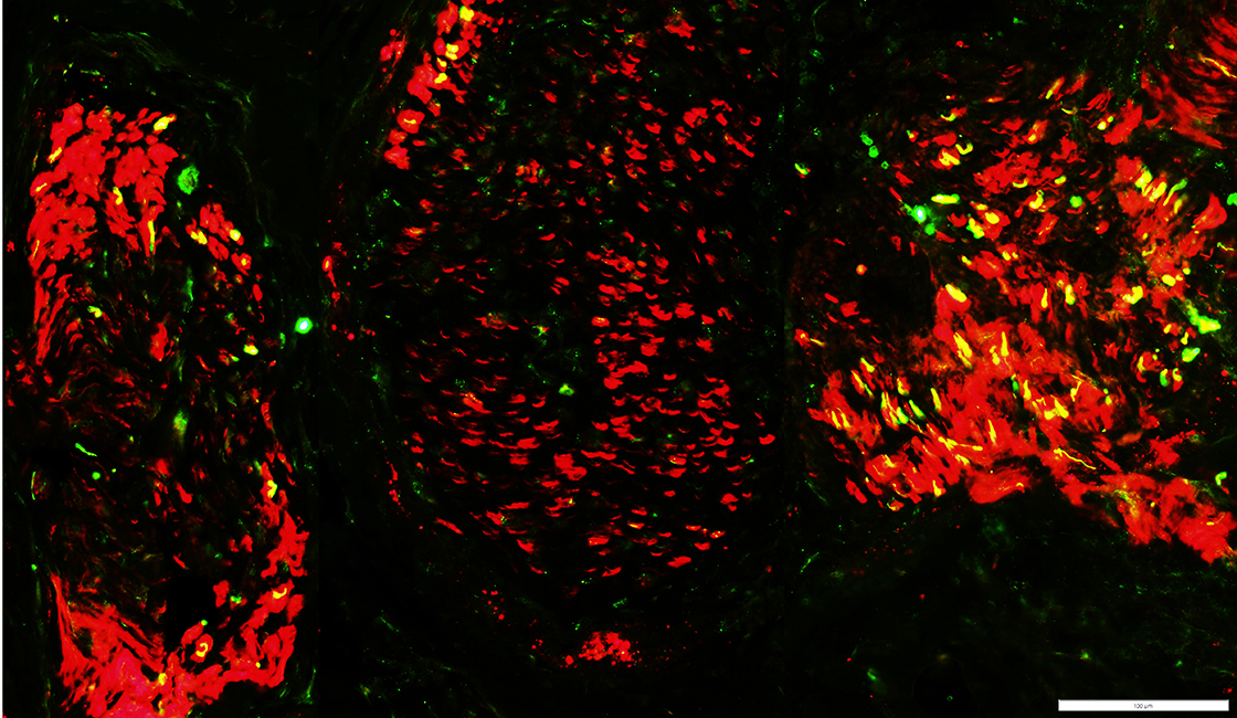 Neurofilament (Green) + NCAM (Red) |
Schwann cell & Myelin Degeneration
Histiocytic cells: Many; Scattered in endoneuriumMyelin sheaths: Abnormal, strong acid phosphatase stain
 Acid phosphatase stain |
Nerve Infarction: Schwann cell loss in focal regions
Schwann cells, Non-myelinating: Reduced in region of endoneurium
 NCAM stain |
Schwann cells (Green & Yellow)
Reduced numbers in region of endoneurium (Left)
Chronic pathology
"Denervated"" SC type: Büngner bands; Most SC co-stain for P0 & NCAM (Yellow)
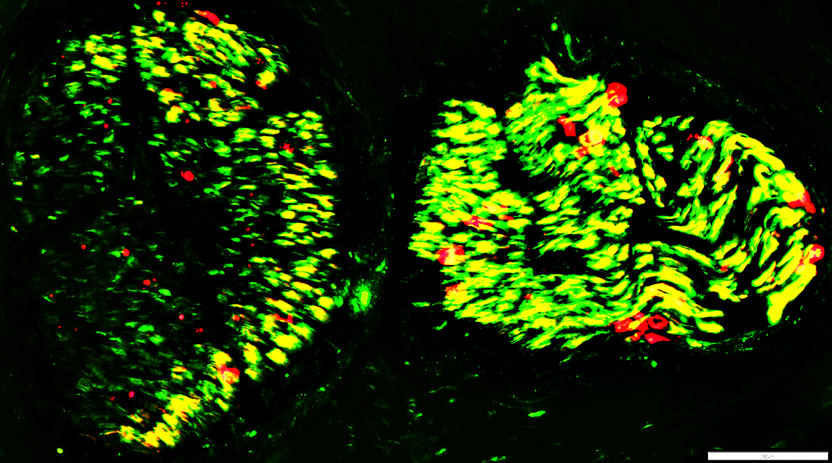 NCAM (Green) + P0 (Red) |
Endoneurial Damage: C5b-9 staining of Endoneurium
C5b-9 stainsEndoneurium: Cells & Connective tissue
Perineurium: Irregular structure; Thick
 C5b-9 stain |
Endoneurial Damage: Hemorrhage
 Toluidine blue stain |
Endoneurial Cells after Axon loss
 From: R Schmidt |
Schwann cells: With myelin & lipid debris
Mast cell
Fibroblast processes
Schwann cells
With Myelin & Lipid debris
Büngner bands
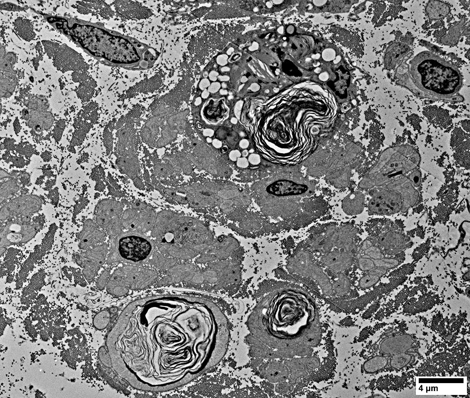 From: R Schmidt |
Epineurial Damage
 Alkaline phosphatase stain |
Stains: Connective tissue around nerve; Epineurium
 Alkaline phosphatase stain |
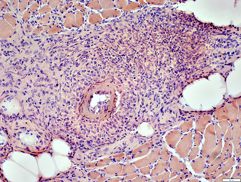
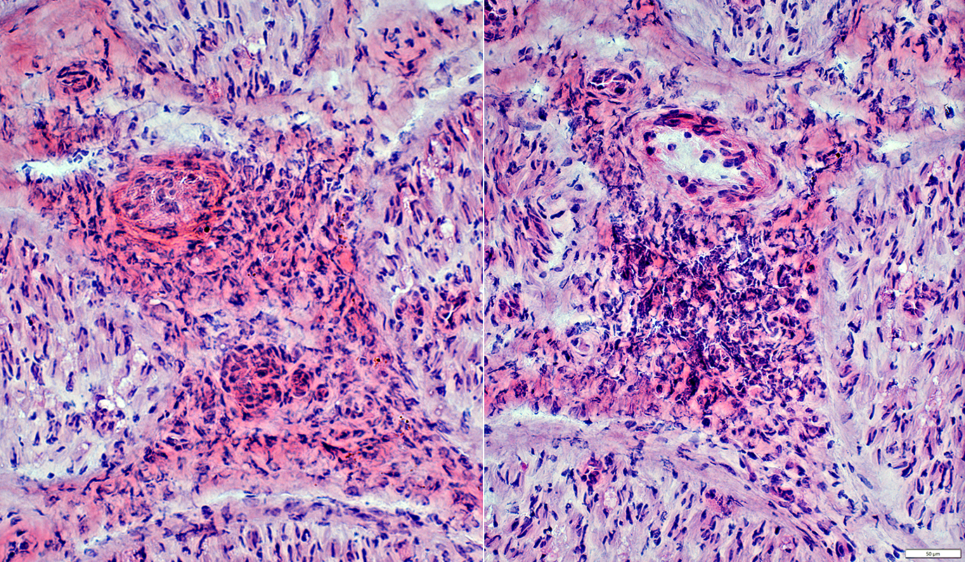
Histiocytic Vasculopathy: Rheumatoid Disorder/IgMκ M-protein/Cryoglobulinemia in Muscle
Vessel Structure: DamagedSee: Perimysial vessels, control
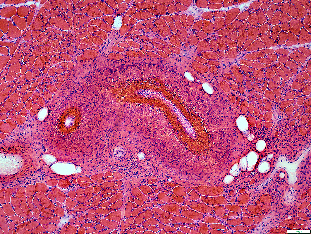 H&E stain |
Size: Very large
Wall Structure
Inner layer: Fibrinoid necrosis
Outer layer: Replaced by large, "palisaded" cells
Lumen: Large cells, possibly endothelium, adherent to inner wall
 H&E stain |
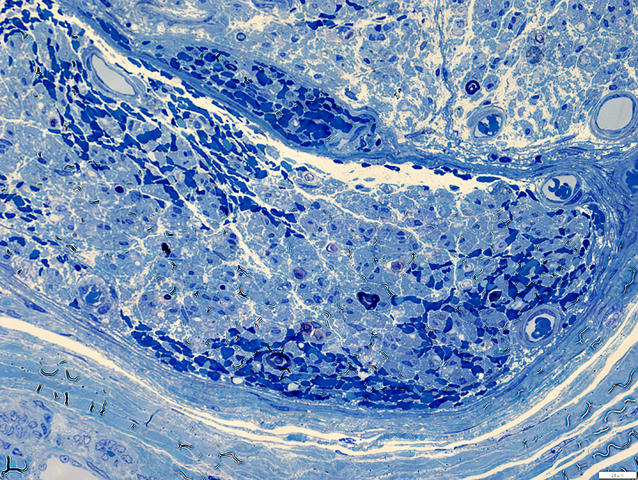
 Gomori trichrome stain |
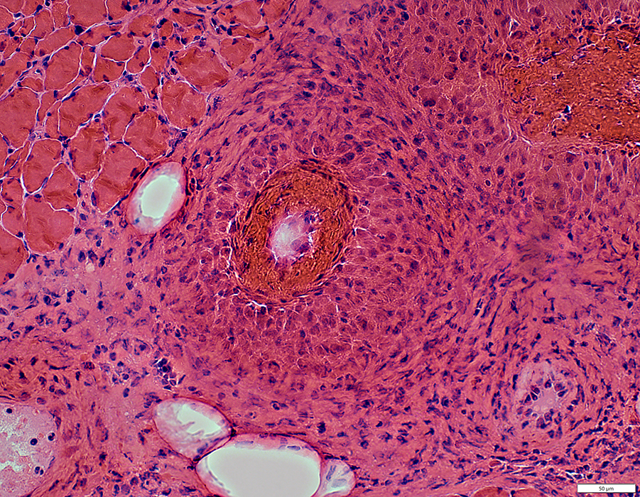
Large cells: Replace wall structure
 Congo red stain |
 AMPDA stain |
Fibrinoid Necrosis: Inside fibril layer
Fibril layer of vessel wall: Irregular
Outer layer: Replaced by large cells
 VvG stain |
Fibrinoid Necrosis: C5b-9 stain
 C5b-9 stain |
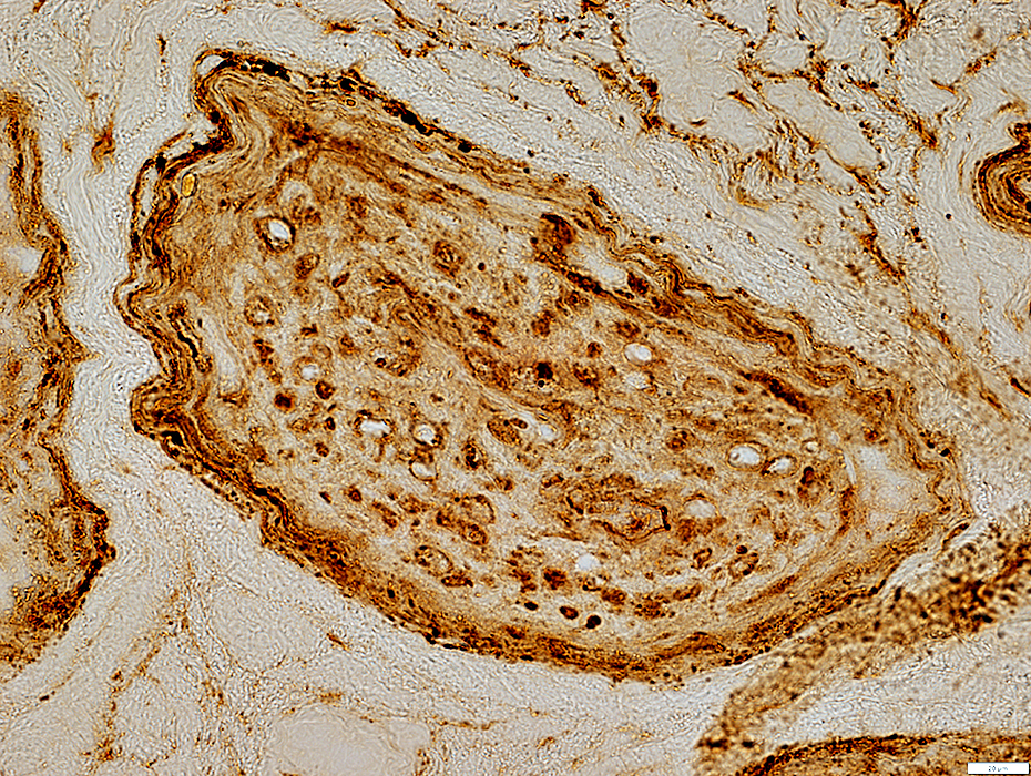
Histiocytic Arteritis
 Acid phosphatase stain |
Replace vessel wall
Nearly all cells are stained
 Acid phosphatase stain |
Esterase stains
Clusters of cells in vessel wall
? Early granulomatous change
 Esterase stain |
Arterial Smooth Muscle Layer: Incomplete & Damaged
 ATPase pH 4.3 stain |
Perimysial Connective Tissue: Alkaline phosphatase stain around damaged artery
 Alkaline phosphatase stain |
Perimysial connective around, & extending away from, damaged artery
 Alkaline phosphatase stain |
Vessel Endothelium Pathology
Strong endothelial stain by NADH
Also some endothelial cell staining on Esterase & Acid phosphatase
 NADH stain |
Vasculopathic Neuropathy with Endothelial Cell Pathology
83 year old female with asymmetric distal weakness & sensory loss in legs and arms progressive over 3 months.Lab testing: Cryoglobulinemia type 2 (Mixed), CREST, IgMκ M-protein; Rheumatoid factor; NfL = 434.
 H& E stain |
 H& E stain |
 Gomori trichrome & Congo red stains |
Return to Neuromuscular Home Page
Return to Pathology index
6/2/2026