Denervated Schwann Cells
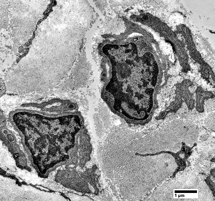
Büngner bands
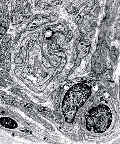
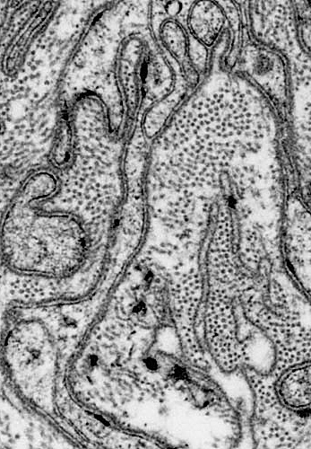
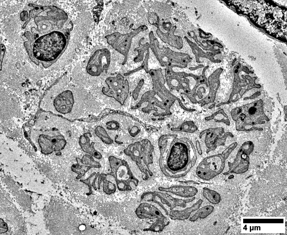
- Bands of interdigitating Schwann cell processes
- Surrounded by basal lamina (Schwann tube)
- Size: > 3 μm
- Schwann cells: Molecular markers
- P0 protein + NCAM: Co-expressed in same Schwann cell processess
- Neurofilament protein (Axon): Not present
- No associated axons
- Basement membrane: Irregular
- Due to
- Loss of Myelinated axons
- Proliferation of associated Schwann cells
- Axon regeneration: Often occurs within Büngner bands
- Course with no axon regeneration
- Schwann cells
- Atrophy
- May develop characteristics of perineurial cells
- Pinocytotic vesicles
- Divide endoneurium into microfascicles
- Denervated: Also present in onion bulbs
- Difference from non-myelinating Schwann cells: Contain P0
- Collagen accumulates
- Schwann cells
- Collagen fibers
- May surround Schwann cell tube
- Thinner (23 to 30 μM than normal endoneurial collagen
- Some nerves with axon loss: Do not develop Büngner bands
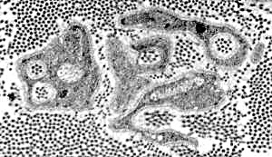
Collagen Pockets
- Due to loss of: Unmyelinated axons
- Non-myelinating Schwann cells (Remak cells) surround: Collagen fibrils
- Proliferation of Remak cells: Response to degeneration of myelinated axons
Büngner Bands
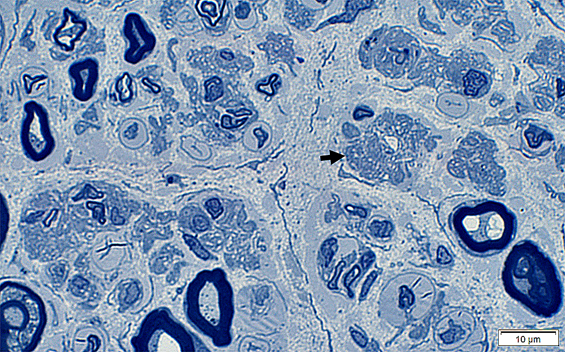 Toluidine blue |
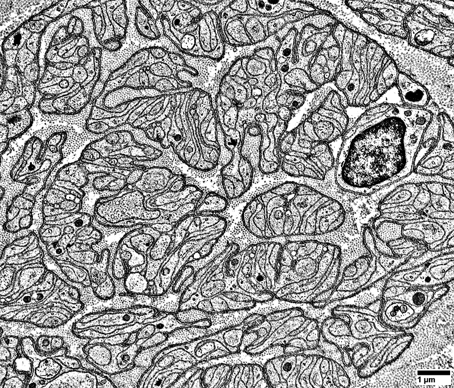 From: R Schmidt |
Clusters of Schwann cell processes are contained within a layer of basal lamina
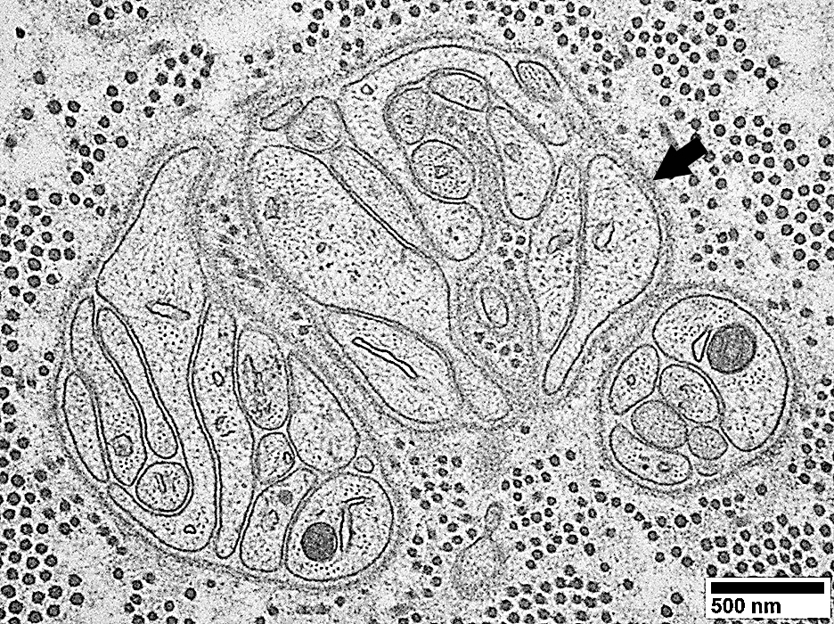 From: R Schmidt |
Schwann cell processes are contained within a layer of basal lamina (Arrows)
Schwann cell processes: Multiple collections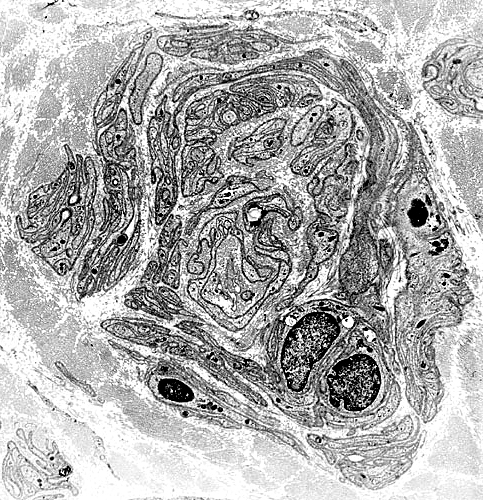
|
Schwann cell processes: Within layer of basal lamina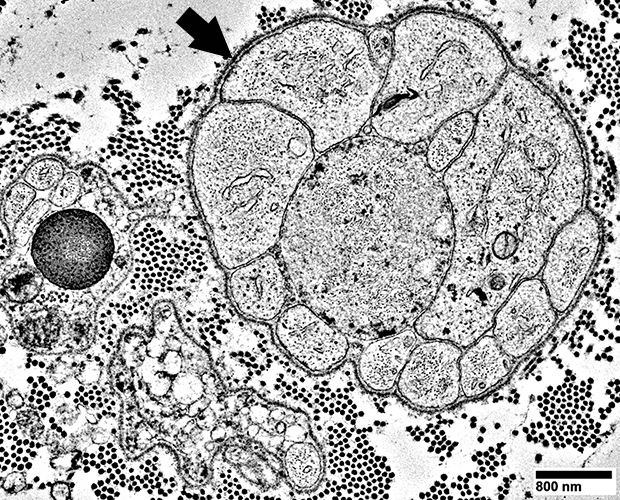
|
|
|
 From: R Schmidt |
 |
Small & Large Axon loss: Severe
 Neurofilaments (Green); P0 (Red) |
Axon loss, Severe: Little staining for large or small axons (Green)
Büngner bands
p0 & NCAM co-localize in Schwann cells with no associated axons (Yellow)
Small NCAM positive cells (Green) are associuated with remaining small, unmyelinated axons
Normal: Many p0 positive myelin sheaths with no NCAM
In nerves with axon loss but no Büngner bands, remaining denervated Schwann cells express NCAM but not P0.
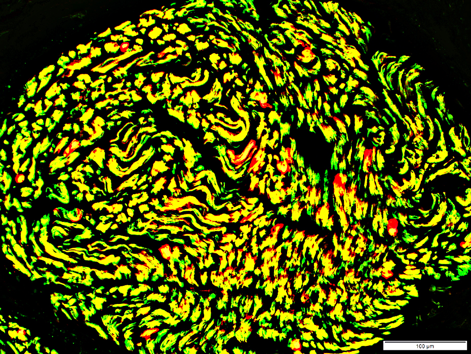 NCAM (Green) & p0 (Red) stains |
Loss of all myelinated axons
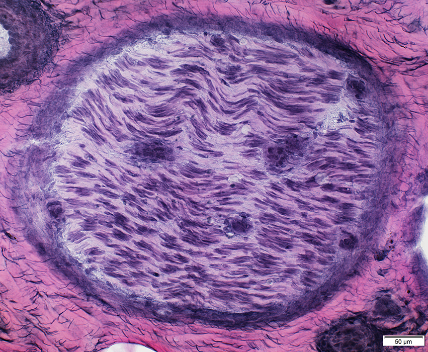
VvG stain |
Loss of all axons
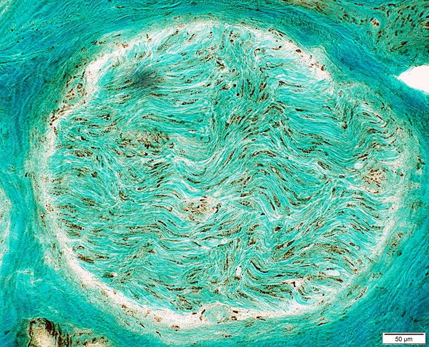 Neurofilament + Gomori stain |
Non-Myelinating Schwann cells (NCAM stain): Remain present all through endoneurium
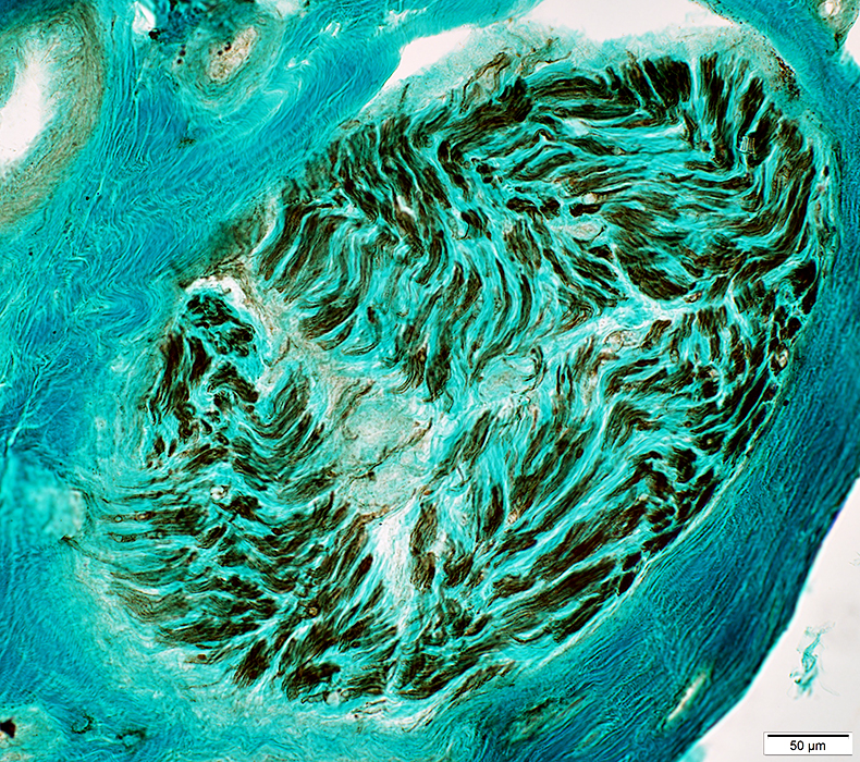 NCAM + Gomori |
Collagen pockets
 |
Return to Axonal loss
Go to Normal nerve
Return to Pathology & Illustrations
Return to Neuromuscular Home Page
2/2/2026