Muscle Fiber Types
Abnormal muscle with many scattered Immature muscle fibers
|
2C fibers Disorders MYH components Control muscle ATPase stains ALS Staining Histochemistry |
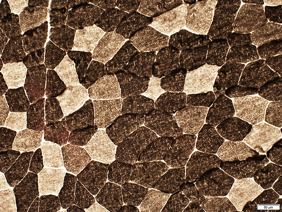
Muscle Fiber Types
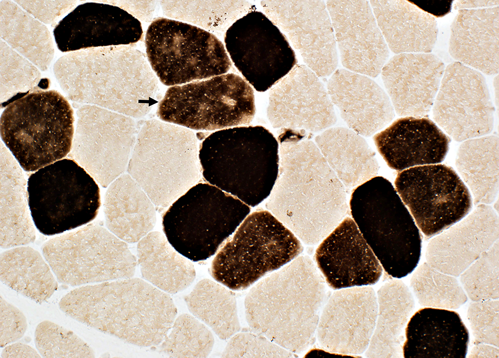 ATPase pH 4.3 |
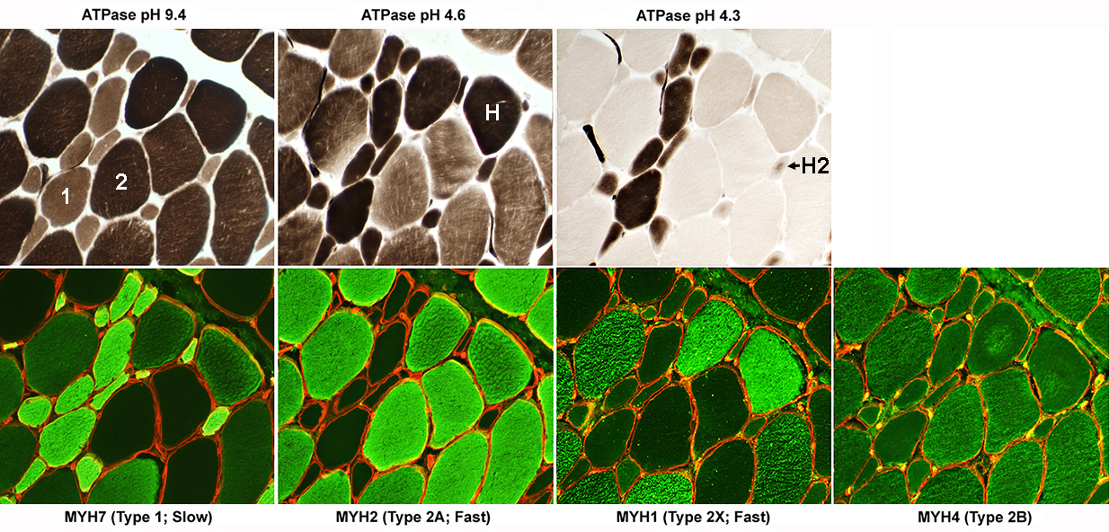
- Type 1 fibers: Dark; Fibers contain MYH7
- Type 2 fibers (Usually): Very pale; Fibers contain MYH2 + other MYH types
- Type 2C fibers (Immature): Intermediate stained (Arrow); Fibers contain MYH7 > MYH2
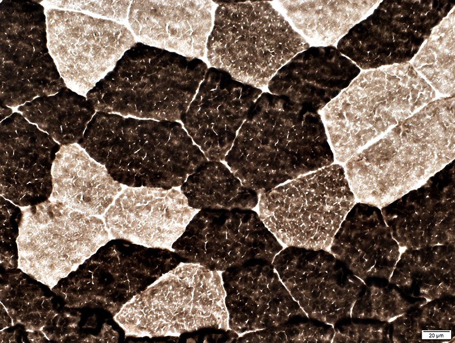
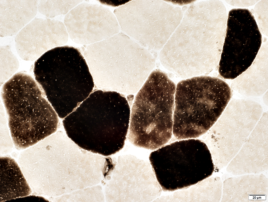 ATPase pH 4.3 stain |
 ATPase pH 4.6 stain |
- Type 1 fibers: Dark
- Type 2A fibers: Pale
- Type 2B fibers: Intermediate
- Type 2C fibers (Immature): Intermediate; Darker than type 2B
 ATPase pH 4.6 stain |
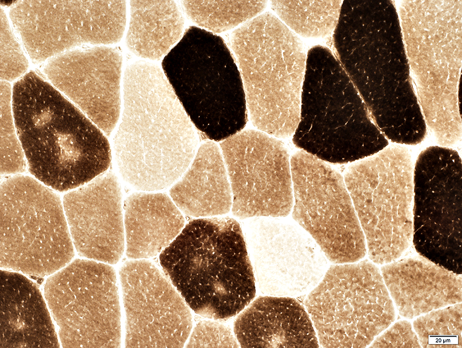
 ATPase pH 9.4 stain |
- Type 1 fibers: Intermediate
- Type 2 fibers: Dark
- Type 2C fibers (Immature): Intermediate; Darker than type 1
 ATPase pH 9.4 stain |
Type 2C (Immature) muscle fibers
- Myosin heavy chain contents: Type 1; Type 2A
- May be associated with
- Development & Young age
- Excess 2C fibers with normal morphology: Coenzyme Q10 deficiency in infancy
- Regeneration
- Reversion of mature muscle fibers to immature state
- CoQ10 deficiency in young children
- Development & Young age
- Morphology: Variable
- Normal size & Internal architecture
- Small size: May occur with denervation & reinnervation
- Muscle fibers with other histological features of immaturity: Regeneration after muscle fiber damage
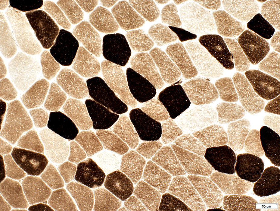
Type 2C muscle fibers: Increased numbers (Same muscle biopsy as above) with normal fiber morphology
Fiber size: Mild variation
No regenerating, or morphologically immature, muscle fibers
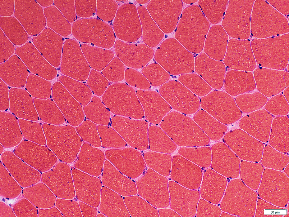 H&E stain |
 VvG stain |
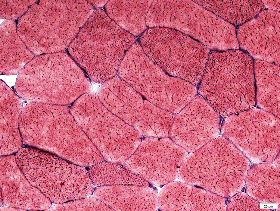
Unremarkable
Rare internal nucleus
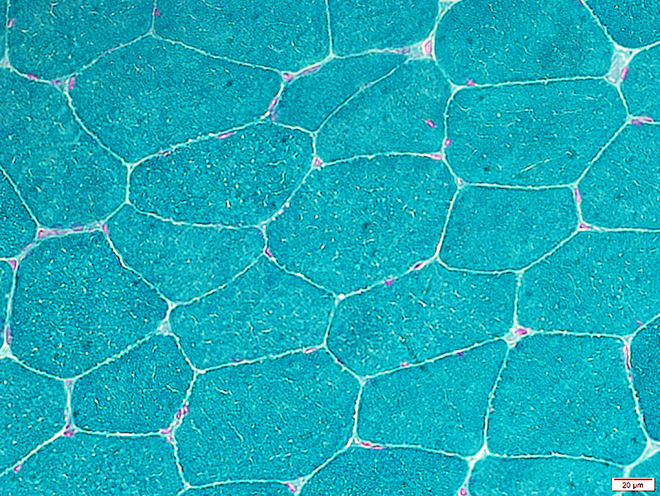 Gomori trichrome stain |
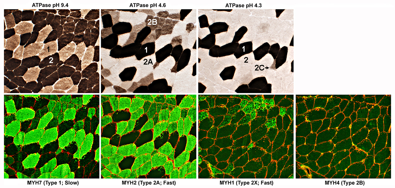
MYH stains = Green; Collagen IV = Red
ALS Muscle: Fiber Types
Type 1 fibers: NormalATPase: Intermediate color at pH 9.4 & Dark at pH 4.6 & 4.3
All have MYH7, but not MYH2 or MYH1
Type 2 fibers: Variant
All have MYH2 & moderate MYH7
Type 2A fibers
All have MYH2 & Moderate MYH7, but no MYH1
ATPase: Dark at pH 9.4; Pale at pH 4.6 & 4.3
Type 2B fibers
All have MYH2 & Moderate MYH7 & MYH1
ATPase: Dark at pH 9.4; Intermediate at 4.6; Pale at pH 4.3
Type 2C fibers
All have MYH2 & MYH7, but no MYH1
ATPase: Moderately Dark at pH 9.4 & 4.6; Internediate at pH 4.3
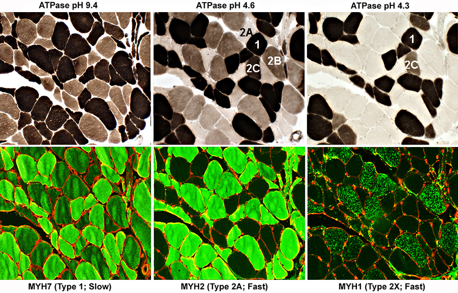 MYH stains = Green; Collagen IV = Red |
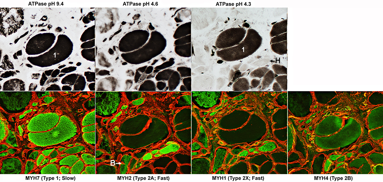 MYH stains = Green; Collagen IV = Red |
 MYH stains = Green; Collagen IV = Red |
Return to Neuromuscular Home Page
Return to Pathology index
2/18/2025