Coenzyme Q10 Deficiency: Childhood Pathology
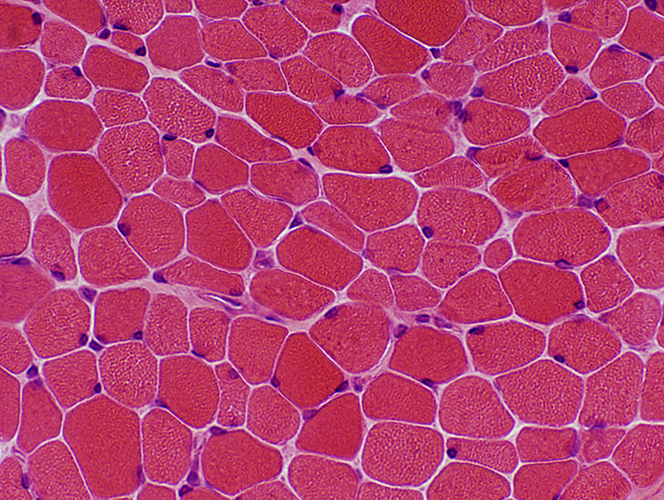 H & E stain Muscle fiber sizes: Varied 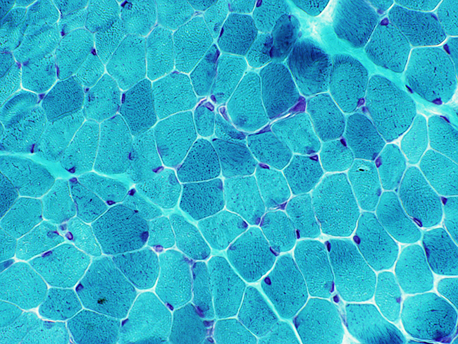 Gomori trichrome stain Muscle fiber morphology: Normal 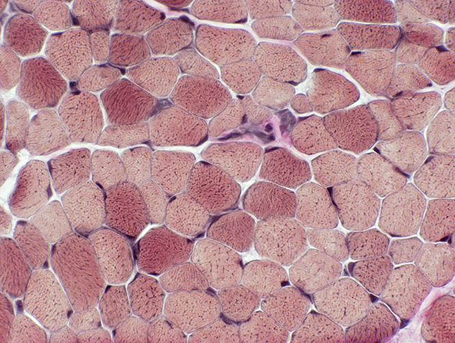 VvG stain |
|
|
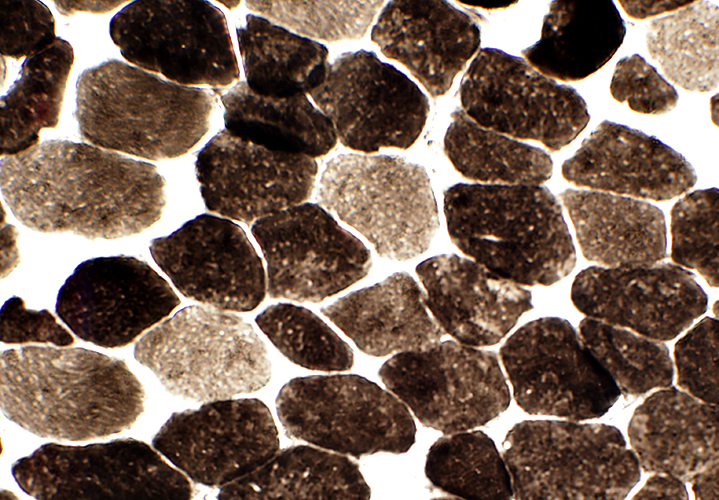
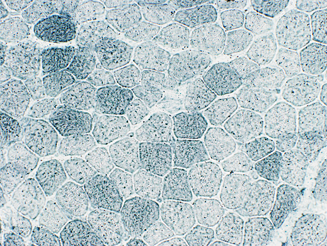
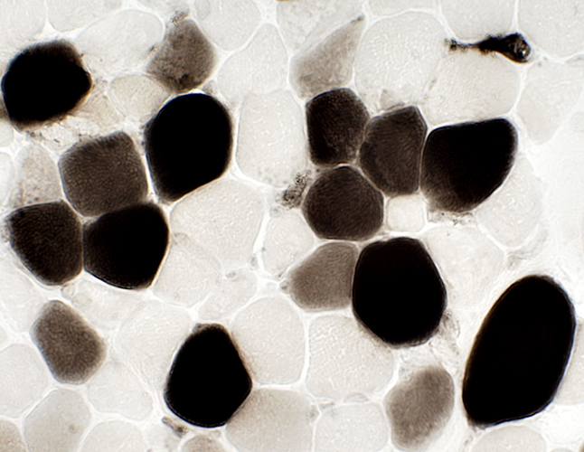 ATPase pH 4.3 stain Muscle fiber types: Excessive numbers of type 2C (Intermediate stained, Above) 1 |
|
|
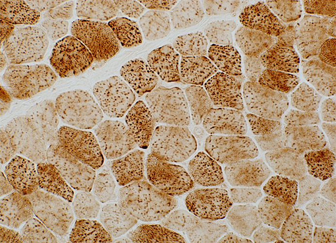
 ATPase pH 9.4 stain Muscle fiber types: Mild type 2 predominance |
|
|
 COX stain Mitochondria: Mildly enlarged |
|
|
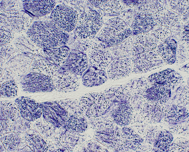 SDH stain Mitochondria: No proliferation |
|
|
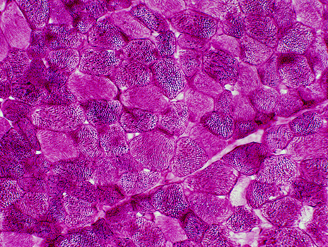 PAS stain Glycogen: Increased in muscle fibers |
|
|
 Sudan stain Lipid: Normal in muscle fibers |
Return to Mitochondrial pathology
Return to Mitochondrial syndromes
Return to Muscle biopsies
References
1. Muscle Nerve 2013 Mar 14
8/21/2013